The Success of Topical Treatment of Onychomycosis Seems to Be Influenced by Fungal Features
←
→
Page content transcription
If your browser does not render page correctly, please read the page content below
Hindawi
Evidence-Based Complementary and Alternative Medicine
Volume 2021, Article ID 5553634, 7 pages
https://doi.org/10.1155/2021/5553634

Research Article
The Success of Topical Treatment of Onychomycosis Seems to Be
Influenced by Fungal Features

          Vanessa Vasconcellos-Pontello, Flávia Franco Veiga , Marina Cristina Gadelha,
          Marielen Ribeiro , Melyssa Negri , and Terezinha Inez Estivalet Svidzinski
          Departamento de Análises Clı́nicas e Biomedicina, Centro de Ciências da Saúde, Universidade Estadual de Maringá (UEM),
          Avenida Colombo, 5790, Maringá, PR, CEP 87020-900, Brazil

          Correspondence should be addressed to Terezinha Inez Estivalet Svidzinski; tiesvidzinski@uem.br

          Received 21 January 2021; Revised 20 April 2021; Accepted 3 July 2021; Published 10 July 2021

          Academic Editor: Yoshiki Mukudai

          Copyright © 2021 Vanessa Vasconcellos-Pontello et al. This is an open access article distributed under the Creative Commons
          Attribution License, which permits unrestricted use, distribution, and reproduction in any medium, provided the original work is
          properly cited.

          Aim. To evaluate the topical treatment of onychomycosis using a 10% hydroalcoholic propolis extract (PE) in two aleatorily
          chosen patients and analyze possible risk factors from hosts including some particularities of the isolated fungi that may justify
          the outcomes achieved. Materials and Methods. A topical treatment, with PE, was started in two cases of toe onychomycosis due
          to T. rubrum. The in vitro PE antifungal activity against these isolates was confirmed. Moreover, the ability of the fungi to infect
          the human nail was evaluated also in an ex vivo study, analyzed by histopathology. Results. Within four months, both patients
          showed evident improvement, but with different outcomes. The possible host-related risk factors justifying the poorer outcome
          in patient 1 include a longer duration time of onychomycosis (50 years). Some particularities in the T. rubrum strain isolated
          from this patient in relation to that found in patient 2 were observed: (1) the hypha morphology suggesting a major adaptation
          of the fungus to the host; (2) a 16 times greater propolis concentration was required in vitro; and (3) a faster ability to start a
          growth using the nail as the only nutritional source. Additionally, this isolate was more efficient in producing a biofilm on the
          nail surface. Conclusions. A partial clinical and complete mycological cure for the two patients was achieved after four months
          of PE daily use. Despite a complete recovery, a different outcome was observed between both cases. A more persistent
          onychomycosis, added to greater fungal potential to produce biofilm on the nail, seems to influence greatly the success of a
          topical treatment with PE.

1. Introduction                                                             In this context, new topical compounds have been de-
                                                                        veloped and tested, but cure rates are still less than desirable
Onychomycosis is the most common nail pathology in                      [8]. Thus, it is necessary to develop a safe topical treatment,
the world [1] mainly affecting people aged over 60 years                 which has few side effects, has good nail permeation ca-
[2, 3] and significantly impacting the quality of life [4].              pacity, and is effective against the main onychomycosis-
Despite its great relevance, the therapeutic options                    causing microorganisms [9]. Recently, propolis has been
available for onychomycosis are limited, have few                       considered among the new therapeutic options in the
pharmacological alternatives, and are slow and costly.                  management of superficial fungal diseases including ony-
Moreover, cure rates for onychomycosis are often low                    chomycosis [10]. In fact, propolis has exhibited suitable
and relapses are frequent [2]. Systemic terbinafine is                   results in this context, and the authors proved that ethanol
considered the gold standard for onychomycosis treat-                   propolis extract (PE) is a topical therapeutic option for
ment [5, 6]. According to some authors, the topical                     onychomycosis, in a translational study from in vitro to the
administration of drugs is preferred over the systemic                  clinics. Antifungal activity against the planktonic cells and
route as it has fewer side effects [3, 7].                               biofilm formed by Trichophyton spp. was clearly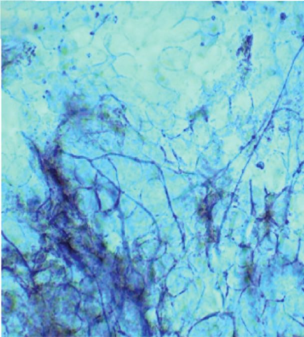
2                                                                   Evidence-Based Complementary and Alternative Medicine

demonstrated, besides the low cytotoxicity and the capacity        Collections of the Paraná Network (Taxonline) at the Federal
of PE to penetrate human nails. However, the performance           University of Paraná as T. rubrum CMRP2912 and
of this product in patients needs to be further investigated       T. rubrum CMRP2918, respectively.
since the results, although promising, were heterogeneous,
providing from partial or complete cure to no improvement
                                                                   2.3. Treatment Application and Follow-Up. The first orien-
[11]. Thus, the objective of the current study was to evaluate
                                                                   tation was made just after the laboratory conclusion when
the topical treatment of onychomycosis using PE in two
                                                                   the treatment was started. The lesions in the affected area
aleatorily chosen new patients and analyze possible risk
                                                                   were treated topically, exclusively with two drops, twice a
factors from hosts besides some particularities of the isolated
                                                                   day of PE. Both patients were instructed to clean their nails
fungi that may justify the outcomes achieved.
                                                                   with soap, water, and a brush daily and to polish the affected
                                                                   areas of the nails weekly.
2. Materials and Methods                                               The follow-up included monthly interviews besides nail
                                                                   samples collection being conducted 1 month and 4 months
2.1. Ethical Aspects and Patients’ History. This study was         later aiming to evaluate the clinical and laboratory outcomes.
approved by the Ethics Committee of the State University of
Maringa (approval number 1246516), according to resolu-
tion no. 466/12 of research involving human beings and with        2.4. Propolis In Vitro Susceptibility Testing. Some laboratory
the Declaration of Helsinki under ethical principles for           tests were performed on the clinical isolates obtained from
medical research involving human beings.                           the two patients, T. rubrum CMRP2912 and T. rubrum
    Patient 1 is a 69-year-old male with no known under-           CMRP2918, using the reference T. rubrum ATCC 40051
lying disease; his medical history includes abnormal fasting       strain. Each test was performed in duplicate and in two
glycemia and, therefore, did not receive medication. He has        independent experiments.
had nail injury in both feet compromising one hallux and the           The in vitro susceptibility test with the PE used in the
adjacent skin (tinea pedis) for approximately 50 years; his        patients was performed by the microdilution broth method
nail bed was thickened and associated with nail dystrophy.         described previously [11]. The serial dilutions of PE ranged
He had been treated with oral medication in the past but           from 17,500.00 to 34.17 μg/mL of total polyphenols present
without clinical cure. Patient 2 was a 65-year-old female with     in PE. Three controls were included: positive (fungal ino-
chronic hypertension on continuous medication. She pre-            culum + culture medium), negative (culture medium only),
sented a toe-nail damage with keratosis and associated             and alcohol (fungal inoculum + ethyl alcohol at a concen-
dystrophy for at least six years. This patient had been treated,   tration equivalent to that in PE). All preparations were
in another service, with topical terbinafine but without            incubated at 37°C for 48 h to determine the MIC for PE
improvement.                                                       against the fungal strains. To complement this result, the
    These patients were selected from a bigger project that        minimum fungicide concentration (MFC) was determined
included our previous publication [11]. Both gave their            by placing aliquots of MIC test preparations (fungi + PE) on
written consent for the treatment of onychomycosis only by         SDA plates.
a topical application of a 10% hydroalcoholic propolis ex-
tract, made in ethanol (PE), provided by the UEM school            2.5. The Nail Invasion by an Ex Vivo Study. The ability of each
pharmacy.                                                          of the fungal isolates to invade the nail plate was evaluated
                                                                   using nails obtained from healthy adult volunteers. The nails
                                                                   were manually cut into fragments of similar size and then
2.2. Nail Sampling and Microbiological Analysis. After
                                                                   autoclaved at 121°C for 20 min. An inoculum of 1 × 10⁷ CFU/
clinical evaluation by a dermatologist, nail samples with
                                                                   mL (colony forming units per milliliter) of each fungus was
clinically suspected onychomycosis were collected by
                                                                   prepared, and 500 µL of each suspension was incorporated in
scraping. This material was sent to the Laboratory of Medical
                                                                   a proportion of 1 : 100 into the yeast nitrogen base without
Mycology, UEM, for processing.
                                                                   amino acids agar (YNB agar, BD DifcoTM Detroit) while it
    Mycological diagnosis was performed in two stages:
                                                                   was still liquefied. This preparation did not contain any
direct mycological examination with 20% KOH and 0.5%
                                                                   nutritional source. After agar solidification, sterilized
Evans blue to clarify the material and observation of the
                                                                   healthy nail fragments were introduced. The cultures were
sample through light microscopy. The samples were also
                                                                   incubated at 25°C and inspected daily for three weeks. The
                                                    ™
spread on sabouraud dextrose agar (SDA) (Difco Detroit)
                                                                   time needed for each isolate to grow was evaluated, and the
                                                   ™
and selective agar for pathogenic fungi (Difco Detroit),
with nine inoculations in each type of culture medium. The
                                                                   macroscopic and microscopic aspects of the growing col-
                                                                   onies were studied.
cultures were incubated at 25°C for 30 days to allow fungus
growth, with daily evaluation to verify the growth rate. The
identification of each isolated fungus considered growth            2.6. Histopathologic Analyses on the Fungal Growth into the
time, uniformity of the colonies grown, macro and micro-           Nail. The infected nail fragments were embedded in par-
morphology, and biochemical test results [12]. In both cases,      affin, and continuous 4 micrometer sections were made.
T. rubrum, an anthropophilic dermatophyte fungus, was              They were placed on glass slides and analyzed by histopa-
identified. The isolates were deposited in the Microbial            thology processing methods. The sections were stained with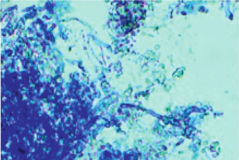
Evidence-Based Complementary and Alternative Medicine                                                                           3

periodic acid–Schiff (PAS) and Grocott’s methenamine                fasting glycemia (prediabetic status), another risk factor
silver (GMS) followed by the analysis by light microscopy in       associated with low therapeutic response of onychomycosis.
100× and 400× magnifications over the entire length of the              The current study investigated, for the first time, some
nail.                                                              possible intrinsic differences between the fungal isolates
                                                                   from the two patients, which could explain the differences
3. Results and Discussion                                          found in the therapeutic outcomes.
                                                                       First, differences in the morphology of the fungi were
The treatment of onychomycosis is still a challenge [8] and        observed through direct mycological examination. Samples
needs to be improved since it significantly influences               obtained through scrapings of the nails of patient 1 showed
people’s quality of life [4]. Studies have shown that the          long, thin, regular hyphae in large quantities (Figure 1(c));
combination of topical and oral treatment provides the             while, in samples from patient 2, there were shorter hyphae
best results [13], but in some groups, such as older people,       and in small quantities (Figure 1(d)). These differences in the
an effective topical treatment would be more interesting.           fungal morphology could be considered as a result from
The current research searched for some possible variables          different levels of adaptation of the fungus to the host,
that could interfere with the outcome of a treatment of            influencing the outcome of topical treatment. The hyphal
onychomycosis exclusively made with topical PE. The                morphology in patient 1 suggests a complete adaptation of
clinical and direct mycological findings for onychomy-              the fungus to the host, with possible development of tol-
cosis in the two patients before treatment with PE are             erance. In contrast, the hyphal morphology in material from
shown in Figure 1.                                                 patient 2 suggests a host resistance against the fungus, which
    After one month, none of the patients complained of            would contribute to greater fungal vulnerability to topical
discomfort with the medication (burning, erythema, or ir-          medication.
ritation). Only nail appearance was affected by a dark-yel-             The in vitro susceptibility of fungal isolates to propolis
lowish color of the extract (Figure 2). However, little clinical   also was investigated, as shown in Figure 4; the MFC of PE
improvement was observed, and upon direct mycological              was 546.87 μg/mL for the fungus isolated from patient 1 and
examination, many fungal structures were still observed in         34.17 μg/mL for the fungus from patient 2. Interestingly, the
the lesion scrapings. After four months of treatment, there        in vitro response of each strain was consistent with the
were mycological cure and partial clinical improvement of          respective performance by the each patient, i.e., the fungus
the nail lesions (approximately 60% and 70%, respectively)         found to be more sensitive in vitro was more responsive to
with apparent growth of a healthy underlying nail in both          therapeutic treatment. Therefore, these results confirm that
patients (Figure 3). These cure rates are close to those re-       these two fungal isolates had different responses to propolis,
ported with synthetic drugs [5, 13, 14], and therefore, our        similarly to those found with classic antifungals [16].
results were considered promising.                                     Additionally, an ex vivo study was addressed aiming to
    Given the different clinical responses, we decided to look      better understand the interaction between the fungi and nail
in depth searching among the characteristics of the two            simulating a fungus/host relationship. Figure 5 shows that,
patients and their respective fungal isolates possible expla-      within 10 days, the three isolates (two clinical and one
nations for the initial therapeutic response. T. rubrum is the     reference strain) started growing using the nail as the only
most frequent agent of onychomycosis [15]; it does not             nutritional source. Histological sections of the infected nails
respond well to conventional antifungals [16]. It may cause        in this time, stained with GMS, confirmed the presence of
both uncomplicated and complicated cases, including the            hyphae inside the nails. T. rubrum isolated from patient 1
dermatophytomas [17], making difficult in achieving satis-           grew faster on the nail fragments and, interestingly, pro-
factory clinical treatment [6].                                    duced a component suggestive of an extracellular matrix
    During the four months of follow-up, the two patients          (ECM) and therefore a supposed biofilm (arrow). Biofilm
adhered to the treatment, but the response of patient 2 was        formation would probably be related to a dermatophytoma
better. Indeed, women usually achieve more success in the          found in some patients [17].
treatment of onychomycosis [3]. However, this study pro-               This finding is fundamental to understand the com-
poses to investigate other variables besides sex, which might      plexity of case 1. Onychomycosis has been associated with
result in different outcomes of treatment with PE.                  the ability of fungi to naturally organize themselves into a
    Initially, patient 1 also presented tinea pedis, a known       biofilm [18] and produce an ECM, as suggested in Figure 5,
complication that may also influence onychomycosis cure             which makes the permeation of antifungal drugs difficult. In
[3]. This issue was resolved with the use of the same PE           this case, the longer onychomycosis time of patient 1 (50
applied on the affected skin, resulting in complete resolution      years) could explain its more difficult eradication. Of the
of tinea pedis (Figure 3). The too long duration of ony-           note, T. rubrum isolated from patient 2 (better outcome) was
chomycosis patient 1 (50 years) must also have negatively          not able to produce biofilm on the nail in 10 days.
impacted the therapeutic response. Patient 2 also had                  Another issue would be the fungal ability to invade the
onychomycosis for a long time (6 years), but significantly          nail; after three weeks of incubation, all fungi showed visible
shorter. Although there are few reports in this regard, it is      macroscopic growth on the nails. There was increased and
reasonable to imagine that lesions existing for such long          consolidated mycelial growth with time (Figure 6). Histo-
durations are difficult to eradicate. In addition, patient 1,        pathological analysis by PAS confirmed that all fungi pen-
although not yet diagnosed with diabetes, had impaired             etrated the nail fragments and caused an ex vivo infection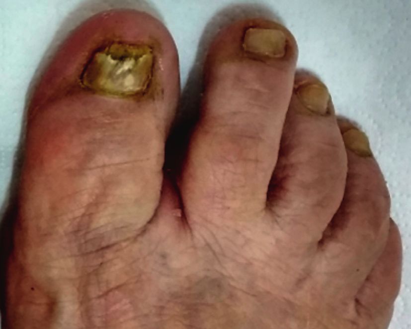
4 Evidence-Based Complementary and Alternative Medicine
(a) (b)
(c) (d)
Figure 1: Clinical and laboratory findings (obtained through direct mycological examination) for onychomycosis in the two patients before
treatment with propolis extract. Patient 1 (a) and (c). Patient 2 (b) and (d).
(a) (b)
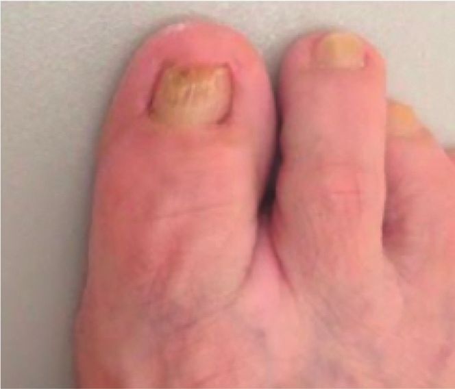
Figure 2: Clinical findings regarding the nails of the two patients after one month olf treatment.
(a) (b)
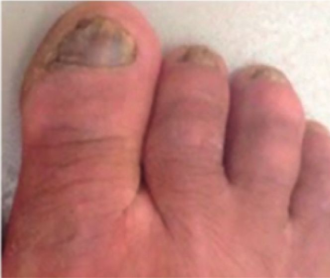
Figure 3: Nails treated with propolis extract for four months. (a) Patient 1 showed a clinical response of around 60%, and (b) patient 2
showed a better clinical response than patient 1, approximately 70%. Nail scrapings of both patients no longer showed the presence of fungal
structures (indicating mycological cure).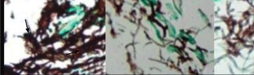
Evidence-Based Complementary and Alternative Medicine 5
μg/mL
C
34.17
68.35
136.71
273.43
546.87
1093.75
2187.50
4375.00
8750.00
17500.00
Patient 02
alcohol
Patient 01
propolis
extract
Patient 02
propolis
extract
Figure 4: Minimum fungicidal concentration (MFC) of the propolis extract (PE) for Trichophyton rubrum isolated from two onycho-
mycosis cases. Line 1 shows that ethanol (used as a propolis diluent) did not interfere with fungal growth. Line 2 shows the performance of
PE against the isolate from patient 1 (MFC � 546.87 μg/mL of total polyphenols present in PE). Line 3 shows the performance of the isolate
from patient 2 (MFC � 34.17 μg/mL of total polyphenols present in PE). C (control) � fungal inoculum without propolis or alcohol.
T. rubrum
CMRP2912 CMRP2918 ATCC 40051
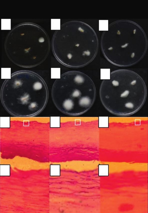
Figure 5: Illustration of the invasiveness of Trichophyton rubrum on a healthy human nail in ten days. CMRP2912: isolated from patient 1;
CMRP2918: isolated from patient 2. T. rubrum ATCC 40051. Topline: macroscopic aspects of fungal growth in nail fragments. Bottom line:
features of fungal invasion as seen under a light microscope. Histological sections of nails stained by GMS, at 400 ×.
(Figures 6(g)–6(i)). It was also clear that the macroscopic Finally, despite all the factors associated with the poorer
growth, in the third week, was similar for the three strains outcome of patient 1, it is important to highlight a sig-
evaluated. The results shown in Figures 5 and 6 allow us to nificant improvement after four months of exclusively
conclude that the two fungal isolates have the ability to grow topical treatment with PE. In fact, the capacity of PE to act
on the nail without another nutritional source. Furthermore, effectively on human pathogenic fungi [19, 20] has been
the fungus from patient 1 interacted more easily with the previously confirmed for fungal biofilms [21, 22] and for
substrate (nail), probably explaining the lower therapeutic the treatment of onychomycosis caused by opportunistic
response. fungi [23].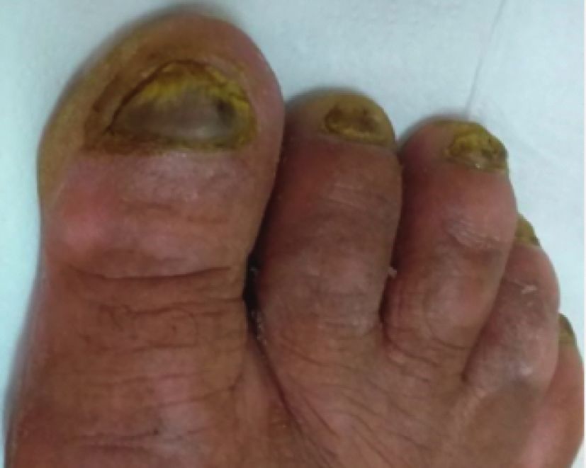
6                                                                            Evidence-Based Complementary and Alternative Medicine

                                                                 T. rubrum
                             CMRP2912                            CMRP2918                               ATCC 40051

                     (a)                                  (b)                               (c)

                     (d)                                  (e)                                 (f)

                    (g)                                  (h)                                (i)

                     (j)                                 (k)                                (l)

Figure 6: Macroscopic aspects of fungal growth on healthy nail fragments over two (a), (b), (c) and three weeks (d), (e), (f ). Histological
sections of ex vivo infected nails and PAS-stained nails, showing that hyphae penetrated the nails, at 400x (g), (h), (i) and 1000x (j), (k), (l).

4. Conclusions                                                             cure and partial clinical cure of the two patients with mild to
                                                                           moderate onychomycosis were observed after four months
This study confirmed the efficiency of a hydroalcoholic                       of daily use, twice a day.
extract of propolis for the topical treatment of onychomy-                     The different performance of PE seems to be related to
cosis, without the need for facilitating vehicles. Mycological             factors of the host such as lesion time and comorbidities.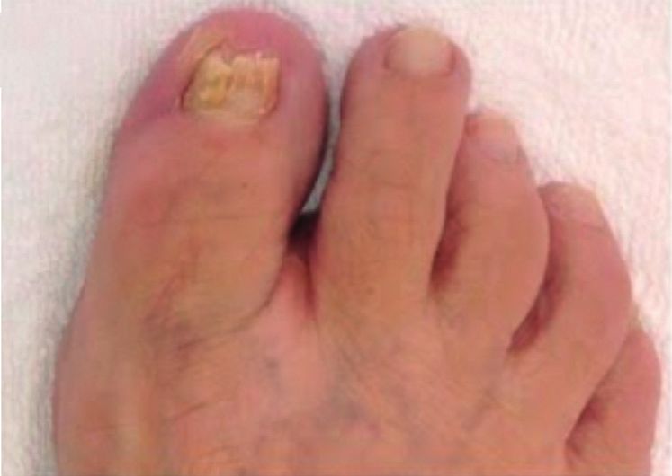
Evidence-Based Complementary and Alternative Medicine                                                                                        7

Moreover, it suggests the involvement of intrinsic factors related      [8] A. K. Gupta, M. Paquet, and F. C. Simpson, “Therapies for the
to the fungus as a greater ability to invade the nail and to form           treatment of onychomycosis,” Clinics in Dermatology, vol. 31,
biofilm, besides natural different susceptibility to propolis.                no. 5, pp. 544–554, 2013.
                                                                        [9] A. C. P. Oliveira, C. S. Shinobu, R. Longhini, S. L. Franco, and
                                                                            T. I. E. Svidzinski, “Antifungal activity of propolis extract
Data Availability                                                           against yeasts isolated from onychomycosis lesions,”
                                                                            Memórias Do Instituto Oswaldo Cruz, vol. 101, no. 5,
The data are available from the corresponding author upon
                                                                            pp. 493–497, 2006.
reasonable request.                                                    [10] D. D. Demirseren, “New therapeutic options in the man-
                                                                            agement of superficial fungal diseases,” Dermatologic Therapy,
Conflicts of Interest                                                       vol. 33, no. 6, Article ID e12855, 2020.
                                                                       [11] F. F. Veiga, M. C. Gadelha, M. R. T. Da Silva et al., “Propolis
The authors declare that they have no conflicts of interest.                 extract for onychomycosis topical treatment: from bench to
                                                                            clinic,” Frontiers in Microbiology, vol. 9, p. 779, 2018.
Authors’ Contributions                                                 [12] G. S. De Hoog, “Atlas of clinical fungi,” in Mycoses, J. Guarro,
                                                                            Ed., vol. 39, no. 7-8, p. 323, 1995.
The first two authors Vanessa Vasconcellos-Pontello &                   [13] B. M. Piraccini, M. Iorizzo, A. Lencastre, P. Nenoff, and
Flávia Franco Veiga contributed equally to this study. VV-P                D. Rigopoulos, “Ciclopirox hydroxypropyl chitosan (HPCH)
was the dermatologist responsible for the follow-up with the                nail lacquer: a review of its use in onychomycosis,” Derma-
patients. FFV performed all laboratory experiments. MCG                     tology and Therapy, vol. 10, no. 5, pp. 917–929, 2020.
and MR performed the collection of nail material, identi-              [14] S. Jinna and J. Finch, “Spotlight on tavaborole for the
                                                                            treatment of onychomycosis,” Drug Design, Development and
fication of fungi, and MIC. MN and TIES designed and
                                                                            Therapy, vol. 9, pp. 6185–6190, 2015.
formulated the study and guided the biological tests and the           [15] P. Nenoff, C. Krüger, G. Ginter-Hanselmayer, and H.-J. Tietz,
manuscript, as well as its correction. All the authors have                 “Mycology-an update. Part 1: dermatomycoses: causative
contributed to the research, writing, and approval of the final              agents, epidemiology and pathogenesis,” JDDG: Journal der
version of the manuscript.                                                  Deutschen Dermatologischen Gesellschaft, vol. 12, no. 3,
                                                                            pp. 188–210, 2014.
Acknowledgments                                                        [16] C. V. D. A. Azambuja, L. A. Pimmel, G. B. Klafke, and
                                                                            M. O. Xavier, “Onychomycosis: clinical, mycological and in
This study was financed by the Coordenação de Aperfei-                       vitro susceptibility testing of isolates of Trichophyton
çoamento de Pessoal de Nı́vel Superior-Brasil (CAPES)-                      rubrum,” Anais Brasileiros de Dermatologia, vol. 89, no. 4,
Finance Code 001, Conselho Nacional de Desenvolvimento                      pp. 581–586, 2014.
Cientı́fico e Tecnológico (CNPq), Fundação Araucária, and             [17] C. Wang, W. Cantrell, T. Canavan, and B. Elewski, “Successful
                                                                            treatment of dermatophytomas in 19 patients using efina-
(FINEP/COMCAP).
                                                                            conazole 10% solution,” Skin Appendage Disorders, vol. 5,
                                                                            no. 5, pp. 304–308, 2019.
References                                                             [18] A. K. Gupta and K. A. Foley, “Evidence for biofilms in
                                                                            onychomycosis,” Giornale italiano di dermatologia e venere-
 [1] M. A. Bodman and K. Krishnamurthy, Onychomycosis,” in                  ologia : organo ufficiale, Societa italiana di dermatologia e
     StatPearls, StatPearls Publishing, Treasure Island, FL, USA,           sifilografia, vol. 154, no. 1, pp. 50–55, 2019.
     2020.                                                             [19] F. K. Tobaldini-Valerio, P. S. Bonfim-Mendonça,
 [2] D. P. Westerberg and M. J. Voyack, “Onychomycosis: current             H. C. Rosseto et al., “Propolis: a potential natural product to
     trends in diagnosis and treatment,” American Family Physi-             fightCandidaspecies infections,” Future Microbiology, vol. 11,
     cian, vol. 88, no. 11, pp. 762–70, 2013.                               no. 8, pp. 1035–1046, 2016.
 [3] A. K. Gupta, S. G. Versteeg, N. H. Shear, V. Piguet, A. Tosti,    [20] K. Gucwa, B. Kusznierewicz, S. Milewski, P. Van Dijck, and
     and B. M. Piraccini, “A practical guide to curing onycho-              P. Szweda, “Antifungal activity and synergism with azoles of
     mycosis: how to maximize cure at the patient, organism,                polish propolis,” Pathogens (Basel, Switzerland), vol. 7, no. 2,
     treatment, and environmental level,” American Journal of               2018.
     Clinical Dermatology, vol. 20, no. 1, pp. 123–133, 2019.          [21] J. Galletti, F. K. Tobaldini-Valerio, S. Silva et al., “Antibiofilm
 [4] C. R. Stewart, L. Algu, R. Kamran et al., “Effect of onycho-            activity of propolis extract on Fusarium species from ony-
     mycosis and treatment on patient-reported quality-of-life              chomycosis,” Future Microbiology, vol. 12, no. 14,
     outcomes: a systematic review,” Journal of the American                pp. 1311–1321, 2017.
     Academy of Dermatology, 2020.                                     [22] I. R. Capoci, P. S. Bonfim-Mendonça, G. S. Arita et al.,
 [5] T. Auvinen, R. Tiihonen, M. Soini, M. Wangel, A. Sipponen,             “Propolis is an efficient fungicide and inhibitor of biofilm
     and J. J. Jokinen, “Efficacy of topical resin lacquer, amorolfine         production by vaginal Candida albicans,” Evidence-based
     and oral terbinafine for treating toenail onychomycosis: a              Complementary and Alternative Medicine: ECAM, vol. 2015,
     prospective, randomized, controlled, investigator-blinded,             Article ID 287693, 2015.
     parallel-group clinical trial,” British Journal of Dermatology,   [23] F. F. Veiga, M. I. Costa, ÉS. K. Cótica, T. I. E. Svidzinski, and
     vol. 173, no. 4, pp. 940–948, 2015.                                    M. Negri, “Propolis for the treatment of onychomycosis,”
 [6] L. Maxfield, C. V. Preuss, and R. Bermudez, Terbinafine,                 Indian Journal of Dermatology, vol. 63, no. 6, pp. 515–517,
     StatPearls Publishing, Treasure Island, FL, USA, 2021.                 2018.
 [7] M. Ghannoum and N. Isham, “Fungal nail infections (ony-
     chomycosis): a never-ending story?” PLoS Pathogens, vol. 10,
     no. 6, Article ID e1004105, 2014.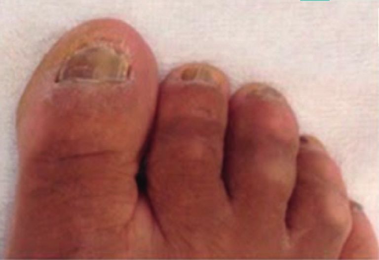
You can also read