Scapular body fractures-should we be fixing more of these?
←
→
Page content transcription
If your browser does not render page correctly, please read the page content below
Review Article
                                                                                                                                      Page 1 of 7

Scapular body fractures—should we be fixing more of these?
Austin Heare1, Stephen M. Oleszkiewicz1, Roberto Carlos Hernández Irizarry2, Peter A. Cole3,4,5
1
    Department of Orthopedics, University of Miami, Miami, FL, USA; 2Department of Orthopedics, Grady Memorial Hospital, Atlanta, GA, USA;
3
Department of Orthopaedic Surgery, University of Minnesota, Minneapolis, MN, USA; 4Department of Orthopaedic Surgery, Regions Hospital, St.
Paul, MN, USA; 5HealthPartners Orthopaedics & Sports Medicine, Bloomington, MN, USA
Correspondence to: Austin Heare, MD. Assistant Professor Orthopaedic Surgery, University of Miami Miller School of Medicine, Department of
Orthopaedics, Don Soffer Clinical Research Center, 1120 NW 14th Street, Room 1263Z, Miami, FL 33136, USA. Email: HeareMD@gmail.com.

                  Abstract: The question of whether we should be fixing more scapular body fractures originates from the
                  historical preference for nonoperative management of these fractures. Recently, there has been a renewed
                  interest in operative management due to the recognition that scapular malunion can cause significant
                  disability. While the treatment pendulum has shifted away from benign neglect, finding the right balance
                  of surgical aggression remains controversial. In general, the majority of scapula fractures can successfully be
                  treated nonoperatively with excellent functional results. However, numerous case studies exist demonstrating
                  poor outcomes of scapular body or neck fractures with increased deformity. The literature suggests that
                  a glenopolar angle (GPA) less than 20 degrees can lead to a significant decrease in shoulder function.
                  Additionally, retrospective studies using lateral border offset (LBO) greater than >2 cm and angulation >45
                  degrees as a surgical indication demonstrate functional outcomes with near normal strength and range of
                  motion and low complication rates. While numerous cut-offs for surgical indications have been recommended,
                  all indications are considered relative and treatment should be individualized based on patient characteristics
                  and goals.

                  Keywords: Extraarticular scapula fracture; scapula fracture; scapula body fracture; glenopolar angle (GPA)

                  Received: 21 February 2020; Accepted: 16 April 2020; Published: 15 April 2021.
                  doi: 10.21037/aoj-20-46
                  View this article at: http://dx.doi.org/10.21037/aoj-20-46

Introduction                                                                   treatment gained traction due to a few inherent properties
                                                                               of the scapula. First, the robust muscular envelope and
The question of whether we should be fixing more scapular
                                                                               bloody supply of the scapula ensure that the majority of
body fractures originates from the historical preference
                                                                               these fractures will proceed to union. Additionally, the
for nonoperative management of these fractures. Recently,
                                                                               glenohumeral and scapulothoracic joints maintain a large
there has been a renewed interest in operative management                      capacity for compensatory motion if the scapula heals in
due to the recognition that scapular malunion can cause                        a malunited position. For these reasons, coupled with the
significant disability. While the treatment pendulum has                       fact that the scapula has complex bony anatomy with little
shifted away from benign neglect, surgical indications                         area for screw purchase, nonoperative management was the
remain controversial.                                                          prevailing treatment until the turn of the twenty first century.
   The concept of surgical fixation for scapula fractures is                      The treatment strategy for these fractures changed as the
not novel. The first publication of internal fixation for a                    understanding of the fracture patterns, surgical approaches,
scapula fracture was in 1913 by Albin Lambotte from a case                     fixation technology improved and the consequences of
performed in 1910 (1). Despite numerous surgical case series                   scapular malunion were realized. Initial efforts for operative
from surgeons such as Judet (1), it was the nonoperative                       treatment of scapula fractures largely revolved around intra-
opinion of Rowe (2) and Schnepp (3) that dictated how these                    articular glenoid fractures due to well accepted principles of
fractures would be treated for ensuing decades. Nonoperative                   restoring articular congruity and preventing post-traumatic

© Annals of Joint. All rights reserved.                                                  Ann Joint 2021;6:22 | http://dx.doi.org/10.21037/aoj-20-46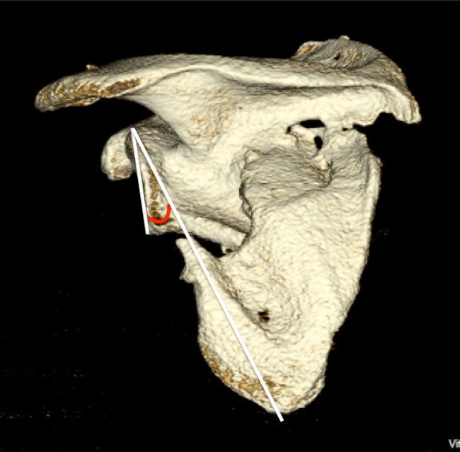
Page 2 of 7 Annals of Joint, 2021
irritation to the patient (9). Alternatively, scapular fracture
diagnosis can be missed or delayed due to the presence of
these more significant injuries. Any patient with multiple rib
fractures or hemopneumothorax on a chest radiograph should
draw the attention to the scapula to ensure it was not injured.
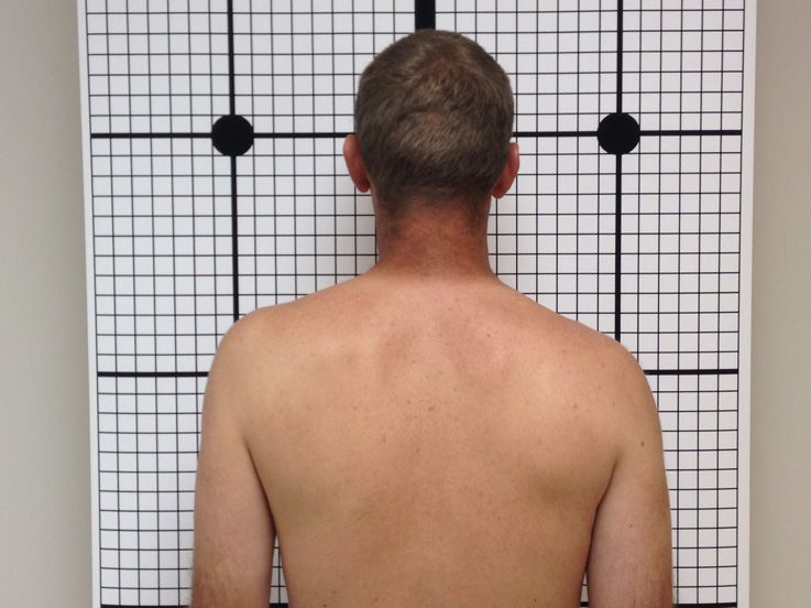
Physical examination of the patient should include
gross inspection of the shoulder for significant deformity.
This deformity is often characterized by marked shoulder
drooping or ptosis on the injured side (Figure 1). The
integrity of the skin and assessment for abrasions over
the shoulder should be noted. Palpation of the acromion,
clavicle and sternoclavicular/acromioclavicular joints
can alert the physician to associated fractures that would
constitute a double disruption of the shoulder suspensory
Figure 1 A patient with a right scapular fracture displaying the complex. Range of motion of the shoulder will undoubtedly
characteristic ptosis or drooping of the shoulder on clinical exam. be painful, but if near full active forward elevation is
present it can let the provider know the fracture is likely
nondisplaced and can safely be treated conservatively.
osteoarthritis (4). More recently, it has been recognized If a scapular fracture is identified on a chest radiograph,
that certain patients with extra-articular scapula fractures a standard shoulder radiographic series including true
can have poor functional outcomes related to altered AP shoulder (Grashey), axillary, and scapular Y views to
rotator cuff biomechanics, scapulothoracic dyskinesia, and assess for displacement should be obtained. If marked
impingement syndrome (5). displacement is identified, a dedicated computed
Even though surgical intervention has become increasingly tomography scan with 3D reconstructions can help define
recognized as appropriate treatment for certain patients, the displacement indicative of potential surgical benefit.
identifying these patients still remains elusive. The recent
standardization of radiographic parameters for displacement
Classification/radiological evaluation
of scapular body fractures has led to improved guidelines
for surgical intervention (6). However, even to this date, Numerous classification systems exist for scapular body
high quality evidence comparing nonoperative to operative fractures, most notably including those of Hardegger (4),
treatment of scapular body fractures does not exist. Ada and Miller (10), as well as the OTA/AO fracture and
dislocation classification (11). Both the Hardegger and
Ada and Miller classifications were based on radiographic
Patient evaluation
appearance of fracture lines rather than CT scans. These
Scapula fractures are rare injuries, comprising only about classification systems broadly group fractures into distinct
0.5% of all fractures. However, fractures of the scapular categories such as scapular spine (IB), scapular neck (IIA,
body or glenoid neck account for approximately 66–98% of B, C), glenoid (III), and body (IV) fractures for the Ada and
scapula fractures making them the most common fracture Miller classification (10). More recently, with the advent of
pattern (7). CT scans, detailed mapping of scapular body fractures was
Scapula fractures are usually the result of high-energy able to be performed. Based on a series of ninety patients
mechanisms and patients frequently have associated injuries. with operative scapular body fractures, 3-dimensional
These injuries include thorax, ipsilateral upper extremity, mapping of the fractures revealed the vast majority of
head, and spine injuries in 80%, 50%, 48%, and 26%, scapular body fractures followed predictable patterns (7).
respectively (8). Scapula fractures should alert the treating These patterns included a high rate of those with a fracture
orthopaedic surgeon to the potential presence of these other of the lateral border just inferior to the glenoid, often with
injuries so they can be treated appropriately. Displaced rib extension to the medial border (7). Early classifications
fractures can have a significant effect on the outcome of systems laid the groundwork for contemporary surgical
patients with scapular body fractures due to prominence and indications while three-dimensional mapping helps dictate
© Annals of Joint. All rights reserved. Ann Joint 2021;6:22 | http://dx.doi.org/10.21037/aoj-20-46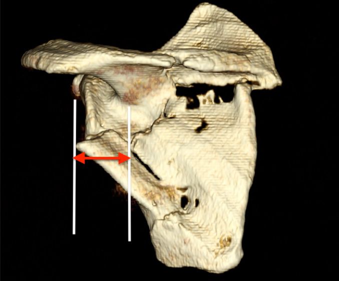
Annals of Joint, 2021                                                                                                            Page 3 of 7

Figure 2 Measurement of the lateral border offset is performed
on a three-dimensional computed tomography (3D CT) oriented              Figure 4 Measurement of the glenopolar angle performed on a
in the semicoronal scapula plane. The common displacement                three-dimensional computed tomography (3D CT) oriented in the
pattern is for the inferior scapula fragment to lateralize in relation   coronal scapula plane. The glenopolar angle measurement is made
to the superior fragment. The lateral border offset is measured          by drawing a line from the inferior to the superior pole of the
drawing a line from the lateralmost extent of the inferior fragment      glenoid fossa and another line from the superior pole to the apex
and another perpendicular line from the lateralmost aspect of the        of the scapula’s body’s inferior angle.
superior fragment.

                                                                         surgical approaches and fixation strategies.
                                                                             Current radiographic parameters indicating surgical
                                                                         fixation of scapular body fractures include lateral border
                                                                         offset (LBO), angulation, and glenopolar angle (GPA).
                                                                         LBO refers to the amount of displacement of the glenoid
                                                                         component in relation to the lateral border of the scapula.
                                                                         As described by Anavian et al. (6), this is found by measuring
                                                                         the distance between vertical lines drawn from the
                                                                         superolateral tip of the lateral border fracture inferiorly and
                                                                         its corresponding fracture bed at the base of the scapular
                                                                         neck superiorly, as seen on an AP radiograph or three-
                                                                         dimensional CT scan (Figure 2). Angulation of scapular
                                                                         body fractures is best determined on a scapular Y view by
                                                                         measuring the angle formed between parallel lines drawn
                                                                         down the proximal and distal segment cortices (6) (Figure 3).
                                                                         Finally, the GPA refers to the obliquity of the glenoid
                                                                         in respect to the scapular body (12). It is formed by first
Figure 3 Angulation deformity is measured on a three-dimensional         drawing a line from the superior point of the glenoid cavity
computed tomography (3D CT) oriented in the transscapular                to the inferior aspect compared with a second line drawn
Y orientation. The angle is formed by the intersection of a line         from the superior point of the glenoid to the inferior angle
running parallel to the proximal fragment and a line running             of the scapula body (6) (Figure 4).
parallel to the distal fragment. Note that even though the inferior
teardrop forms a concave surface over the rib cage, it is the more       Nonoperative treatment
proximal straight portion of the intramedullary canal that is used
for the measurement.                                                     Scapula fractures have classically been treated without

© Annals of Joint. All rights reserved.                                             Ann Joint 2021;6:22 | http://dx.doi.org/10.21037/aoj-20-46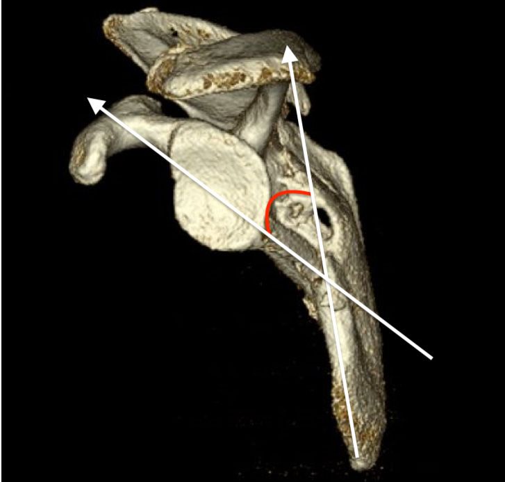
Page 4 of 7                                                                                                   Annals of Joint, 2021

surgery. Research has shown successful outcomes with this         the triceps and provides the most important bony fixation
strategy (13-19). The usual treatment is a sling for about        point for stable internal fixation.
3 weeks to allow subsidence of pain and swelling. During             As with all operations in orthopedic surgery, the surgical
this time, close vigilance is important, as some patients with    approach is of utmost importance in order to access the
extraarticular variants were found to develop increasing          area of interest and apply hardware safely. The classic Judet
deformity in the first weeks of treatment (20). Because of        approach is an extensile approach and exposes the entire
the unique anatomy of the periscapular region, many factors       posterior scapula by elevating the infraspinatus and teres
can lead to increased displacement and deformity, such            minor, but this is rarely necessary for acute extraarticular
as gravity, asymmetric muscle contractions, and fracture          fractures (1). Typically, the Judet curvilinear skin incision
instability. Deforming forces can result in displacement,         is utilized and muscular intervals are exploited for fracture
leading to pain, weakness and glenohumeral imbalance              reduction and fixation with less soft tissue disruption (26).
(12,14,15,21,22). Patients must therefore be monitored               Depending on the fracture pattern, different
radiographically to detect early displacement and deformity.      intermuscular windows can be employed. A more limited
Because the scapula is 30 degrees off the plane of the thorax,    technique can be used to access portions of the lateral
fractures can be easily missed even in experienced centers        border, scapular spine, and vertebral border. To do this, the
(23,24).                                                          muscle plane entered at the spine of the scapula is between
   The rehabilitation process is an important component           the trapezius, originating at the superior margin, and the
regardless of treatment choice. It is important to keep           deltoid at the inferior margin, which is elevated (27,28). The
in mind that patients may develop pseudoparalysis and             majority of extraarticular fractures can be accessed with little
dyskinesia in the early stages of rehabilitation. Additionally,   or no release of the deltoid, but the medial portion of the
treating surgeons must be aware that these injuries tend to       deltoid origin can be released if needed and later repaired.
be the result of high energy trauma, and associated injuries         The acromion can be accessed with an extension of
to the limb and chest wall must be addressed and accounted        the posterior Judet incision laterally along the posterior
for in the rehabilitation process. Associated injuries are        acromion. At the vertebral border of the scapula, the
reported in up to 90% of cases and can serve as distracting       intramuscular plane is between the infraspinatus which
injuries to prompt diagnosis.                                     is elevated, and the rhomboids, which are left attached.
                                                                  The most important window is between the infraspinatus
                                                                  and teres minor for access to the lateral border of the
Surgical treatment
                                                                  scapula and the posterior glenoid rim. Once this interval is
The scapula is an integral part of the superior shoulder          developed, the lateral border of the scapula can be exposed.
suspensory mechanism, which attaches the upper extremity          This is a critical location for fracture reduction, which is
to the axial skeleton. Seventeen muscular attachments             typically anteversion of the glenoid and medialization of
and insertions provide a robust platform from which               the lateral border fragment. Once reduced, typical fixation
glenohumeral motion occurs (25). In the setting of a              consists of low-profile, relatively flexible implants (Figure 5),
fracture, these can also be sources of deformity and              such as 2.7 mm locking compression or reconstruction
displacement. The levator scapulae muscle, which inserts          plates.
on the superomedial border of the scapula can medialize
the cranial fragment but most commonly the lateral border
                                                                  Outcomes
is translated laterally by its attached musculature. This can
be correlated radiographically as LBO and decreased GPA.          The surgical treatment of extraarticular scapular fractures
Posterior surgical approaches are generally safe in regards       remains controversial due to the acceptable results of
to vascularity, since the subscapularis muscle originates from    nonoperative treatment and the lack of high-quality
the anterior surface of the scapula and provides the blood        evidence comparing operative to nonoperative treatment.
supply to fracture fragments. The acromion and clavicle           However, not all extraarticular scapular fractures can
serve as the origin for the deltoid muscle. The lateral border    uniformly be treated nonoperatively with reproducibly
of the scapula is the origin of the teres major and minor         excellent clinical outcomes.
muscles, as well as the insertion point for the long head of         Nonoperative treatment of minimally displaced scapular

© Annals of Joint. All rights reserved.                                     Ann Joint 2021;6:22 | http://dx.doi.org/10.21037/aoj-20-46
Annals of Joint, 2021 Page 5 of 7
A B
Figure 5 AP radiographs of extra-articular scapula body fracture comparing pre-fixation and post-fixation. Ipsilateral minimally displaced
clavicle fracture treated nonoperatively. (A) AP radiograph of right sided scapular body fracture showing lateral border offset and decreased
glenopolar angle; (B) postoperative radiograph showing reduction and stable fixation of scapular body fracture with flexible 2.7 mm plates
and screws with restoration of normal bony anatomy.
body fractures can reliably produce acceptable clinical Constant scores at follow-up. GlenopolarPage 6 of 7 Annals of Joint, 2021
Conclusions PAC reports grants from Stryker, grants from Depuy-
Synthes, other from KLS Martin, grants from AONA,
The management of extraarticular scapular fractures
COTA, OMeGA Depuy-Synthes, Stryker, Zimmer-Biomet,
remains controversial due to the lack of prospective,
BoneFoam, Acumed, KLSMartin, Exactech, other from
randomized controlled trials justifying surgical intervention.
BoneFoam, personal fees from Exactech, Depuy-Synthes,
Plain radiographs with evidence of scapular displacement
personal fees from AO International, outside the submitted
should warrant a CT scan with 3D reconstruction, paying
work. The authors have no other conflicts of interest to
careful attention to LBO, angulation, and the GPA.
declare.
In general, the majority of scapula fractures can
successfully be treated nonoperatively with excellent
Ethical Statement: The authors are accountable for all
functional results. However, numerous case studies
aspects of the work in ensuring that questions related
exist demonstrating poor outcomes of scapular body or
to the accuracy or integrity of any part of the work are
neck fractures with increased deformity (12,14,15). The
appropriately investigated and resolved.
literature suggests that a GPA less than 20 degrees can
lead to significantly worse shoulder function (12,15,30). Open Access Statement: This is an Open Access article
Additionally, retrospective studies using LBO greater than distributed in accordance with the Creative Commons
>2 cm and angulation >45 degrees as a surgical indication Attribution-NonCommercial-NoDerivs 4.0 International
demonstrate functional outcomes with near normal License (CC BY-NC-ND 4.0), which permits the non-
strength and range of motion and low complication rates. commercial replication and distribution of the article with
While numerous cut-offs for surgical indications have been the strict proviso that no changes or edits are made and the
recommended (5,33,34), all indications are considered original work is properly cited (including links to both the
relative and treatment should be individualized based on formal publication through the relevant DOI and the license).
patient characteristics and goals. See: https://creativecommons.org/licenses/by-nc-nd/4.0/.
Should we be fixing more of these? Perhaps, but more
importantly, we should be diagnosing more of these and
presenting our patients with all of their treatment options, References
surgical and non-surgical. 1. Judet R. Surgical Treatment of Scapular Fractures. Acta
Orthop Belg 1964;30:673-8.
Acknowledgments 2. Rowe CR. Fractures of the Scapula. Surg Clin North Am
1963;43:1565-71.
The authors would like to give special thanks to Lavi 3. Schnepp J, Comtet JJ, Cetre J, et al. Value of
Mattingly for his tireless work editing and formatting this nonsurgical treatment of omoplata fractures. Lyon Med
article for publication. 1968;220:809-13.
Funding: None. 4. Hardegger FH, Simpson LA, Weber BG. The operative
treatment of scapular fractures. J Bone Joint Surg Br
1984;66:725-31.
Footnote
5. Jones CB, Sietsema DL. Analysis of operative versus
Provenance and Peer Review: This article was commissioned nonoperative treatment of displaced scapular fractures.
by the Guest Editor (Adam Seidl) for the series Clin Orthop Relat Res 2011;469:3379-89.
“Management of Fractures Around the Shoulder” published 6. Anavian J, Conflitti JM, Khanna G, et al. A reliable
in Annals of Joint. The article has undergone external peer radiographic measurement technique for extra-
review. articular scapular fractures. Clin Orthop Relat Res
2011;469:3371-8.
Conflicts of Interest: All authors have completed the ICMJE 7. Armitage BM, Wijdicks CA, Tarkin IS, et al. Mapping
uniform disclosure form (available at http://dx.doi. of scapular fractures with three-dimensional computed
org/10.21037/aoj-20-46). The series ““Management of tomography. J Bone Joint Surg Am 2009;91:2222-8.
Fractures Around the Shoulder” was commissioned by 8. Cole PA, Gauger EM, Schroder LK. Management
the editorial office without any funding or sponsorship. of scapular fractures. J Am Acad Orthop Surg
© Annals of Joint. All rights reserved. Ann Joint 2021;6:22 | http://dx.doi.org/10.21037/aoj-20-46Annals of Joint, 2021 Page 7 of 7
2012;20:130-41. Biomechanical analysis of scapular neck malunion-
9. Yadav V, Khare GN, Singh S, et al. A prospective study -a simulation study. Clin Biomech (Bristol, Avon)
comparing conservative with operative treatment in 2004;19:906-12.
patients with a 'floating shoulder' including assessment of 22. Pace AM, Stuart R, Brownlow H. Outcome of glenoid
the prognostic value of the glenopolar angle. Bone Joint J neck fractures. J Shoulder Elbow Surg 2005;14:585-90.
2013;95-B:815-9. 23. Herrera DA, Anavian J, Tarkin IS, et al. Delayed operative
10. Ada JR, Miller ME. Scapular fractures. Analysis of 113 management of fractures of the scapula. J Bone Joint Surg
cases. Clin Orthop Relat Res 1991;(269):174-80. Br 2009;91:619-26.
11. Meinberg EG, Agel J, Roberts CS, et al. Fracture and 24. Tadros AM, Lunsjo K, Czechowski J, et al. Causes
Dislocation Classification Compendium-2018. J Orthop of delayed diagnosis of scapular fractures. Injury
Trauma 2018;32 Suppl 1:S1-170. 2008;39:314-8.
12. Romero J, Schai P, Imhoff AB. Scapular neck fracture-- 25. van der Helm FC, Pronk GM. Three-dimensional
the influence of permanent malalignment of the glenoid recording and description of motions of the shoulder
neck on clinical outcome. Arch Orthop Trauma Surg mechanism. J Biomech Eng 1995;117:27-40.
2001;121:313-6. 26. Obremskey WT, Lyman JR. A modified Judet approach to
13. McGinnis M, Denton JR. Fractures of the scapula: a the scapula. J Orthop Trauma 2004;18:696-9.
retrospective study of 40 fractured scapulae. J Trauma 27. Braun C, Wirbel R, Mutschler W. The two-portal
1989;29:1488-93. approach for internal fixation of scapular fractures. Eur J
14. Nordqvist A, Petersson C. Fracture of the body, neck, or Trauma 2005;31:186-93.
spine of the scapula. A long-term follow-up study. Clin 28. Gauger EM, Cole PA. Surgical technique: a minimally
Orthop Relat Res 1992;(283):139-44. invasive approach to scapula neck and body fractures. Clin
15. Bozkurt M, Can F, Kirdemir V, et al. Conservative Orthop Relat Res 2011;469:3390-9.
treatment of scapular neck fracture: the effect of stability 29. Edwards SG, Whittle AP, Wood GW 2nd. Nonoperative
and glenopolar angle on clinical outcome. Injury treatment of ipsilateral fractures of the scapula and clavicle.
2005;36:1176-81. J Bone Joint Surg Am 2000;82:774-80.
16. Wilber MC, Evans EB. Fractures of the scapula. An 30. Gosens T, Speigner B, Minekus J. Fracture of the scapular
analysis of forty cases and a review of the literature. J Bone body: functional outcome after conservative treatment. J
Joint Surg Am 1977;59:358-62. Shoulder Elbow Surg 2009;18:443-8.
17. Oh W, Jeon IH, Kyung S, et al. The treatment of double 31. Kim KC, Rhee KJ, Shin HD, et al. Can the glenopolar
disruption of the superior shoulder suspensory complex. angle be used to predict outcome and treatment of the
Int Orthop 2002;26:145-9. floating shoulder? J Trauma 2008;64:174-8.
18. Schofer MD, Sehrt AC, Timmesfeld N, et al. Fractures of 32. Schroder LK, Gauger EM, Gilbertson JA, et al. Functional
the scapula: long-term results after conservative treatment. outcomes after operative management of extra-articular
Arch Orthop Trauma Surg 2009;129:1511-9. glenoid neck and scapular body fractures. J Bone Joint
19. van Noort A, van Kampen A. Fractures of the scapula Surg Am 2016;98:1623-30.
surgical neck: outcome after conservative treatment in 13 33. Cole PA, Gauger EM, Herrera DA, et al. Radiographic
cases. Arch Orthop Trauma Surg 2005;125:696-700. follow-up of 84 operatively treated scapula neck and body
20. Anavian J, Khanna G, Plocher EK, et al. Progressive fractures. Injury 2012;43:327-33.
displacement of scapula fractures. J Trauma 34. Jones CB, Cornelius JP, Sietsema DL, et al. Modified Judet
2010;69:156-61. approach and minifragment fixation of scapular body and
21. Chadwick EK, van Noort A, van der Helm FC. glenoid neck fractures. J Orthop Trauma 2009;23:558-64.
doi: 10.21037/aoj-20-46
Cite this article as: Heare A, Oleszkiewicz SM, Irizarry RCH,
Cole PA. Scapular body fractures—should we be fixing more of
these? Ann Joint 2021;6:22.
© Annals of Joint. All rights reserved. Ann Joint 2021;6:22 | http://dx.doi.org/10.21037/aoj-20-46You can also read