Structural and Physicochemical Characteristics of Rice Bran Dietary Fiber by Cellulase and High-Pressure Homogenization - MDPI
←
→
Page content transcription
If your browser does not render page correctly, please read the page content below
applied
           sciences
Article
Structural and Physicochemical Characteristics of
Rice Bran Dietary Fiber by Cellulase and
High-Pressure Homogenization
Fengying Xie 1,2,† , Tian Zhao 1,† , Hongchen Wan 1 , Miao Li 1 , Lina Sun 1 , Zhongjiang Wang 1, *
and Shuang Zhang 1, *
 1    College of Food Science, Northeast Agricultural University, 600 Changjiang Road, Harbin 150030, China;
      spxfy@163.com (F.X.); ztian597@163.com (T.Z.); 15070927087@163.com (H.W.); li_miao216@163.com (M.L.);
      minzh@163.com (L.S.)
 2    Harbin Food Industry Research Institute, Harbin 150028, China
 *    Correspondence: wzjname@126.com (Z.W.); szhang@neau.edu.cn (S.Z.)
 †    These authors contributed equally to this work.

 Received: 28 February 2019; Accepted: 22 March 2019; Published: 27 March 2019

 Abstract: The present paper aims to study the effect of cellulase hydrolysis and high-pressure
 homogenization on the structural and physicochemical properties of rice bran dietary fiber (RB-DF).
 Scanning electron microscopy showed that cellulase treatment led to the formation of a porous
 structure on RB-DF surface. High-pressure homogenization affected the laminated microstructure
 of RB-DF, leading to the formation of an irregular and loose surface structure. X-ray diffraction
 demonstrated that joint processing destroyed the amorphous hemicellulose and cellulose regions,
 and changed the crystallinity of RB-DF, albeit with a minor impact on the crystalline region of cellulose.
 Fourier transform infrared spectroscopy indicated that combined processing promoted dissociation of
 some glycosidic bonds in fiber structure, exposing the hydroxyl groups in cellulose, thus improving
 their ability to bind water molecules. Thermogravimetric analysis showed a significant decrease in
 the thermal decomposition temperature of RB-DF (p < 0.05) as well as a decrease in thermal stability
 after combined processing. Cellulase hydrolysis and high-pressure homogenization treatment did
 not improve their oil holding capacity, but significantly increased water holding capacity, swelling
 capability, and cation exchange capacity of RB-DF. Thus, enzymatic hydrolysis and high-pressure
 homogenization treatment can change the structure of RB-DF, exposing a large number of hydrophilic
 groups and enhancing hydration, obtaining uniform RB-DF particle.

 Keywords: rice bran; enzymatic hydrolysis; cellulase; high-pressure homogenization

1. Introduction
     Dietary fiber (DF) is the general term for polysaccharide and lignin that cannot be digested and
absorbed by biological enzymes in human small intestine [1]. As the seventh most important nutrients
for organisms [2], DF is an important component of healthy diet, offering various physiological benefits,
including body weight control, serum lipid and cholesterol reduction, controlled postprandial glucose
responses, and colon cancer prevention [3].
     Rice bran is the outer layer of rice derived as a by-product of milling process and is an ideal
source of DF [4,5]. However, the major components of rice bran DF (RB-DF) are insoluble, whereas
soluble dietary fiber is of greater benefit to human health [6]. Thus, modification of the insoluble DF
to increase its solubility and water holding capacity has become an important research direction for
functional properties improvement of RB-DF in food industry. Insoluble DF modification methods
include chemical, biological, and physical routes [7]. Chemical methods lead to low yields of soluble

Appl. Sci. 2019, 9, 1270; doi:10.3390/app9071270                                 www.mdpi.com/journal/applsci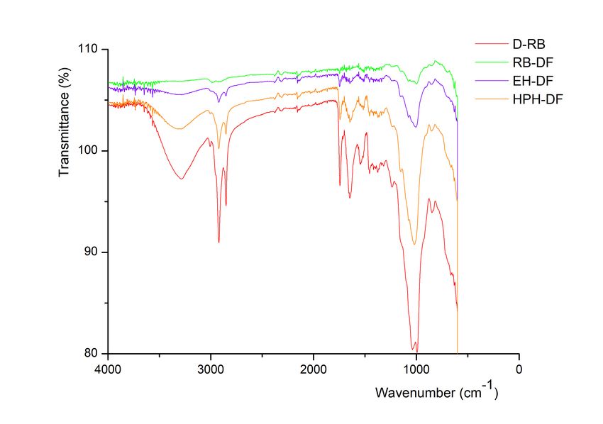
Appl. Sci. 2019, 9, 1270                                                                           2 of 10

DF, in which harmful chemical groups being easily introduced [8,9]. Biological methods are mild, yet
the enzymes used for catalysis are expensive. Thus, the focus is now on physical methods, including
high-pressure homogenization (HPH). HPH is a refinement and dispersion technology that can be
used for liquid–liquid and solid–liquid systems [10]. The high pressures used lead to a series of
changes in the physical, chemical, and structural properties of food products, effectively increasing the
soluble fraction of dietary fiber [11,12]. However, few studies have assessed the effect of HPH on the
functionality or structure of DF [13,14]. Insoluble DF has a compact polymeric structure that is formed
by stiff cellulose and flexible hemicellulose components, which makes it a highly tough structure that
is difficult to disintegrate. Breaking the dense structure of insoluble DF is difficult to achieve using
a single HPH process, but the use of enzymatic hydrolysis allows the lignocellulose to be cleaved
into partial monomer units [15,16]. Additionally, supplementing DF into food requires a substantial
understanding of its chemical structure, since the interactions between DF and other ingredients can
considerably alter the microstructure and properties of the final products.
      Herein, cellulase enzymatic hydrolysis and HPH were combined to treat RB-DF.
The characterization and physicochemical properties of RB-DF were determined at different treatment
stages. This research should contribute towards the development of a novel and effective method for
the modification and application of DF.

2. Materials and Methods

2.1. Materials
     Brown rice was purchased from Wugu Xinhe Agricultural Development Co., Ltd. (Heilongjiang,
China). Crude protein, fat, carbohydrate, dietary fiber of brown rice were 7.9 g/100 g, 3.1 g/100 g,
67.6 g/100 g and 5.3 g/100 g respectively. Alkaline protease (enzyme activity ≥200 U/mg), α-amylase
(enzyme activity ≥50 U/mg), and cellulase (enzyme activity ≥50 U/mg) were obtained from Yuanye
Biological Technology Co., Ltd. (Shanghai, China). All other chemicals were of analytical grade.

2.2. Rice bran Preparation
    Rice bran from brown rice was obtained by polishing the rice using a laboratory rice mill
(LTJM-2099, Tuopu Yunnong Technology Co. Ltd., Zhejiang, China) at 2200 rmp for 90 min. The bran
samples were stored at 0 ◦ C until further analysis.

2.3. Purification of DF
      The isolation and purification of DF were performed as described by [17]. The isolation and
purification of DF were performed as follows: Rice bran (500 g) soaked in deionized water (1 L, 25 ◦ C,
30 min, 3 times) was centrifuged at 3524 g for 10 min. The insoluble part was dried at 50 ◦ C for 12 h
in an air-drying oven, followed by grinding and sieving through a 250 mm mesh and degreasing by
washing three times with petroleum ether at a ratio of 1:3. Defatted rice bran (D-RB) mixed (1:6, w/v)
with 0.2 M phosphate buffer solution (PBS, pH 6.5) was treated with α-amylase 24 U/g (D-RB) at 60 ◦ C
for 2 h to remove all starch. The slurry was adjusted to pH 10 and 60 U/g (D-RB) alkaline protease was
added (2.0 g/100 mL, w/v) at 45 ◦ C for 2 h to remove all proteins. The mixture was heated in a boiling
water bath for 10 min, and then centrifuged at 3524 g for 10 min. The RB-DF was retained without
the supernatant.

2.4. Cellulase Enzymatic Treatments
     DF mixed (1:6, w/v) with 0.2 M acetic acid-sodium acetate buffer (pH 5.5) was treated with 24 U/g
(DF) cellulase (2.0 g/100 mL, w/v) at 55 ◦ C for 2 h. The treated slurry was centrifuged at 3524 g for
10 min after inactivation of enzyme in boiling water bath for 10 min. The insoluble part was washed
thrice with deionized water and centrifuged (3524 g, 10 min).
Appl. Sci. 2019, 9, 1270 3 of 10
2.5. High-Pressure Homogenization (HPH)
The cellulase enzymatically digested sample (5.0 g) hydrated in deionized water (100 mL) in
a plastic bottle (200 mL) was subjected to HPH treatment (FB-110T, Reed Machinery Equipment
Engineering Co. Ltd., Shanghai China) for three loop processing with each 30 s at 25 ◦ C using
treatment pressure of 120 MPa. Finally, the obtained D-RB, RB-DF, cellulase enzymatic treatment
RB-DF (EH-DF), and HPH treatment RB-DF (HPH-DF) were freeze-dried at −50 ◦ C for 24 h, vacuum
degree 0.969 mbar using a Freeze dryer (LGJ-10N Sihao Technology Co., Ltd. Hubei, China), and the
obtained powders were collected for further analysis.
2.6. Scanning Electron Microscopy (SEM)
The surface and microstructure of D-RB, RB-DF, EH-DF, and HPH-DF were observed using
a scanning electron microscope (S-3400, Hitachi, Ltd., Tokyo, Japan). Freeze-dried samples were
mounted on an aluminum stub with double-sided stick tape, coated with gold, and then examined at
an accelerating voltage of 5 kV, according to the method of [18]. Representative images were taken at
500× magnification.
2.7. X-Ray Diffraction (XRD)
XRD analysis and crystallinity assessment of D-RB, RB-DF, EH-DF, and HPH-DF were performed
as described by [19] with slight modifications. The XRD patterns were obtained using a diffractometer
(TTRAX3, theta-theta gonio, USA) with Cu Kα radiation (λ = 0.15418 nm) over a 2θ range of 5–60◦ ,
step of 0.02◦ /step, and incident current of 150 mA. The degree of crystallinity was determined by
calculating area under the curve using Peak Fit v4.12 according to the following equation:
AC
Dc (%) = × 100 (1)
Ac + Aa
where Dc is the degree of crystallinity, Ac is crystallized area, and Aa is amorphous area on the
X-ray diffractogram.
2.8. Fourier Transform Infrared (FT-IR) Spectroscopy
The structural changes of D-RB, RB-DF, EH-DF, and HPH-DF were measured with a FT-IR
spectrometer (MAGNA-IR 560, Nicoet, Ltd., USA), as previously described [20]. Freeze-dried samples
(2 mg) were ground with KBr (200 mg, spectroscopic grade), sheeted to one slice, and scanned with a
blank KBr background. The FT-IR spectra in a resolution range from 400 to 4000 cm−1 . A total of 32
scans were collected.
2.9. Thermogravimetric Analysis (TGA)
The thermal stability of D-RB, RB-DF, EH-DF, and HPH-DF was measured with a TGA
spectrometer (DTG-60, Shimadzu, Ltd., Japan) in a N2 atmosphere. The sample pans were heated from
35 ◦ C to 550 ◦ C, at a rate of 15 ◦ C /min.
2.10. Physicochemical Properties
2.10.1. Water and Oil Holding Capacities
The water and oil holding capacities were determined according to the method described by [21],
and calculated as follows:
W − W0
WHC(g/g) = 1 (2)
W0
W1 − W0
OHC(g/g) = (3)
W0Appl. Sci. 2019, 9, 1270 4 of 10
where W0 and W1 are the weight of the sample before and after oil adsorption, respectively.
2.10.2. Swelling Capacity
The swelling capacity was measured according to the method described in Reference [22], and
calculated as follows:
V − V0
SC(mL/g) = 1 (4)
W
where V1 and V2 are the volume of the sample before and after water adsorption, respectively, and W
is the weight of the sample.
2.10.3. Cation-Exchange Capacity
The cation-exchange capacity was determined following the method of [23] with slight
modifications. Briefly, an accurately weighed 1.0 g dried sample was placed in a 250 mL Erlenmeyer
flask and mixed with 50 mL of 1 mol/L HCl, and kept at 25 ◦ C for 24 h. The mixture was washed with
distilled water to remove all the Cl– and then dried in an oven until a constant mass was obtained.
The dried samples were then accurately weighed to 0.2 g and mixed with 50 mL of 5 g/100 mL NaCl,
followed by the addition of two drops of phenolphthalein indicator. Distilled water was used as a
blank sample, titrated with a 0.01 mol/L NaOH solution to the end point, and the volume of consumed
NaOH solution was recorded. The cation-exchange capacity was then calculated as follows:
( V 1 − V0 ) × C
CEC(mmol/g) = (5)
W
where V1 and V0 are the volumes of NaOH solution consumed by titrating the sample and blank (mL),
C is the concentration of NaOH solution used for titration (mol/L), and W is the mass of the dried
sample (g).
2.11. Statistical Analysis
The average values obtained from three parallel samples were used as experimental data. Data
were analyzed using SPSS software (SPSS for Windows, 22.0, 2012, SPSS Inc., USA) and Origin 8.0
(Origin Institute Inc, USA). Significant differences were determined using ANOVA test; a p value of
less than 0.05 was considered statistically significant.
3. Results and Discussion
3.1. Structural Properties of RD-DF
3.1.1. SEM
The particle morphology of RB-DF at different processing stages was observed by SEM (Figure 1).
D-RB showed a laminated fiber structure, with granular particles and smooth spheres that were likely
composed of protein polymers and small starch granules. Following amylase and protease digestion,
the number of granular particles and smooth spheres on the surface of RB-DF was significantly reduced,
showing an irregular, large, dense, and compact laminated microstructure. Subsequently, cellulase
hydrolysis led to changes in the RB-DF laminated structure (Figure 1B,C). This result is consistent with
the study by [24], wherein cellulase treatment is shown to be beneficial for the formation of a rough
and porous structure in DF. HPH treatment uses water as transmission medium, with its powerful
mechanical force breaking the regular fiber structure and forming deep grooves [25]. Thus, the loose
fiber structure with small particles and irregular flakes was attributed to the combination effect of
cellulase hydrolysis and HPH treatment (Figure 1D).Subsequently, cellulase hydrolysis led to changes in the RB‐DF laminated structure (Figure
1B,C). This result is consistent with the study by [24], w
Appl. Sci. 2019, 9, 1270 5 of 10
A B
C D
Figure
Figure Scanning
1. 1. electron
Scanning microscopy
electron microscopyimages
imagesofof
rice bran
rice dietary
bran fiber
dietary at at
fiber different treatment
different stages
treatment stages
(A) defatted rice bran, (B) rice bran dietary fiber, (C) cellulase enzymatic treatment rice bran dietary
(A) defatted rice bran, (B) rice bran dietary fiber, (C) cellulase enzymatic treatment rice bran dietary
fiber, (D) high pressure treated rice bran dietary fiber.
fiber, (D) high pressure treated rice bran dietary fiber.
3.1.2. Crystalline and Molecular Structure
3.1.2. Crystalline and Molecular Structure
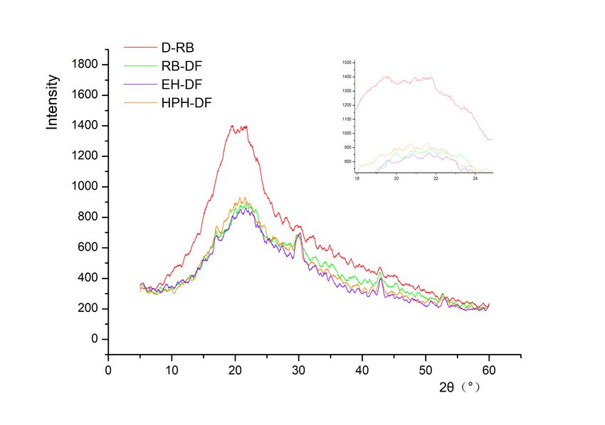
The XRD patterns for D-RB, RB-DF, EH-DF, and HPH-DF (Figure 2) showed a main diffraction
peak atThe XRD
22.5 patterns
◦ , which for D‐RB, RB‐DF,
is attributed EH‐DF,
by cellulose andhemicellulose
and HPH‐DF (Figure (2θ 2)
is showed
15–25◦ ) a[26].
mainHowever,
diffraction
peak at 22.5,
diffraction peak which is attributed
intensity values dobychangecellulose and hemicellulose
significantly (2θ is 15–25)
during sample processing[26].(pHowever,cleavage of hydrogen bonds between cellulose molecular chains and destroyed the amorphous
regions of cellulose. With the dissociation of the amorphous region, the arrangement regularity of
cellulose molecule increased, resulting in an increase in crystallinity. Thus, HPH treatment leads to
cellulose destruction of the connection between crystals, but does not affect the structural
framework of the cellulose polymer backbone.
Appl. Sci. 2019, 9, 1270 6 of 10
Figure 2. X-ray diffraction patterns of rice bran dietary fiber at different treatment stages. Inset shows
main diffraction peaks about rice bran in four treatment stages. D-RB defatted rice bran, EH-DF
Figure 2. X‐ray
cellulase enzymatic diffraction
treatment rice patterns of rice fiber,
bran dietary bran dietary
HPH-DFfiberhigh-pressure
at different treatment stages.
treatment Inset
rice shows
bran dietary
main diffraction peaks about rice bran in four treatment stages. D‐RB defatted rice bran, EH‐DF
fiber, RB-DF rice bran dietary fiber.
cellulase enzymatic treatment rice bran dietary fiber, HPH‐DF high‐pressure treatment rice bran
dietary fiber, RB‐DF rice bran dietary fiber.
3.1.3. FTIR Spectra
In the3.1.3. FTIRspectrum
infrared Spectra of RB-DF at various treatment stages (Figure 3), absorbance at 3323 cm−1
and 2921 cm-1 In were assigned
the infrared to hydroxyl
spectrum groups
of RB‐DF and C-H
at various bonds,
treatment respectively
stages [28]. The absorption
(Figure 3), absorbance at 3323
intensity ofcmrice bran was decreased after being treated with amylase and protease, and the absorption
−1 and 2921 cm‐1 were assigned to hydroxyl groups and C‐H bonds, respectively [28]. The
intensity ofabsorption
RB-DF was intensity of riceafter
increased bran enzymatic
was decreased after beingand
hydrolysis treated with amylasetreatment.
high-pressure and protease,
Thisandresult
the absorption intensity of RB‐DF was increased after enzymatic hydrolysis and high‐pressure
indicates that the infrared absorption peak intensity of RB-DF is greatly reduced with the dissociation
treatment. This result indicates that the infrared absorption peak intensity of RB‐DF is greatly
of starch from fat-removing D-RB. The widening and smoothing absorption peak of EH-DF and
reduced with the dissociation of starch from fat‐removing D‐RB. The widening and smoothing
HPH-DF indicates
absorptionthat
peakcellulase
of EH‐DF hydrolysis
and HPH‐DF andindicates
HPH treatment exposed
that cellulase hydroxyl
hydrolysis groups
and HPH promotes
treatment
disintegration
exposedof glycosidic bonds promotes
hydroxyl groups in DF structure [29]. Although
disintegration of glycosidicthebonds
intensity
in DFof the characteristic
structure [29].
absorptionAlthough
peak of the intensityofofcellulose
carbonyl the characteristic
uronic absorption
acid at 1743 peak
cm of–1carbonyl of cellulosethe
was weakened, uronic acid at
characteristic
absorption1743peakscm–1
ofwas weakened,
cellulose and the characteristic absorption
oligosaccharide at 1647 cm peaks
–1 andof cellulose
1018 cm–1 and[30]
oligosaccharide
were enhanced at in
1647 cm–1 and 1018 cm–1 [30] were enhanced in EH‐DF and HPH‐DF. This phenomenon also
EH-DF and HPH-DF. This phenomenon also indicates that cellulase hydrolysis and HPH treatment
indicates that cellulase hydrolysis and HPH treatment dissociate the non‐crystalline regions in rice
dissociate bran
the non-crystalline
DF, promoting theregions in rice
degradation bran DF, promoting
of small‐molecule theand
saccharides, degradation of small-molecule
effectively changing RB‐DF
saccharides, and effectively changing RB-DF microstructure.
microstructure.
3.1.4. Thermal stability analysis
The differential TGA curves for RB-DF at various treatment stages were obtained from the first
derivatives of weight loss rate (Figure 4). D-RB, RB-DF, EH-DF, and HPH-DF showed a weight
loss peak (~3%) at 100 ◦ C, attributed to the evaporated water. The TGA curves of the four samples
had significant differences, whereas that for D-RB showed only one significant weight loss peak
at 320–390 ◦ C, and the corresponding weight loss weight was 42%, where 350 ◦ C was its thermal
decomposition temperature. For RB-DF, two weight loss peaks were observed at 290–360 ◦ C and
380–500 ◦ C, with thermal decomposition at 330 ◦ C. The weight loss region for EH-DF was 278–397 ◦ C,
with thermal decomposition at 320 ◦ C. Finally, for HPH-DF, the thermal decomposition temperature
was 315 ◦ C, with weight loss area at 260–380 ◦ C corresponding to a 55% thermal weight loss rate, and
a second weight loss area at 410–500 ◦ C, where thermal loss weight loss rate reached 74%. An increase
in weight loss rate is often accompanied by a decrease in thermal decomposition temperature [31,32].
In agreement with this, the weight loss rate of the four samples changed from 42–74%, and the thermalAppl. Sci. 2019, 9, 1270 7 of 10 decomposition temperature decreased from 315–350 ◦ C. According to the abovementioned data, the thermal decomposition temperature and thermal stability of RB-DF decreased significantly (p
Appl. Sci. 2019, 9, 1270                                                                                                 8 of 10

3.2. Physicochemical Properties of RB-DF
     The physical and chemical properties of RB-DF at different stages of cellulase hydrolysis and HPH
are shown in Table 1. With the removal of starch and protein by hydrolysis, RB-DF showed a significant
change in oil and water holding capacity, expansion capacity, and cation-exchange capacity (p
Appl. Sci. 2019, 9, 1270 9 of 10
References
1. Lattimer, J.M.; Haub, M.D. Effects of Dietary Fiber and Its Components on Metabolic Health. Nutrients 2010,
2, 1266–1289. [CrossRef]
2. Chen, J.; Zhao, Q.; Wang, L.; Zha, S.; Zhang, L.; Zhao, B. Physicochemical and functional properties of dietary
fiber from maca (Lepidium meyenii Walp.) liquor residue. Carbohydr. Polym. 2015, 132, 509–512. [CrossRef]
[PubMed]
3. Chater, P.I.; Wilcox, M.D.; Pearson, J.P.; Brownlee, I.A. The impact of dietary fibres on the physiological
processes governing small intestinal digestive processes. Bioact. Carbohydr. Diet. Fibre 2015, 6, 117–132.
[CrossRef]
4. Ryan, E.P. Bioactive food components and health properties of rice bran. J. Am. Vet. Med. Assoc. 2011, 238,
593–600. [CrossRef]
5. Kahlon, T.S.; Chow, F.I.; Knuckles, B.E.; Chiu, M.M. Cholesterol-lowering effects in hamsters of beta-gluca
n-enriched barley fraction, dehulled whole barley, rice bran, and oat bran and their combinations. Cereal
Chem. 1993, 70, 435–440.
6. Huang, S.; He, Y.; Zou, Y.; Liu, Z. Modification of insoluble dietary fibres in soya bean okara and their
physicochemical properties. Int. J. Food Sci. Technol. 2016, 50, 2606–2613. [CrossRef]
7. Ma, M.; Mu, T. Modification of deoiled cumin dietary fiber with laccase and cellulase under high hydrostatic
pressure. Carbohydr. Polym. 2016, 136, 87–94. [CrossRef]
8. Mateosaparicio, I.; Mateospeinado, C.; Rupérez, P. High hydrostatic pressure improves the functionality of
dietary fibre in okara by-product from soybean. Innov. Food Sci. Emerg. Technol. 2010, 11, 445–450. [CrossRef]
9. Sangnark, A.; Noomhorm, A. Effect of particle sizes on functional properties of dietary fibre prepared from
sugarcane bagasse. Food Chem. 2003, 80, 221–229. [CrossRef]
10. Alba, K.; Macnaughtan, W.; Laws, A.P.; Foster, T.J.; Campbell, G.M.; Kontogiorgos, V. Fractionation and
characterisation of dietary fibre from blackcurrant pomace. Food Hydrocoll. 2018, 81, 398–408. [CrossRef]
11. Chau, C.F.; Wang, Y.T.; Wen, Y.L. Different micronization methods significantly improve the functionality of
carrot insoluble fibre. Food Chem. 2007, 100, 1402–1408. [CrossRef]
12. Guo, X.; Zhao, W.; Pang, X.; Liao, X.; Hu, X.; Wu, J. Emulsion stabilizing properties of pectins extracted
by high hydrostatic pressure, high-speed shearing homogenization and traditional thermal methods: A
comparative study. Food Hydrocoll. 2014, 35, 217–225. [CrossRef]
13. Mudgil, D.; Barak, S. Composition, properties and health benefits of indigestible carbohydrate polymers as
dietary fiber: A review. Int. J. Boil. Macromol. 2013, 61, 1–6. [CrossRef]
14. Palmero, P.; Colle, I.; Lemmens, L.; Panozzo, A.; Nguyen, T.T.; Hendrickx, M.; Van Loey, A. Enzymatic cell
wall degradation of high-pressure-homogenized tomato puree and its effect on lycopene bioaccessibility. J.
Sci. Food Agric. 2016, 96, 254–261. [CrossRef]
15. Zhao, X.; Dong, C. Extracting xylooligosaccharides in wheat bran by screening and cellulase assisted
enzymatic hydrolysis. Int. J. Biol. Macromol. 2016, 92, 748–752. [CrossRef]
16. Paz, A.; Outeiriño, D.; Guerra, N.P.; Domínguez, J.M. Enzymatic hydrolysis of brewer’s spent grain to obtain
fermentable sugars. Bioresour. Technol. 2019, 275, 402–409. [CrossRef]
17. Wen, Y.; Niu, M.; Zhang, B.; Zhao, S.; Xiong, S. Structural characteristics and functional properties of rice
bran dietary fiber modified by enzymatic and enzyme-micronization treatments: Food science + technology.
Science + technologie alimentaire. LWT 2017, 75, 344–351. [CrossRef]
18. Zhang, M.; Bai, X.; Zhang, Z. Extrusion process improves the functionality of soluble dietary fiber in oat
bran. J. Cereal Sci. 2011, 54, 98–103. [CrossRef]
19. Ullah, I.; Yin, T.; Xiong, S.; Huang, Q.; Din, Z.U.; Zhang, J.; Javaid, A.B. Effects of thermal pre-treatment on
physicochemical properties of nano-sized okara (soybean residue) insoluble dietary fiber prepared by wet
media milling. J. Food Eng. 2018, 237, 18–26. [CrossRef]
20. Shen, X.L.; Wu, J.M.; Chen, Y.; Zhao, G. Antimicrobial and physical properties of sweet potato starch films
incorporated with potassium sorbate or chitosan. Food Hydrocoll. 2010, 24, 285–290. [CrossRef]
21. Wang, L.; Xu, H.; Yuan, F.; Fan, R.; Gao, Y. Preparation and physicochemical properties of soluble dietary
fiber from orange peel assisted by steam explosion and dilute acid soaking. Food Chem. 2015, 185, 90–98.
[CrossRef]Appl. Sci. 2019, 9, 1270 10 of 10
22. Sowbhagya, H.B.; Suma, P.F.; Mahadevamma, S.; Tharanathan, R.N. Spent residue from cumin: A potential
source of dietary fiber. Food Chem. 2007, 104, 1220–1225. [CrossRef]
23. Chau, C.F.; Cheung, C.K. Effects of the physico-chemical properties of three legume fibers on cholesterol
absorption in hamsters. Nutr. Res. 1999, 19, 257–265. [CrossRef]
24. Wang, Y.; Zhou, Y.L.; Cheng, Y.K.; Jiang, Z.Y.; Jin, Y.; Zhang, H.S.; Liu, D.; Teng, L.R.; Zhang, G.R.
Enzymo-chemical preparation, physico-chemical characterization and hypolipidemic activity of granular
corn bran dietary fibre. J. Food Sci. Technol. 2015, 52, 1718–1723. [CrossRef] [PubMed]
25. Floury, J.; Desrumaux, A.; Legrand, J. Effect of Ultra-high-pressure Homogenization on Structure and on
Rheological Properties of Soy Protein-stabilized Emulsions. J. Food Sci. 2010, 67, 3388–3395. [CrossRef]
26. Chen, D.; Lawton, D.; Thompson, M.R.; Liu, Q. Biocomposites reinforced with cellulose nanocrystals derived
from potato peel waste. Carbohydr. Polym. 2012, 90, 709–716. [CrossRef] [PubMed]
27. Karaman, E.; Yılmaz, E.; Tuncel, N.B. Physicochemical, microstructural and functional characterization of
dietary fibers extracted from lemon, orange and grapefruit seeds press meals. Bioact. Carbohydr. Diet. Fibre
2017, 11, 9–17. [CrossRef]
28. Xu, Y.X.; Kim, K.M.; Hanna, M.A.; Nag, D. Chitosan–starch composite film: Preparation and characterization.
Ind. Crops Prod. 2005, 21, 185–192. [CrossRef]
29. Chylińska, M.; Szymańska-Chargot, M.; Kruk, B.; Zdunek, A. Study on dietary fibre by Fourier
transform-infrared spectroscopy and chemometric methods. Food Chem. 2016, 196, 114–122. [CrossRef]
30. SzymaåńSka-Chargot, M.; Cybulska, J.; Zdunek, A. Sensing the structural differences in cellulose from apple
and bacterial cell wall materials by Raman and FT-IR spectroscopy. Sensors 2011, 11, 5543–5560. [CrossRef]
31. Vanderghem, C.; Brostaux, Y.; Jacquet, N.; Blecker, C.; Paquot, M. Optimization of formic/acetic acid
delignification of Miscanthus×giganteus for enzymatic hydrolysis using response surface methodology. Ind.
Crops Prod. 2012, 35, 280–286. [CrossRef]
32. Ma, S.; Han, W.; Li, L.; Zheng, X.; Wang, X. The thermal stability, structural changeability, and aggregability of
glutenin and gliadin proteins induced by wheat bran dietary fiber. Food Funct. 2019, 10, 172–179. [CrossRef]
[PubMed]
33. Fan, X.; Li, M.; Lan, X.; Zhang, W.; Gong, S.; Wu, J.; Wang, Z. Modification of dietary fibers from
purple-fleshed potatoes (Heimeiren) with high hydrostatic pressure and high pressure homogenization
processing: A comparative study. Innov. Food Sci. Emerg. Technol. 2017, 42, 157–164.
34. Lan, G.; Chen, H.; Chen, S.; Tian, J. Chemical composition and physicochemical properties of dietary fiber
from Polygonatum odoratum as affected by different processing methods. Food Res. Int. 2012, 49, 406–410.
[CrossRef]
35. Tejada-ortigoza, V.; Garcia-Amezquita, L.E.; Serna-Saldívar, S.O.; Welti-Chanes, J. Advances in the Functional
Characterization and Extraction Processes of Dietary Fiber. Food Eng. Rev. 2015, 8, 1–21. [CrossRef]
© 2019 by the authors. Licensee MDPI, Basel, Switzerland. This article is an open access
article distributed under the terms and conditions of the Creative Commons Attribution
(CC BY) license (http://creativecommons.org/licenses/by/4.0/).You can also read