Hypoxia is a key mechanism for regulating inflammation in ulcerative colitis
←
→
Page content transcription
If your browser does not render page correctly, please read the page content below
ISSN 2304-3415, Russian Open Medical Journal                                                                               1 of 5
                     2020. Volume 9. Issue 1 (March). Article CID e0101
                                                                                                                 Physiology and Pathophysiology
                     DOI: 10.15275/rusomj.2020.0101

Review

                    Hypoxia is a key mechanism for regulating inflammation in ulcerative colitis

                   Ekaterina A. Postovalova, Olga V. Makarova, Anna M. Kosyreva, Dzhuliia S. Dzhalilova

                                        Science Research Institute of Human Morphology, Moscow, Russia

                                                 Received 04 September 2019, Accepted 13 January 2020

© 2019, Postovalova E.A., Makarova O.V., Kosyreva A.M., Dzhalilova D.S.
© 2019, Russian Open Medical Journal

Abstract: Intestinal bowel diseases (IBD), including ulcerative colitis (UC), is the group of difficult to diagnose widespread among the
population diseases. Pathogenesis of the disease is associated with a complex interaction of the genetic factors, the environment, the
microbiome and the unpredicted reaction of the immune system, and the existing treatment methods are not effective enough. It is
known, that hypoxia plays a key role in both system and local inflammatory reactions, mainly due to microcirculatory disorders and
disseminated intravascular coagulation. Therefore a lot of studies have demonstrated that severity of any inflammatory diseases, including
Crohn's disease (CD) and UC depends on hypoxia resistance. In this review we discussed microcirculation of blood and physiological
hypoxia in the intestine, the role of hypoxia-inducible factors in the development of IBD and UC, as well as their influence on the severity of
the inflammatory process. Authors described the protective effect of various PHD inhibitors and its benefits and disadvantages, so as new
approaches of searching of very specific low molecular weight substanses as drugs for the control of IBD and UC.

Keywords: intestinal bowel diseases, HIF, treatment, inflammation, epithelial barrier

Cite as Postovalova EA, Makarova OV, Kosyreva AM, Dzhalilova DS. Hypoxia is a key mechanism for regulating inflammation in ulcerative colitis. Russian
Open Medical Journal 2020; 9: e0101.

Correspondence to Anna M. Kosyreva. Address: Tsyurupa str., 3, Moscow, 117418, Russia. Phone: +79269501760. Email: kosyreva.a@list.ru.

     Introduction                                                                  The factors contributing the IBD development include
     Hypoxia, on the one hand, can lead to the development of                 environmental influence, tobacco smoke, oral contraceptives,
inflammation, but on the other hand, any inflammatory process                 titanium dioxide in toothpastes, microplastics particles etc. [4-5].
especially with pronounced system manifestations is accompanied               However accurate evidences about relation of these factors with
by oxygen deficiency [1-2]. It is known, that hypoxia plays a key             mortality have not been revealed. It is currently believed, that the
role in both the system and local inflammatory reactions, mainly              onset of IBDs associates with genetic predisposition [5]. The key
due to microcirculatory disorders and disseminated intravascular              loci associated with UC and CD development were identified; this
coagulation [3]. In this regard a number of studies have shown                is nucleotide-binding oligomerization domain containing 2 (NOD2),
that severity of any inflammatory diseases, including inflammatory            which is an intracellular receptor of immune cells involved in
diseases of the gut such as Crohn's disease (CD) and ulcerative               innate immunity reactions [11], macrophage-stimulating protein-1
colitis (UC) depends on hypoxia resistance [4-7].                             (MST1), that regulates expression of Foxp3 and, therefore, the
                                                                              expression and development of regulatory T-lymphocytes (Tregs)
     UC, CD and other intestinal diseases of unknown etiology on
                                                                              [12], and finally the major histocompatibility complex (MHC),
the clinical manifestation, mechanisms of their development and
                                                                              which is central for the presentation of peptide antigens to T-cells
treatment approaches are combined into the group of
                                                                              [13]. Mutations in loci of NOD2, 3p21 (MST1) and MHC are also
inflammatory bowel diseases (IBD) [4,5,8]. In CD, all parts of the
                                                                              detected in patients with such immune related diseases as
gastrointestinal tract may be affected, while in UC, pathological
                                                                              ankylosing spondylitis and psoriasis [8].
process develops first in the rectum and then spread proximally,
affecting all part of the colon [9].                                               During the last few years, important roles in the development
                                                                              of IBD play the changes in microflora composition. It was
     IBD is widespread throughout the world, and UC happens
                                                                              demonstrated that Clostridium difficile lead to a fulminant course
more often than CD. According to the statistic, the highest rate of
                                                                              of pseudomembranous colitis in humans, because they produce
UC cases is in North Europe and North America, where they range
                                                                              toxins, such as A-enterotoxin, which disturbed the barrier function
from 156 to 291 on 100,000 people [10]. The lowest rate is
                                                                              of the intestinal mucosa, and B-cytotoxin [14]. Due to the increase
determined in African and Asian countries. In Russia frequency of
                                                                              in mucosal permeability, obligate and conditionally pathogenic
UC is 20 cases per 100,000 people, CD is 3.5 per 100,000 people. In
                                                                              microflora translocates to the intestinal wall, lymph nodes and
IBD the risk of colorectal cancer increases and its frequency is 0.4-
                                                                              other organs, which results in the increase of lipopolysaccharides
0.8% [4].
                                                                              level, which are the classic pro-inflammatory endotoxins and,
                                                                              which through TLR4 and NF-kB increase the production of pro-
[

© 2020, LLC Science and Innovations, Saratov, Russia                                                                                     www.romj.org
ISSN 2304-3415, Russian Open Medical Journal 2 of 5
2020. Volume 9. Issue 1 (March). Article CID e0101
Physiology and Pathophysiology
DOI: 10.15275/rusomj.2020.0101
inflammatory cytokines, so that the cellular and humoral immune contours of the villi [23]. The consequence of this is a high pO 2
responses are activated [15]. gradient in the villi in the zone of the crypts and the apex of the
Thus, IBD is a group of difficult to diagnose widespread among villi. The blood supply decrement of the mucosa may contribute to
the population disorders. Now there are about 1-1.4 million a more pronounced decrease in pO2 on the surface of the
people with IBD in the USA. Pathogenesis of the disease is intestine.
associated with a complex interaction of the genetic factors, the
environment, the microbiome and the uncontrolled reaction of the Physiological hypoxia of the intestine
immune system, and the existing treatment methods are not
The epithelial barrier is located on the border between the
effective enough.
internal and external environment of the organism and consists of
the mucus layer, the glycocalyx and the epithelial lining [24]. The
Hypoxia-inducible factor epithelial lining is formed by a single layer of cylindrical cells
During hypoxia, to prevent the lack of oxygen in the organism, represented by colonocytes or columnar absorptive cells, the
a complex repertoire of transcriptional changes is being realized, apical part of which forms the lining of the luminal surface of the
which, on the one hand, is targeted to the reduction of oxygen intestine, by the goblet cells, enteroendocrine and M-cells [25].
consumption, and on the other hand, to enhancement its delivery Intestinal epithelial cells (IECs) are interconnected by a complex of
to cells or tissue. Hypoxia-inducible transcription factor (HIF) has a intercellular contacts that maintain the integrity of the epithelial
central role in the regulation of this transcriptional response to a lining and prevent the paracellular transport of bacteria and
decrease in oxygen content. HIF consists of an α-subunit sensitive macromolecules [24]. IECs are covered with mucus, which
to oxygen (HIF-1α, HIF-2α, HIF-3α) and a constitutive co-activator provides protection from physical and chemical damage.
HIF-1β. HIF-1α and HIF-2α can regulate both common genes and IECs are supplied with oxygen from the vessels of the
also have pro- and anti-inflammatory effects on the expression of microvasculature, depending on their localization on the surface of
different genes [16]. HIF is responsible for the activation of more the villus or crypt. In addition, the oxygenation of ESCs is
than 200 genes involved in erythropoiesis, angiogenesis, intestinal depended on their location relative to the oxygen environment of
barrier integrity, iron homeostasis and glycolysis [17]. the intestinal lumen.
Under normoxic conditions, mRNA of HIF-α is expressed The role of the intestinal microbiota in the conditions of the
constitutively, but the HIF protein is rapidly and efficiently physiological norm and in the mechanisms of the development of
disintegrates due to the activity of the prolyl hydroxylase (PHD) a wide range of diseases, such as IBDs, autism, obesity, Alzheimer's
related to the 2-oxoglutarate-dependent dioxygenase family. If disease and others, is being actively studied [25-28]. The main
there is a sufficient level of oxygen, PHDs hydroxylate specific functions of the intestinal microbiota include competition with
proline residues in the oxygen-dependent domain of HIF-α [18-19], pathogenic bacteria (for example, Clostridium difficile), synthesis
directing the protein to VHL-dependent (tumor suppressor Von of vitamins (for example, vitamin K), modification of bile acids, and
Hippel– Linda) ubiquitination and proteasome degradation [20]. the production of short-chain fatty acids (SCFA), such as butyrate,
During hypoxia, the ability of PHD to hydroxylate HIF-α is propionate and acetate [26]. According to the literature, butyrate
impaired, since atomic oxygen is used in the reaction. Then HIF-α increases the microbiota's oxygen consumption and HIF
can accumulate and translocate into the nucleus, where it stabilization in mouse IECs, while HIF expression in IECs of
activates gene expression as a result of stabilization with HIF-1β gnotobiotic mice is reduced and hypoxia-sensitive dyes in these
and p300. mice are delayed [29]. All of these indicates the important role of
microbiota in the establishment of "physiological" hypoxia,
depending on the content of butyrate. F. Rivera-Chávez et al. [30]
Microcirculation of blood in the intestines observed an increase in colonocyte oxygenation in response to
Oxygenated blood enters the intestine from the celiac trunk, depletion of producing butyrate Clostridia pool. Surprisingly the
the upper and lower mesenteric arteries which makes up 20-25% elimination of commensal anaerobic Clostridia with antibiotics
of cardiac output in fasting conditions, but increases dramatically contributes to the enhanced growth of Salmonella in the intestine
in response to food intake. Nutrients such as glucose, peptides and [30].
lipids can increase total intestinal blood flow by more than 200%. Thus, the intestinal mucosa appears to be in conditions of
However the partial pressure of oxygen (pO2) in the perivascular "physiological hypoxia". The degree of intestinal oxygenation is
zones at the apex of the villus can be reduced by about half under influenced by the oxygen diffusion from subepithelially located
the same conditions [21]. The intestinal mucosa is well capillaries, the localization of IECs in the villi and crypts, the
vascularized, however, the villi of the small intestine and the crypt change in the anaerobes and aerobes ratio, and the production of
of the large intestine receive a disproportionate amount of blood SCFA (for example, butyrate) by microorganisms.
flow — 60% in the villi of the small intestine, 40% in the colon
crypts [22].
HIF and ulcerative colitis
The villi of the small intestine are perfused by the bringing and
outflowing vessels, which form a capillary loop at the top of the There is a disturbance of the tight junctions integrity in IBDs,
villi, before blood enters the venule. Considering the size of the that increases the permeability of the epithelial barrier, and a
villus (ISSN 2304-3415, Russian Open Medical Journal 3 of 5
2020. Volume 9. Issue 1 (March). Article CID e0101
Physiology and Pathophysiology
DOI: 10.15275/rusomj.2020.0101
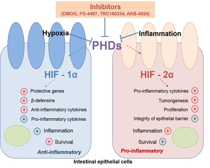
the production of cytokines and leads to the increase in demonstrated the induces of a spontaneous inflammatory process
production of β-defensins (Figure 1), that play a key rolein development in the intestine.
antimicrobial immunity [31]. HIF-2α directly regulates the Thus, to sum up, HIF-1α is an anti-inflammatory agent in IBD
production of some pro-inflammatory cytokines, including tumor and it ensures the integrity of the epithelial barrier and
necrosis factor-α, which leads to HIF-2α-induced inflammation antimicrobial immunity by increasing the synthesis of β-defensins,
[32]. In addition, a prolonged increase in the level of HIF-2α results while HIF-2α activates pro-inflammatory cytokine production and
in a high turnover of tight junction proteins, such as occludin, epithelial proliferation.
which leads to a decrease in the integrity of the epithelial barrier
[33] (Figure 1). It was shown that HIF-2α plays an important role in
reparative processes in wound inflammation [32-33]. It is Perspectives for the treatment of ulcerative colitis
important to note that overexpression of both HIF-1α and HIF-2α On the one hand, “physiological” hypoxia is a marker of the
genes enhances the inflammatory response, which suggests that normal functioning of the intestine and, on the other hand,
activation of HIF-1α does not prevent the pro-inflammatory oxygen-dependent pathways and HIF play important role in the
response, induced by HIF-2α [32]. development of IBDs. Obviously, the search for new molecular
According to the literature, in patients with CD and UC, targets for the pharmacological correction of oxygen-dependent
expression of HIF-1α and HIF-2α is increased in colonocytes [32]. signaling pathways attracts the attention of researchers in terms
Moreover, a positive correlation was detected between the of developing new drugs for the treatment of IBDs.
expression of HIF-1α and the severity of the disease course in Independent studies revealed the protective effect of various
patients with UC during remission and in the acute phase of PHD inhibitors in many experimental models of colitis in 2008. To
diseases [34]. date, some pharmacological “hypoxia mimetics” have been
The most comprehensive study of the expression of HIF-1α developed, used to activate the HIF pathway for therapeutic
and HIF-2α, as well as the effect on the organism of PHD inhibitors effect. Many of these drugs today are non-specific hydroxylase
in connection with systemic disorders in humans, is difficult for inhibitors structurally related to 2-hydroxyglutarate [8, 38]. PHD
several reasons; therefore, these studies are widely conducted in inhibitors such as DMOG, FG-4497 and TRC160334, AKB-4924
experimental models. were tested as therapeutic agents in colitis experimental models
[38]. DMOG and FG-4497 are panhydroxylase inhibitors capable of
The so-called “chemical” models of IBD are most often used in
inhibiting the activity of HIF-PHD and asparaginyl hydroxylase.
experimental studies. Oral or rectal administration of specific
chemical agents results in intestine inflammation that is similar in The authors first identified the protective effects of
clinical and morphological manifestations to UC or CD. Such hydroxylase inhibition in models of colitis, in which the HIF-
chemical agents are, for example, sodium dextran sulfate (Dextran signaling pathways and the regulating inflammation nuclear factor
Sodium Sulfate, DSS), TNBS (2,4,6-Trinitrobenzenesulfonic acid), NF-κB, probably plays an important role [39-42]. These two factors
oxazolone [35]. are interconnected, thus, on the one hand, in the proximal part of
the HIF-1α gene promoter is the NF-κB-binding site [43] and, on
The role of hypoxia-inducible factors in the development of
the other hand, mRNA and HIF-1α protein levels increase
IBD and UC, as well as their influence on the severity of the
dependent on NF-κB activation (the data obtained on the system
inflammatory process, is primarily investigated in experimental
inflammatory response induced by lipopolysaccharide) [44].
models. HIF-1α knockout mice have been shown to be more
susceptible to TNBS colitis [36], but knockout of this gene does not
affect the severity of DSS colitis. The low expression of HIF-2α
reduces the severity of the DSS colitis, despite the increase in the
production of pro-inflammatory cytokines [32]. While
overexpression of HIF-2α leads to a more severe course of UC,
spontaneous inflammation of the intestine [32], as well as
progression of cancer in experimental models [37]. An increase in
the severity of DSS colitis was shown in mice with the deficiency of
VHL that stabilizes HIF-1α and HIF-2α [38].
A lot of articles were devoted to the understanding the role of
mechanisms of oxidative stress, in particular, PHD inhibitors, in
search for the new drugs for the IBD treatment. Chemical
inhibition of PHD, which both results in HIF-1α and HIF-2α subunits
stabilization, does not affect the severity of UC, but instead leads
to its remission through the HIF-1α-dependent pathway [31].
These data may seem contradictory; we discussed above, the
activation of HIF-1α does not reduce the severity of the pro-
inflammatory response induced by HIF-2α [32]. However it is
possible to select a dose of dimethyloxalylglycine (DMOG),
sufficient to activate HIF-1α, but not inducing inflammation along
the HIF-2α-dependent pathway [32]. Also, usage of PHD inhibitors Figure 1. The role of HIFs in intestinal inflammation. PHDs – Prolyl
leads to non-selective, temporary activation of HIF-1α and HIF-2α, Hydroxylase Domain Proteins, Using of PHDs inhibitors leads to non-
selective, temporary activation of HIF-1α and HIF-2α.
but not to prolonged overexpression of HIF-2α, that was
[
© 2020, LLC Science and Innovations, Saratov, Russia www.romj.orgISSN 2304-3415, Russian Open Medical Journal 4 of 5
2020. Volume 9. Issue 1 (March). Article CID e0101
Physiology and Pathophysiology
DOI: 10.15275/rusomj.2020.0101
However therapeutic agents mentioned above are not specific 10. Ordas I, Eckmann L, Talamini M, Baumgart DC, Sandborn WJ.
and the result of their action is not only in HIF-1α subunit Ulcerative colitis. Lancet 2012; 380(9853): 1606-1619.
stabilization, but also in HIF-2α, which can enhance the https://doi.org/10.1016/S0140-6736(12)60150-0.
inflammatory response. Recently it was investigated that, unlike 11. Maeda S, Hsu LC, Liu H, Bankston LA, Iimura M, Kagnoff M. et al. Nod2
HIF-1α, in the structure of the HIF-2α subunit there is a ligand- mutation in Crohn's disease potentiates NF-kappaB activity and IL-
1beta processing. Science 2005; 307(5710): 734-738.
binding cavity. Despite the fact that endogenous substrates have
https://doi.org/10.1126/science.1103685.
not yet been identified, this difference in the structure of subunits
12. Du X, Shi H, Li J, Dong Y, Liang J, Ye J, et al. Mst1/Mst2 regulate
is considered as a promising line of research for highly specific low
development and function of regulatory T cells through modulation of
molecular weight inhibitors as drugs for the control of IBDs in Foxo1/Foxo3 stability in autoimmune disease. J Immunol 2014; 192(4):
general and in particular UC [45]. 1525-1535. https://doi.org/10.4049/jimmunol.1301060.
13. Neefjes J, Jongsma ML, Paul P, Bakke O. Towards a systems
Conclusion understanding of MHC class I and MHC class II antigen presentation.
Nat Rev Immunol 2011; 11(12): 823-836.
Thus, UC is a disease, characterized by the interraption of the https://doi.org/10.1038/nri3084.
intestinal barrier, a disorder of immunity and microbial dysbiosis.
14. Chyornenkaya TV. Pseudomembranous colitis: diagnosis, treatment and
Hypoxia is a key feature of the normal physiology of intestine, and prevention. Russian Sklifosovsky Journal "Emergency Medical Care" 2016;
in inflammatory conditions, such as UC, tissue hypoxia may be (1): 33-39. Russian. https://www.elibrary.ru/item.asp?id=25778788.
aggravated. HIF plays a central role in the normal function of a 15. Trivedi PP, Jena GB. Ulcerative colitis-induced hepatic damage in mice:
healthy intestinal mucosa, and using of PHD inhibitors are studies on inflammation, fibrosis, oxidative DNA damage and GST-P
protective in experimental models of colitis. However, they are not expression. Chem Biol Interact 2012; 201(3): 19-30.
specific and can enhance the inflammatory response in UC. In this https://doi.org/10.1016/j.cbi.2012.12.004.
case, it is important to continue to develop additional therapeutic 16. Keith B, Johnson RS, Simon MC. HIF1alpha and HIF2alpha: sibling
strategies that use the protective potential of oxygen-sensitive rivalry in hypoxic tumour growth and progression. Nat Rev Cancer
methods, taking into account differences in spatial organization of 2011; 12(1): 9-22. https://doi.org/10.1038/nrc3183.
HIF-1α and HIF-2α subunits. 17. Schödel J, Mole DR, Ratcliffe PJ. Pan-genomic binding of hypoxia-
inducible transcription factors. Biol Chem 2013; 394(4): 507-517.
https://doi.org/10.1515/hsz-2012-0351.
Conflict of interest
18. Ivan M, Kondo K, Yang H, Kim W, Valiando J, Ohh M, et al. HIFalpha
The authors declare that they have no conflict of interest. targeted for VHL-mediated destruction by proline hydroxylation:
implications for O2 sensing. Science 2001; 292(5516): 464-468.
References https://doi.org/10.1126/science.1059817.
1. Lukyanova LD. Cellular mechanism responsible for beneficial effects of 19. Jaakkola P, Mole DR, Tian YM, Wilson MI, Gielbert J, Gaskell SJ, et al.
hypoxic therapy. In: Adaptation biology and Medicine, Volume 3. J. Targeting of HIF-alpha to the von Hippel-Lindau ubiquitylation complex
Мoravec, et al., eds. New Dehli: Narosa Puplishing House, 2002: 290- by O2-regulated prolyl hydroxylation. Science 2001; 292(5516): 468-
303. 472. https://doi.org/10.1126/science.1059796.
2. Lyzhko NA. Molecular-genetic mechanisms of initiation, promotion 20. Maxwell PH, Wiesener MS, Chang GW, Clifford SC, Vaux EC, Cockman
and progression of tumors. Russian Journal of Biotherapy 2017; 16(4): ME, et al. The tumour suppressor protein VHL targets hypoxia-
7-17. Russian. https://doi.org/10.17650/1726-9784-2017-16-4-7-17. inducible factors for oxygen-dependent proteolysis. Nature 1999;
399(6733): 271-275. https://doi.org/10.1038/20459.
3. Cinel I, Opal CM. Molecular biology of inflammation and sepsis: a
primer. Crit Care Med 2009; 37(1): 291-304. 21. Bohlen HG. Intestinal tissue pO2 and microvascular responses during
https://doi.org/10.1097/CCM.0b013e31819267fb. glucose exposure. Am J Physiol 1980; 238(2): H164-H171.
https://doi.org/10.1152/ajpheart.1980.238.2.H164.
4. Belousova EA. Ulcerative colitis and Crohn’ disease. Moscow: Triada,
2002; 128 p. Russian. 22. Matheson PJ, Wilson MA, Garrison RN. Regulation of intestinal blood
flow. J Surg Res 2000; 93(1): 182-196.
5. Kapuller LL. Pathological changes of the colon in non-specific
https://doi.org/10.1006/jsre.2000.5862.
inflammatory bowel disease. In: Nonspecific inflammatory bowel
disease. G.I. Vorob’ev, I.L. Khalif, eds. Moscow, Russia: Miklosh, 2008: 23. Hallbäck DA, Hultén L, Jodal M, Lindhagen J, Lundgren O. Evidence for
71-105. Russian. the existence of a countercurrent exchanger in the small intestine in
man. Gastroenterology 1978; 74(4): 683-690.
6. Dzhalilova DS, Polyakova MA, Diatroptov ME, Zolotova NA, Makarova
https://www.ncbi.nlm.nih.gov/pubmed/631505.
OV. Morphological changes in the colon and composition of peripheral
blood lymphocytes in acute colitis in mice with different resistance to 24. Merga Y, Campbell BJ, Rhodes JM. Mucosal barrier, bacteria and
hypoxia. Molecular Medicine 2018; 16(6): 46-50. Russian. inflammatory bowel disease: possibilities for therapy. Dig Dis 2014;
https://doi.org/10.29296/24999490-2018-06-08. 32(4): 475-483. https://doi.org/10.1159/000358156.
7. Kosyreva AM, Dzhalilova DS, Tsvetkov IS, Diatroptov ME, Makarova 25. Ross MH, Pawlina W. Histology: A text and atlas: with correlated cell
OV. Age-specific features of hypoxia tolerance and intensity of and molecular biology. Baltimore, MD: Lippincott Wiliams & Wilkins,
lipopolysaccharide-induced systemic inflammatory response in Wistar 2006; 906 p.
rats. Bull Exp Biol Med 2019; 166(5): 699-703. 26. O'Hara AM, Shanahan F. The gut flora as a forgotten organ. EMBO Rep
https://doi.org/10.1007/s10517-019-04421-3. 2006;7(7): 688-693. https://doi.org/10.1038/sj.embor.7400731.
8. Cummins EP, Crean D. Hypoxia and inflammatory bowel disease. 27. Baumgart DC, Sandborn WJ. Crohn's disease. Lancet 2012; 380(9853):
Microbes Infect 2017; 19(3): 210-221. 1590-1605. https://doi.org/10.1016/S0140-6736(12)60026-9.
https://doi.org/10.1016/j.micinf.2016.09.004. 28. Hsiao EY, McBride SW, Hsien S, Sharon G, Hyde ER, McCue T, et al.
9. Cioffi M, Rosa AD, Serao R, Picone I, Vietri MT. Laboratory markers in Microbiota modulate behavioral and physiological abnormalities
ulcerative colitis: Current insights and future advances. World J associated with neurodevelopmental disorders. Cell 2013; 155(7):
Gastrointest Pathophysiol 2015; 6(1): 13-22. 1451-1463. https://doi.org/10.1016/j.cell.2013.11.024.
[
https://doi.org/10.4291/wjgp.v6.i1.13.
© 2020, LLC Science and Innovations, Saratov, Russia www.romj.orgISSN 2304-3415, Russian Open Medical Journal 5 of 5
2020. Volume 9. Issue 1 (March). Article CID e0101
Physiology and Pathophysiology
DOI: 10.15275/rusomj.2020.0101
29. Kelly CJ, Zheng L, Campbell EL, Saeedi B, Scholz CC, Bayless AJ, et al. Authors:
Crosstalk between microbiota-derived short-chain fatty acids and Ekaterina A. Postovalova – PhD, Researsher, Department of
intestinal epithelial HIF augments tissue barrier function. Cell Host immunomorphology of inflammation, Science Research Institute of Human
Microbe 2015; 17(5): 662-671. Morphology, Moscow, Russia. https://orcid.org/0000-0002-3413-3122.
https://doi.org/10.1016/j.chom.2015.03.005. Olga V. Makarova – DSc, Professor, Head of the Department of
30. Rivera-Chávez F, Zhang LF, Faber F, Lopez CA, Byndloss MX, Olsan EE, immunomorphology of inflammation, Science Research Institute of Human
et al. Depletion of butyrate-producing Clostridia from the gut Morphology, Moscow, Russia. https://orcid.org/0000-0001-8581-107X.
microbiota drives an aerobic luminal expansion of Salmonella. Cell Anna M. Kosyreva – DSc, Leading Researsher, Department of
Host Microbe 2016; 19(4): 443-454. immunomorphology of inflammation, Science Research Institute of Human
https://doi.org/10.1016/j.chom.2016.03.004. Morphology, Moscow, Russia. https://orcid.org/0000-0002-6182-1799.
Dzhuliia S. Dzhalilova – Junior Researcher, Department of
31. Keely S, Campbell EL, Baird AW, Hansbro PM, Shalwitz RA, Kotsakis A,
immunomorphology of inflammation, Science Research Institute of Human
et al. Contribution of epithelial innate immunity to systemic protection
Morphology, Moscow, Russia https://orcid.org/0000-0002-1337-7160.
afforded by prolyl hydroxylase inhibition in murine colitis. Mucosal
Immunol 2014; 7(1): 114-123. https://doi.org/10.1038/mi.2013.29.
32. Xue X, Ramakrishnan S, Anderson E, Taylor M, Zimmermann EM,
Spence JR, et al. Endothelial PAS domain protein 1 activates the
inflammatory response in the intestinal epithelium to promote colitis
in mice. Gastroenterology 2013; 145(4): 831-841.
https://doi.org/10.1053/j.gastro.2013.07.010.
33. Xie L, Xue X, Taylor M, Ramakrishnan SK, Nagaoka K, Hao C, et al.
Hypoxia-inducible factor/MAZ-dependent induction of caveolin-1
regulates colon permeability through suppression of occludin, leading
to hypoxia-induced inflammation. Mol Cell Biol 2014; 34(16): 3013-
3023. https://doi.org/10.1128/MCB.00324-14.
34. Xu C, Dong W. Role of hypoxia-inducible factor-1α in pathogenesis and
disease evaluation of ulcerative colitis. Exp Ther Med 2016; 11(4):
1330-1334. https://doi.org/10.3892/etm.2016.3030.
35. Alex P, Zachos NC, Nguyen T, Gonzales L, Chen TE, Conklin LS, et al.
Distinct cytokine patterns identified from multiplex profiles of murine
DSS and TNBS-induced colitis. Inflamm Bowel Dis 2009; 15(3): 341-352.
https://doi.org/10.1002/ibd.20753.
36. Karhausen J, Furuta GT, Tomaszewski JE, Johnson RS, Colgan SP, Haase
VH. Epithelial hypoxia-inducible factor-1 is protective in murine
experimental colitis. J Clin Invest 2004; 114(8): 1098-1106.
https://doi.org/10.1172/JCI21086.
37. Xue X, Taylor M, Anderson E, Hao C, Qu A, Greenson JK, et al. Hypoxia-
inducible factor-2alpha activation promotes colorectal cancer
progression by dysregulating iron homeostasis. Cancer Res 2012;
72(9): 2285–2293. https://doi.org/10.1158/0008-5472.CAN-11-3836.
38. Shah YM. The role of hypoxia in intestinal inflammation. Mol Cell
Pediatr 2016; 3(1): 1. https://doi.org/10.1186/s40348-016-0030-1.
39. Taylor CT, Colgan SP. Regulation of immunity and inflammation by
hypoxia in immunological niches. Nat Rev Immunol 2017; 17(12): 774-
785. https://doi.org/10.1038/nri.2017.103.
40. Fratantonio D, Cimino F, Speciale A, Virgili F. Need (more than) two to
Tango: Multiple tools to adapt to changes in oxygen availability.
Biofactors 2018; 44(3): 207-218. https://doi.org/10.1002/biof.1419.
41. Krzywinska E, Stockmann C. Hypoxia, metabolism and immune cell
function. Biomedicines 2018; 6(2). pii: E56.
https://doi.org/10.3390/biomedicines6020056.
42. Stothers CL, Luan L, Fensterheim BA, Bohannon JK. Hypoxia-inducible
factor-1α regulation of myeloid cells. J Mol Med (Berl) 2018; 96(12):
1293-1306. https://doi.org/10.1007/s00109-018-1710-1.
43. van Uden P, Kenneth NS, Rocha S. Regulation of hypoxia-inducible
factor-1alpha by NF-kappaB. Biochem J 2008; 412(3): 477-484.
https://doi.org/10.1042/BJ20080476.
44. Frede S, Stockmann C, Freitag P, Fandrey J. Bacterial
lipopolysaccharide induces HIF-1 activation in human monocytes via
p44/42 MAPK and NF-kappaB. Biochem J 2006; 396(3): 517-527.
https://doi.org/10.1042/BJ20051839.
45. Scheuermann TH, Li Q, Ma HW, Key J, Zhang L, Chen R, et al. Allosteric
inhibition of hypoxia inducible factor-2 with small molecules. Nat
Chem Biol 2013; 9(4): 271-276.
https://doi.org/10.1038/nchembio.1185.
[
© 2020, LLC Science and Innovations, Saratov, Russia www.romj.orgYou can also read