Case Series Staphylococcus aureus Infections in the Paediatric Intensive Care Unit: Illustrated Cases
←
→
Page content transcription
If your browser does not render page correctly, please read the page content below
Hindawi
Case Reports in Pediatrics
Volume 2021, Article ID 6661932, 5 pages
https://doi.org/10.1155/2021/6661932

Case Series
Staphylococcus aureus Infections in the Paediatric Intensive Care
Unit: Illustrated Cases

          Kam Lun Hon ,1 Ronald C. M. Fung ,1 Karen K. Y. Leung ,1 Alexander K.C. Leung ,2
          Wun Fung Hui ,1 and Wing Lum Cheung 1
          1
            Paediatric Intensive Care Unit, Department of Paediatrics and Adolescent Medicine, Hong Kong Children’s Hospital,
            Hong Kong, Hong Kong
          2
            Department of Pediatrics, The University of Calgary and The Alberta Children’s Hospital, Calgary, Alberta T2M 0H5, Canada

          Correspondence should be addressed to Kam Lun Hon; ehon@hotmail.com

          Received 24 December 2020; Revised 18 April 2021; Accepted 16 May 2021; Published 4 June 2021

          Academic Editor: Nina L. Shapiro

          Copyright © 2021 Kam Lun Hon et al. This is an open access article distributed under the Creative Commons Attribution License,
          which permits unrestricted use, distribution, and reproduction in any medium, provided the original work is properly cited.

          Staphylococcus aureus is known to be one of the most common gram-positive microorganisms and an important pathogen
          associated with sepsis and toxic shock. We present four anonymized consecutive cases in a paediatric intensive care unit (PICU) to
          illustrate the different clinical manifestations of staphylococcal infections, including local infection versus systemic infection, toxic
          shock versus septic shock, and osteomyelitis. Eczema, short gut syndrome, and scald injury may be associated. Haematologic and
          coagulopathic abnormalities may be present. Prompt diagnosis and use of appropriate antimicrobial treatments is essential to
          reducing mortality and morbidity associated with staphylococcal infections.

1. Introduction                                                            with high fever (40 °C), tachycardia (heart rate 200 per
                                                                           minute), oliguria, and respiratory failure. He was treated
Staphylococcus aureus is known to be one of the most                       with cefotaxime for presumed septic shock. Echocardi-
common gram-positive microorganisms and an important                       ography showed mildly impaired left ventricular func-
pathogen associated with sepsis and toxic shock in paediatric              tion. He was also noted to have hemoglobin 7.7 g/dL,
patients requiring intensive care [1]. There are different                  absolute neutrophil 0.37 × 10⁹/L, and platelet 117,000/
strains of Staphylococcus aureus which can cause a wide                    mm3 . He was suspicious of bone marrow failure, possibly
range of infections due to its extensive virulence factors [2].            consistent with transient erythroblastopenia of child-
We present four consecutive cases to illustrate the different               hood (TEC) or hemophagocytic lymphohistiocytosis
clinical manifestations of staphylococcal infections, in-                  (HLH) or malignancy since he had gross hep-
cluding local infection versus systemic infection, toxic shock             atosplenomegaly and high ferritin (1283 pmol/L), though
versus septic shock, and osteomyelitis [2]. Prompt diagnosis               normal fibrinogen and triglyceride. An urgent bone
and use of appropriate antimicrobial treatments is essential               marrow examination revealed pure red-cell aplasia with
to reducing mortality and morbidity associated with                        granulocytic maturation arrest and reactive changes but
staphylococcus infections.                                                 no malignant infiltration. The patient was stabilized with
                                                                           mechanical ventilation, red-cell transfusion, antibiotics,
2. Case Series                                                             and low-dose adrenaline infusion. He was also noted to
                                                                           have an ulcerated lesion over his left forearm. The family
2.1. Case 1: An Infant with Eczema, Methicillin-Sensitive                  had two cats, and they lived in a village house. Bed bugs
Staphylococcus aureus, and Transient Erythroblastopenia of                 were also found to be present in old furniture. Investi-
Childhood. A 6-month-old boy with eczema presented                         gations did not show evidence of scrub typhus, dengue
2 Case Reports in Pediatrics
fever, or bartonella; but throat swab and left forearm
wound swab revealed heavy growth of methicillin-sen-
sitive Staphylococcus aureus (MSSA) and scant growth of
Staphylococcus epidermidis; the minimum inhibitory
concentrations are 0.25 μg/ml and ≤0.12 μg/ml, respec-
tively. The child’s condition gradually improved after 4
days of treatment. He was able to be extubated and
weaned off inotropic support. Haemoglobin, neutrophil,
and platelet counts normalized over a week.
2.2. Case 2: An Infant with Methicillin-Sensitive Staphylo-
coccus aureus Osteomyelitis. A 6-month-old infant girl, with
a history of eczema and short-gut syndrome due to neonatal
volvulus, requiring parenteral nutrition, was admitted to a
PICU for septic shock and right ankle cellulitis. She had fever
(temperature of 38.1°C), tachycardia (heart beat 190 per
minute), and a red swollen right ankle. Blood culture from
her central venous catheter yielded methicillin-sensitive
Staphylococcus aureus (MSSA). Magnetic resonance imaging Figure 1: Osteomyelitis changes in the right tibial region by MRI.
(MRI) of her right ankle showed right distal tibial osteo-
myelitis, extending into the epiphyseal plate with a large
subperiosteal collection around the distal tibia (Figure 1). impaired left ventricular function. She was intubated for
The patient was treated with vancomycin and clindamycin. suspected acute respiratory distress syndrome (lowest PaO2/
Urgent right tibial debridement was performed. A 6-week FiO2 61.5, FiO2 1.0). Intravenous immunoglobulin (1 g/kg/
course of cloxacillin was added. day) was given for two consecutive days. Multisystem in-
Pus was drained from the subperisosteal space, and volvement was evident from elevated serum creatinine ki-
cultures grew MSSA. She remained hemodynamically stable, nase 5019 IU/L, raised serum alanine aminotransferase
with normal hematologic, renal, and liver functions. After 65 IU/L, lowplatelet count 89,000/mm3 (hematologic), and
the antibiotic therapy and the debridement, she returned to alterations in consciousness (central nervous system).
baseline functioning and was discharged from the PICU 3 Capillary refill time over the right sole and left chest rash was
days later to the surgical team to complete a 6-week course of 10 seconds and 2 seconds, respectively. The capillary refill
antibiotics. became 5 seconds over the chest whilst the rash evolved
(Figure 2). Wound swab culture grew MSSA. Blood cultures
remained negative. This girl met criteria for staphylococcal
2.3. Case 3: Scald Injury Complicated by Staphylococcal Toxic toxic shock syndrome, as per the 2011 case definition from
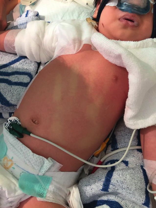
Shock Syndrome. A previously healthy 21-month-old girl the Centers for Disease Control and Prevention (CDC).
presented with fever and shock shortly after wound de- Other blood investigations showed serum lactate
bridement for her scald burn, which was sustained to the 3.8 mmol/L, serum procalcitonin 103.07 ng/ml, C-reactive
right neck, right shoulder, and scapular region down to the protein 246 mg/L, prolonged prothrombin time 25.3 sec-
right arm. The burn, sustained after accidentally pouring a onds, and activated partial thromboplastin time 53 seconds
boiling congee over herself, was of partial thickness (second with normal fibrinogen. The girl’s condition gradually sta-
degree) and covered 5% body surface area. She was initially bilized with mechanical ventilation, inotropic support, and
admitted to the surgical ward, where she had the blisters antibiotic therapy. She was eventually extubated and weaned
deroofed and the wounds dressed in sterile saline gauzes. off all inotropes after 4 days.
Wound debridement was scheduled to be performed in the
operation theatre 3 days later. However, she developed a
fever of up to 38.8°C just before the debridement and the 2.4. Case 4: A teenager with Toxic Shock and Methicillin-
temperature remained elevated (38.7°C) the night after the Resistant Staphylococcus aureus Bacteriaemia. A previously
operation. She had marked tachycardia (150-190/min) and healthy 12-year-old girl presented with a 3-day history of
hypotension (89/40 mm Hg), and capillary refill was 5 fever, menorrhagia, and a painful left submandibular swelling.
seconds. She was lethargic and required 1 L/min nasal ox- She had a mixed picture of hemorrhagic shock and septic
ygen to maintain the saturation. She was noted to have shock, as evidenced by tachycardia, hypotension, and anaemia
diffuse erythroderma over her trunk. She was suspected to (hemoglobin 4.1 g/dL). Respiratory, hepatic, and neurological
have toxic shock syndrome and was transferred to the PICU functions were not affected. She immediately received fluid
for immediate intervention, including fluid resuscitation resuscitation, red-cell transfusion, noradrenaline infusion,
and broad-spectrum antibiotics, cefotaxime, and vanco- and broad-spectrum antibiotic therapy including cefotaxime
mycin. She required inotropic support with dopamine, and vancomycin. Apart from anaemia, blood investigations
adrenaline, and noradrenaline. The vasoactive inotropic revealed neutropenia (zero absolute neutrophil), lymphope-
score was up to 35. Echocardiography revealed mildly nia (absolute lymphocyte 0.1 × 10⁹/L), and thrombocytopeniaCase Reports in Pediatrics 3
atypical as he presented with septic shock, pancytopenia, and
gross hepatosplenomegaly; hence, malignancy was suspected
and supportive treatment for sepsis was started. Although
only the wound swab was positive for Staphylococcus aureus,
the septic shock was likely a result of the systemic effects
from the exfoliative toxins. Transient erythroblastopenia of
childhood (TEC) is an uncommon benign normocytic
anaemia. Although it was observed that TEC often follows a
viral-like illness, no specific aetiologic agent was identified
[6]. We were unable to find any literature reporting the
association of TEC with Staphylococcus aureus infections.
Furthermore, TEC is uncommon in infants less than 1 year
of age, and most patients with TEC are hemodynamically
stable [6]. Therefore, we are of the view that TEC could be a
coincidental finding in our patient with eczema infected by
Staphylococcus aureus. Staphylococcus aureus is an impor-
tant infectious pathogen in children with eczema, and the
clinical features (especially severity and lesion intensity) are
useful indicators in “predicting” moderate-to-heavy
Staphylococcus aureus colonization and infection in children
Figure 2: Deteriorated central capillary refill with palm mark on with eczema [7–9].
the torso.
3.2. Systemic Disseminated Infection and Osteomyelitis. In
3
(platelet 19,000/mm ). Serum amylase was normal (61 IU/L). Case 2, osteomyelitis was immediately suspected as the
The blood culture was positive for methicillin-resistant infant presented with septic shock and right ankle cellulitis.
Staphylococcus aureus (MRSA, Panton–Valentine leucocidin Osteomyelitis was the result of haematogenous dissemina-
negative). Serologic evaluation ruled out Epstein–Barr virus, tion of staphylococcal infection [10]. Early diagnosis is the
cytomegalovirus, measles, varicella-zoster virus, parvovirus, cornerstone of the successful management of this disease
and human immunodeficiency virus. Computed tomography [10]. In view of the history of eczema and need for long-term
of the neck and thorax showed diffuse left parotid swelling central-line use, longer duration of cloxacillin coverage is
with reactive inflammatory features and enlarged left upper required. Empiric antibiotic treatment consists of a short
cervical lymph nodes without abscess. Bone marrow exam- intravenous cycle based on antistaphylococcal penicillin or a
ination revealed a markedly hypocellular marrow without cephalosporin in children aged over 3 months, with the
hemopoietic elements, blasts, or abnormal infiltrates. The addition of gentamicin in infants aged under 3 months. An
girl’s condition stabilized, and she was discharged from the oral regimen may also be an option depending on the
PICU after 4 days. bioavailability of the antibiotic chosen and the assessment of
clinical and laboratory data [10].
3. Discussion
3.3. Local Infection and Toxic Shock Syndrome. For Case 3,
Staphylococcus aureus is the leading cause of infection in
the predisposing factor for toxic shock syndrome (TSS) was
critical-care settings accounting for significant morbidity
the scald injury. TSS is primarily the result of a superantigen-
and mortality [3]. It is capable of causing a wide range of
mediated cytokine storm and M-protein-mediated neutro-
infections in humans, from life-threatening infections in
phil activation, resulting in the release of mediators leading
otherwise healthy individuals to nosocomial infections
to respiratory failure, vascular leakage, and shock [11]. It is
complicating the clinical course of patients with other
caused by the toxin-producing strains of Staphylococcus
primary medical or surgical disease processes [4, 5]. There
aureus and Streptococcus pyogenes [12]. Management of TSS
are many different strains of Staphylococcus aureus, and
should be aggressive. The majority of patients with TSS will
there is controversy over whether all strains are equally
require admission to an intensive care unit for supportive
pathogenic or whether the invasive disease is associated with
care, particularly in the case of multiple organ failure
particular virulent genotypes [5]. The five stages in the
[12–14]. A surveillance study in the UK reported that about
pathogenesis of Staphylococcus aureus infections are colo-
8% of TSS were associated with burns; hence, physicians
nization, local infection, systemic disseminated infection
should be on alert that this can be a possible cause in
and/or sepsis, metastatic infection, and toxinosis. Some of
children with burns who suddenly become unwell [12, 14].
these stages can be clearly identified and delineated in the
Antibiotic treatment should cover both Staphylococcus au-
cases we reviewed (Table 1) [2].
reus and Streptococcus pyogenes, for example, a combination
of cephalosporin, penicillin, and vancomycin. The addition
3.1. Local Infection with Systemic Effects. In the case of the of clindamycin or gentamicin can also reduce toxin pro-
infant with eczema (Case 1), the patient’s presentation was duction and mortality [15, 16]. Furthermore, it is also4 Case Reports in Pediatrics
Table 1: Staphylococcus aureus disease in the PICU.
Case 1 Case 2 Case 3 Case 4
Gender Male Female Female Female
Age 5 months 6 months 21 months 12 years
Short-gut syndrome,
Premorbid Eczema Scald injury Healthy
Broviac line, and eczema
Organ system
Impaired LV function.
Cardiovascular Impaired LV function Normal Low diastolic pressure
Low diastolic pressure
Mechanical ventilation for
Pulmonary Normal Intubated for ARDS Normal
respiratory failure
Pancytopenia with gross Pancytopenia. Aplastic
Thrombocytopenia.
Haematologic hepatosplenomegaly. Normal Normal anaemia. Normal
Prolonged PT and aPTT
clotting clotting
Renal Normal Normal Normal Normal
Hepatic Normal Normal Raised ALT Normal
CNS Normal Normal Lethargic Normal
Sites of Blood culture from Broviac
Throat swab (MSSA) + wound
Staphylococcus line (MSSA), subperiosteal Wound swab (MSSA) Blood culture (MRSA)
swab (S. epidermidis)
aureus abscess (MSSA)
Septic shock and TEC, possibly MRSA septicemia
MSSA, toxic shock
Final diagnosis associated with bed bug bite and MSSA, osteomyelitis associated with aplastic
syndrome following scald
MSSA anaemia
ARDS: acute respiratory distress syndrome. LV: left ventricular. MSSA: methicillin-sensitive Staphylococcus aureus. MRSA: methicillin-resistant Staphy-
lococcus aureus. TEC: transient erythroblastopenia of childhood.
important to remove the foci of bacterial toxin production as including local infection versus systemic infection, toxic
the outcomes are worse in patients who do not have the shock versus septic shock, and osteomyelitis (Table 1). Ec-
source of infection removed [12, 13]. Although the use of zema, short-gut syndrome, and scald injury may be asso-
intravenous immune globulin (IVIG) therapy in children ciated. Haematologic and coagulopathic abnormalities may
with TSS has been controversial, the use of IVIG may be be present. Prompt diagnosis with aggressive support
considered in patients with severe staphylococcal TSS who treatments and antimicrobials are essential in managing and
are unresponsive to other therapeutic measures even though improving clinical outcomes of these critically ill children.
the optimal IVIG regimen has not been sufficiently inves-
tigated [17]. Data Availability
Vital signs and some blood parameters were included in this
3.4. Systemic Infection with MRSA. In Case 4, the teenager retrospective review of anonymized cases.
had hemorrhagic and septic shock due to MRSA bacter-
aemia. Pancytopenia led to a blood marrow examination
that revealed aplastic anaemia. Apart from MRSA, other Ethical Approval
infectious causes for aplastic anaemia were not found. In- Ethical approval to review mortality and morbidity of PICU
fection is a major cause of death for patients with aplastic admissions has been granted.
anaemia, while infections associated with neutropenia are a
major cause of morbidity and mortality in this patient
population [18]. Recovery from neutropenia is directly re- Conflicts of Interest
lated to survival, and therefore, aplastic anaemia patients Professor Alexander KC Leung serves on the editorial board
who are severely neutropenic should ideally be nursed in and is one of the academic editors of Case Report in Pe-
isolation, while prophylactic antibiotics and antifungals diatrics. This manuscript was sent out for independent peer
should also be considered [19, 20]. review.
4. Conclusions References
Staphylococcus aureus is an important human pathogen, and [1] M. Gimenes, T. P. Salci, M. C. B. Tognim, V. L. D. Siqueira,
paediatricians must not overlook its potential to inflict and S. M. Caparroz-Assef, “Treating Staphylococcus aureus
significant morbidity and mortality [21]. The four cases il- infections in an intensive care unit at a University Hospital in
lustrate severe staphylococcal disease affecting children of all Brazil,” International Journal of Clinical Pharmacy, vol. 38,
ages by different pathogenesis and clinical manifestations, no. 2, pp. 228–232, 2016.Case Reports in Pediatrics 5
[2] G. L. Archer, “Staphylococcus aureus: a well-armed pathogen,” American Academy of Pediatrics (AAP), Itasca, IL, USA, 31st
Clinical Infectious Diseases, vol. 26, no. 5, pp. 1179–1181, 1998. edition, 2018.
[3] A. P. Kourtis, K. Hatfield, J. Baggs et al., “Vital signs:epide- [18] H. A. Torres, G. P. Bodey, K. V. I. Rolston, H. M. Kantarjian,
miology and recent trends in methicillin-resistant and in I. I. Raad, and D. P. Kontoyiannis, “Infections in patients with
methicillin-susceptiblestaphylococcus aureusbloodstream in- aplastic anemia,” Cancer, vol. 98, no. 1, pp. 86–93, 2003.
fections-United States,” MMWR. Morbidity and Mortality [19] J. M. Valdez, P. Scheinberg, N. S. Young, and T. J. Walsh,
Weekly Report, vol. 68, no. 9, pp. 214–219, 2019. “Infections in patients with aplastic anemia,” Seminars in
[4] G. R. Sampedro and J. B. Wardenburg, “Staphylococcus aureus Hematology, vol. 46, no. 3, pp. 269–276, 2009.
in the intensive care unit: are these golden grapes ripe for a [20] S. B. Killick, N. Bown, J. Cavenagh et al., “Guidelines for the
new approach?” Journal of Infectious Diseases, vol. 215, no. 1, diagnosis and management of adult aplastic anaemia,” British
pp. S64–S70, 2017. Journal of Haematology, vol. 172, no. 2, pp. 187–207, 2016.
[5] D. C. Melles, R. F. J. Gorkink, H. A. M. Boelens et al., “Natural [21] F. Miles, L. Voss, E. Segedin, and B. J. Anderson, “Review of
population dynamics and expansion of pathogenic clones of Staphylococcus aureus infections requiring admission to a
Staphylococcus aureus,” Journal of Clinical Investigation, paediatric intensive care unit,” Archives of Disease in Child-
vol. 114, no. 12, pp. 1732–1740, 2004. hood, vol. 90, no. 12, pp. 1274–1278, 2005.
[6] M. van den Akker, Y. Dror, and I. Odame, “Transient
erythroblastopenia of childhood is an underdiagnosed and
self-limiting disease,” Acta Paediatrica, vol. 103, no. 7,
pp. e288–e294, 2014.
[7] K. L. Hon, Y. C. Tsang, N. H. Pong, C. Ng, M. Ip, and
T. F. F. Leung, “Clinical features and Staphylococcus aureus
colonization/infection in childhood atopic dermatitis,”
Journal of Dermatological Treatment, vol. 27, no. 3, pp. 1–6,
2015.
[8] K. L. Hon, K. Y. Tsang, J. S. Kung, T. F. Leung, C. W. Lam, and
C. K. Wong, “Clinical signs, Staphylococcus and atopic ec-
zema-related seromarkers,” Molecular Cell, vol. 22, no. 2,
p. E291, 2017.
[9] K. L. Hon, Y. C. K. Tsang, N. H. Pong, T. F. Leung, and M. Ip,
“Exploring Staphylococcus epidermidis in atopic eczema:
friend or foe?” Clinical and Experimental Dermatology, vol. 41,
no. 6, pp. 659–663, 2016.
[10] G. Autore, L. Bernardi, and S. Esposito, “Update on acute
bone and joint infections in paediatrics: a narrative review on
the most recent evidence-based recommendations and ap-
propriate antinfective therapy,” Antibiotics, vol. 9, no. 8,
p. 486, 2020.
[11] D. E. Low, “Toxic shock syndrome,” Critical Care Clinics,
vol. 29, no. 3, pp. 651–675, 2013.
[12] S. Adalat, T. Dawson, S. J. Hackett, and J. E. Clark, “Toxic
shock syndrome surveillance in UK children,” Archives of
Disease in Childhood, vol. 99, no. 12, pp. 1078–1082, 2014.
[13] J. Zimbelman, A. Palmer, and J. Todd, “Improved outcome of
clindamycin compared with beta-lactam antibiotic treatment
for invasive Streptococcus pyogenes infection,” The Pediatric
Infectious Disease Journal, vol. 18, no. 12, pp. 1096–1100, 1999.
[14] A. P. Brown, K. Khan, and S. Sinclair, “Bacterial toxicosis/
toxic shock syndrome as a contributor to morbidity in
children with burn injuries,” Burns, vol. 29, no. 7,
pp. 733–738, 2003.
[15] P. M. Schlievert and J. A. Kelly, “Clindamycin-induced
suppression of toxic-shock syndrome-associated exotoxin
production,” Journal of Infectious Diseases, vol. 149, no. 3,
p. 471, 1984.
[16] P. Van Langevelde, J. T. Van Dissel, C. J. Meurs, J. Renz, and
P. H. Groeneveld, “Combination of flucloxacillin and gen-
tamicin inhibits toxic shock syndrome toxin 1 production by
Staphylococcus aureus in both logarithmic and stationary
phases of growth,” Antimicrobial Agents and Chemotherapy,
vol. 41, no. 8, pp. 1682–1685, 1997.
[17] American Academy of Pediatrics, “Staphylococcus aureus,” in
Red Book: 2018 Report of the Committee on Infectious Diseases,
D. Kimberlin, M. Brady, M. Jackson, and S. Long, Eds.,You can also read