PLACENTA IN SARS-COV-2 INFECTION: A NEW TARGET FOR INAMMATORY AND THROMBOTIC EVENTS - RESEARCH SQUARE
←
→
Page content transcription
If your browser does not render page correctly, please read the page content below
Placenta in SARS-CoV-2 infection: a new target for
in ammatory and thrombotic events
Luca Bertero
Pathology Unit, Department of Medical Sciences, University of Turin, Turin, Italy
Fulvio Borella
Obstetrics and Gynecology 1U, Department of Surgical Sciences, Sant'Anna Hospital, University of Turin, Turin, Italy
Giovanni Botta
Pathology Unit, “Città della Salute e della Scienza di Torino” University Hospital, Turin, Italy
Andrea Carosso
Obstetrics and Gynecology 1U, Department of Surgical Sciences, Sant'Anna Hospital, University of Turin, Turin, Italy
Stefano Cosma
Obstetrics and Gynecology 1U, Department of Surgical Sciences, Sant'Anna Hospital, University of Turin, Turin, Italy
Marialuisa Bovetti
Obstetrics and Gynecology 1U, Department of Surgical Sciences, Sant'Anna Hospital, University of Turin, Turin, Italy
Marco Carosso
Obstetrics and Gynecology 1U, Department of Surgical Sciences, Sant'Anna Hospital, University of Turin, Turin, Italy
Giancarlo Abbona
Pathology Unit, “Città della Salute e della Scienza di Torino” University Hospital, Turin, Italy
Mauro Papotti
Pathology Unit, Department of Oncology, University of Turin, Turin, Italy
Paola Cassoni ( paola.cassoni@unito.it )
Pathology Unit, Department of Medical Sciences, University of Turin, Turin, Italy
Chiara Benedetto
Obstetrics and Gynecology 1U, Department of Surgical Sciences, Sant'Anna Hospital, University of Turin, Turin, Italy
Research Article
Keywords: COVID-19, SARS-CoV-2, pathology, thrombosis, in ammation, obstetrics, gynecology, placenta, preterm
DOI: https://doi.org/10.21203/rs.3.rs-30412/v1
License: This work is licensed under a Creative Commons Attribution 4.0 International License. Read Full License
Page 1/16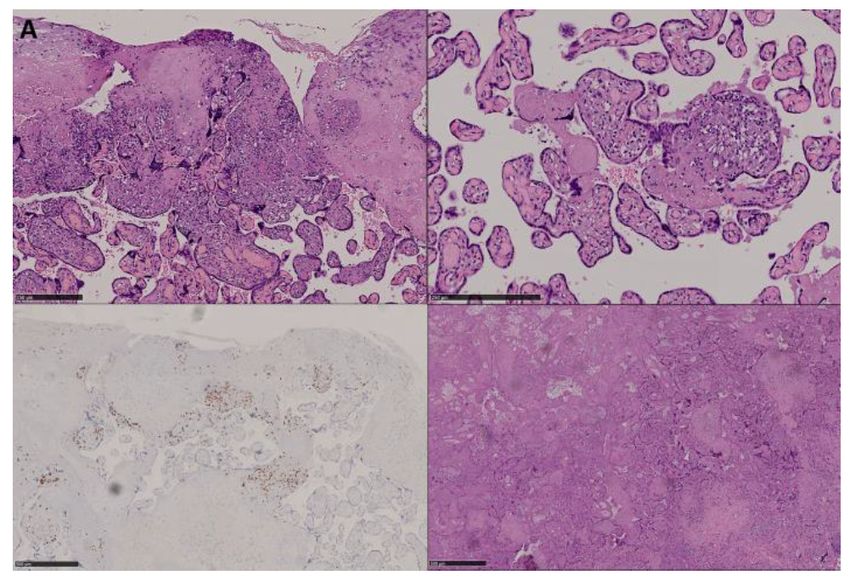
Abstract
Infection by SARS-CoV-2 has been shown to involve a wide range of organs and tissues, leading to a kaleidoscope of clinical
conditions. Within this spectrum, an increased rate of preterm deliveries has been reported in women with COVID-19. High
expression of proteins (ACE2/TMPRSS2) required for SARS- CoV-2 cell entry has been observed in the maternal-fetal tissues,
supporting the possibility of placental viral involvement. A consecutive series of 6 placentas, delivered by 5 women with COVID-
19 have been investigated. Three out of six placentas showed changes consistent with chronic villitis, while in one case chronic
histiocytic intervillositis was diagnosed. Vascular abnormalities consisting of thrombo- hemorrhagic alterations were identi ed
in the three cases with chronic villitis. These pathological ndings: i) provide a basis to explain the higher rate of obstetric
complications in these patients; 2) suggest the need to investigate heparin use in COVID-19 affected pregnant patients to
prevent adverse outcomes.

Background
On March 12, 2020, due do the rapid escalation of the coronavirus disease 2019 (COVID–19) outbreak, the WHO declares a
pandemic.1 COVID–19 is caused by the severe acute respiratory syndrome coronavirus 2 (SARS-CoV–2), a betacoronavirus
similar to SARS-CoV and MERS-CoV, with multiple possible transmission routes and characterized by a high infectivity.2, 3
Emerging data on maternal outcomes of COVID–19 suggest that clinical course is similar to non-pregnant patients4, 5, 6 and
outcomes of perinatal COVID–19 infection are reassuring.7 However, adverse effects on newborns, such as fetal and respiratory
distress, thrombocytopenia accompanied by abnormal liver function and even death have been reported.8 In addition, evidence
of an higher incidence of preterm birth is being increasingly reported.9, 10 Concern is also rising on the possibility that SARS-
CoV–2 may be vertically transmitted,11, 12, 13 but most patient series do not report cases of proved vertical transmission.9, 10,
14

To date, reported data regarding placental histopathology in COVID–19 are extremely limited (four cases in total),15, 16 but high
expression of proteins (ACE2/TMPRSS2) required for SARS-CoV–2 cell entry has been observed in maternal-fetal interface
tissues enabling the possibility of placental viral involvement.17 Moreover, the increased thrombotic risk and the in ammatory
activation described in patients with COVID–1918, 19, 20 make the placenta a potential target of pathophysiological
phenomena which could affect pregnancy outcomes and support the preliminary evidence of increased preterm birth risk.

For all these reasons, histological analysis of placentas delivered by women with COVID–19 could add insightful information.
The aim of this work was to evaluate the consecutive series of placentas delivered at our Institution from women with COVID–
19 infection, investigating the occurrence of pathological changes and the potential correlations with obstetric outcomes.

Case Series
Case 1 - Pathological ndings
Placenta weight was 590 grams before formalin xation, lateral dimensions were 16 cm x 15 cm with thickness ranging from 2
cm to 3 cm. Umbilical cord length was 45 cm, its structure normal and a peripheral insertion was observed. Placental
macroscopic features were normal with translucent membranes. Histological examination showed only mild abnormalities
including a scant lymphocytic in ammation which involved both the decidua and basal placental villi with initial villous
agglutination (Figure 2A–2B). Lymphocytes showed a prevalent T cell cytotoxic (CD8-positive) phenotype (Figure 2C–2D).
These ndings were consistent with chronic villitis (low grade, multifocal since in ammatory foci were present on more than
one slide).21 Focal intervillous hemorrhage was also identi ed, while maturation was normal with features suggesting initial
hypermaturation.

Case 2 - Pathological ndings

                                                           Page 2/16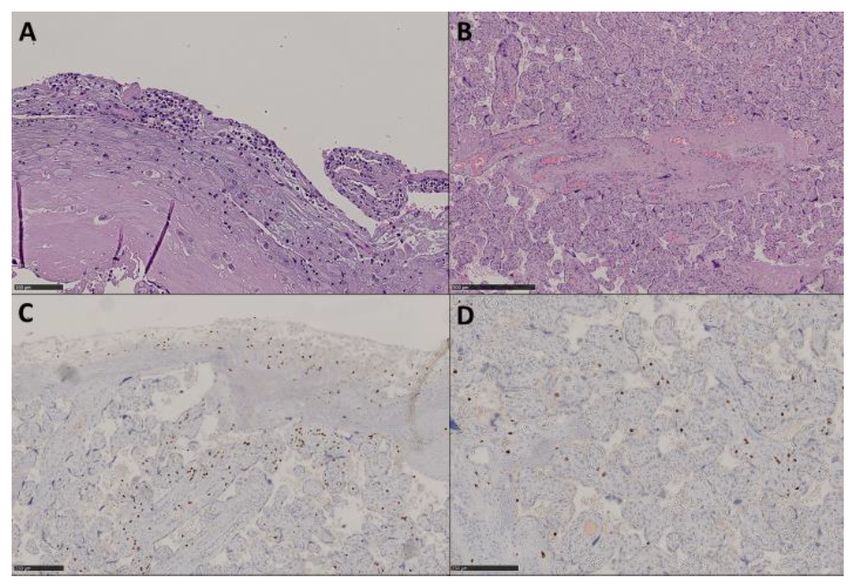
Placenta weighted 429 grams before xation, dimensions were 15.5 cm x 15 cm x 2 to 3.1 cm thickness. Umbilical cord (length:
16 cm) and membranes showed a normal morphology. After sectioning, focal and small whitish areas were observed close to
the chorial surface and multiple, small (largest dimension
In 4 cases, we found microscopic signs of in ammation involving the decidua and the chorial villi: three out of four cases
showed a CD8-positive T cell lymphocytic in ltrate, while in the remaining case an intervillous histiocytic in ltrate was identi ed.

The presence of an in ammatory lymphocytic in ltrate (cytotoxic CD8+ T cell lymphocytes in particular, as observed in our
series) and of villous damage are consistent with the diagnosis of chronic villitis.22, 23 Of note, none of these three patients
had comorbidities that could justify the presence of placental in ammation. Chronic villitis etiology is often related to an
undergoing infection, although in most cases it is not possible to identify the speci c etiopathogenetic agent: these cases are
de ned as “villitis of unknown etiology” (VUE) and can be found in 2–33.8% of placentas.24 Within the group of chronic villitis
secondary to maternal infections, viruses are the most frequently involved pathogens and many speci c agents, such as
varicella zoster, herpes simplex virus, cytomegalovirus, and in uenza A/H1N1, have been found to be associated with this
condition.25, 26, 27, 28 Little is known about the association between placental pathology and coronavirus infections. Seven
placentas from coronavirus-infected women during the 2002–2004 SARS outbreak have been previously described:29
histological examination found no abnormalities in 2/7, an increase in subchorionic and intervillous brin in 3/7, and massive
thrombotic vasculopathy associated with intra-uterine growth restriction (IUGR) in the remaining 2/7 cases. No signs of chronic
villitis were detected in any case and the same is true for MERS infections.30 Based on present knowledge, infection by SARS-
CoV–2 is characterized by an extensive immune response with activation of both CD4+ and CD8+ lymphocytes18, 31, 32, thus
 ndings consistent with chronic villitis could be expected in the presence of placental involvement, also considering the high
expression of ACE2 in decidual cells, villous cytotrophoblast and syncytiotrophoblast cells.17

Moreover, experiences derived from in uenza A/H1N1 infections in pregnancy suggest that activation of cellular immune
response with release of in ammatory cytokines can lead to indirect placental damage.28 Considering this evidence, the
placenta could be potentially harmed because of intrinsic active virus replication and/or through indirect activity of
in ammatory cytokines.33 Viral in situ assessment (by immunohistochemistry for viral proteins and/or in situ hybridization of
viral RNA) will be important to de ne which speci c mechanisms are involved in this setting.

In the fourth case with in ammatory abnormalities, features consistent with chronic histiocytic intervillositis were identi ed.
This rare entity is usually associated with maternal immunologic conditions like systemic lupus erythematosus, lupus
anticoagulant and antiphospholipid syndrome.34, 35 Despite the different type of in ammatory in ltrate (histiocytic versus
lymphocytic) and of etiologies (chronic histiocytic intervillositis is usually not related to viral infections), chronic histiocytic
villositis is frequently associated with chronic villitis (30% to 50% of cases), supporting a potential overlap in terms of involved
immunologic pathways.36, 37 Moreover, alveolar histiocytic in ltration seems to be a frequent autoptic hallmark of SARS-CoV–
2 infection.38 Lastly, it has to be noted that this patient’s signi cant comorbidities (chronic hypertension, severe obesity, and
gestational diabetes) are not usually associated with chronic histiocytic intervillositis.

Our data open up an interesting perspective: the three patients with chronic villitis had a shorter/milder history of COVID–19
(
Regarding our knowledge about the clinical outcomes of pregnancy in COVID–19 patients, it is based on limited series and
systematic reviews. Chen et al.6 reported the outcomes of a relatively large series of pregnant women affected by COVID–19
observing a high rate (14/68, 21%) of preterm delivery. A systematic review evaluating 41 pregnancies affected by COVID–19
con rmed this observation, reporting preterm birth (< 37 weeks) in above 40% of pregnancies.9

Could the poorer obstetrics outcomes reported in women with COVID–19 be related to SARS-CoV–2- induced in ammatory
and/or vascular abnormalities? The ndings of our study provide an initial pathological basis for these clinical ndings: chronic
villitis/VUE as well as vascular abnormalities are associated with several obstetric and neonatal adverse outcomes including
preterm birth.22, 23, 27, 42, 43, 44 Moreover, it is well known that an in ammatory/infectious reaction can lead to preterm
delivery by increasing prostaglandins secretion or by impairing the fetal immune tolerance.

Negative results of SARS-CoV–2 placental swabs observed in our series should not be taken as a conclusive evidence of SARS-
CoV–2 absence in these tissues. This nding could rather depend on compartmentalization of virus replication within speci c
cell types. This hypothesis is supported by the differential expression of ACE2 and TMPRSS2, proteins required for SARS-CoV–2
cell entry, observed in the maternal-fetal interface tissues.17, 45 This open question could also be e caciously addressed by in
situ viral assays which will hopefully become more widely available in the coming weeks.

The main limitation of our observations is that our pathological ndings are shared by multiple conditions and etiologies, which
could have been pre-existent at time of SARS-CoV–2 infection. Nevertheless, some considerations support this relationship: i)
the observed rate of abnormalities is higher compared to what could have been expected in a consecutive unselected series of
placentas; ii) ndings are consistent with other viral infections and with our current knowledge about SARS-CoV–2 pathogenetic
mechanisms; iii) patients’ histories did not include comorbidities justifying the identi ed pathological hallmarks. Analysis of
larger series of cases and in situ viral assays will be needed to de nitively prove and characterize the causative role of SARS-
CoV–2. Nevertheless, our ndings are important to shed a light on the reported associations between COVID–19 infection and
poorer obstetrics outcomes.

Finally, can therapeutic implications be derived from our data? As prophylactic low-molecular-weight heparin is recommended in
pregnant women with high thrombotic risk,46, 47 our ndings provide a pathological basis to consider heparin use in COVID–19
affected pregnant patients to prevent in ammatory and vascular thrombotic complications. This suggestion is in line with the
recent proposal of The Society of Thrombosis and Haemostasis which recommends (if not otherwise contraindicated) the use
of prophylactic low molecular weight heparin in all patients who require hospitalization for COVID–19, regardless of illness
severity.48

Materials And Methods
Our study is based on the pathological analysis of 6 consecutive placentas delivered from 5 women affected by COVID–19
admitted to the Obstetrics and Gynecology clinic of S. Anna Hospital—”Città della Salute e delle Scienza di Torino”, University of
Turin from March 22, 2020 to April 23, 2020. Sample collection was performed within the framework of the “SARS-CoV–2
infection during pregnancy and puerperium: an Italian Obstetric Surveillance System (ItOSS)" study by the Italian National
Institute of Health. All patients showed symptomatic COVID–19 disease con rmed by a nasopharyngeal swab according to
World Health Organization (WHO) guidelines. SARS-CoV–2 RNA was detected using an automated real-time RT-PCR assay
[DiaSorin Molecular Simplexa™ COVID–19 Direct, target genes S and ORF1ab]. Pathological examination was performed
according to routine procedures and immunohistochemistry was set up on a BenchMark ULTRA platform (Ventana Medical
Systems Inc., Tucson, AZ, USA). The study was conducted in accordance with The Code of Ethics of the World Medical
Association (Declaration of Helsinki and following amendments) for studies involving humans and within the guidelines and
regulations de ned by the Research Ethics Committee of the University of Turin. Patients signed a written informed consent.
Data supporting study results are included within the manuscript.

Declarations

                                                            Page 5/16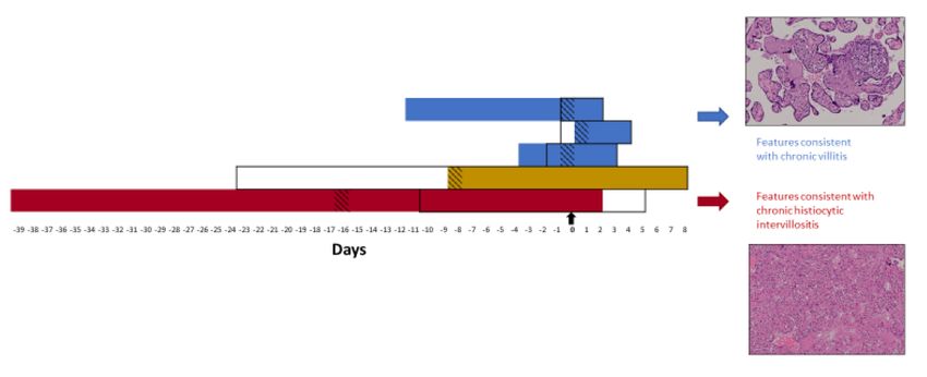
Con icts of Interest
The authors declare they have no con ict of interest.

Acknowledgements
The present study was kindly supported by a grant from “Banca del Piemonte” dedicated to COVID-19 research.

Authorship statement
GB, PC and CB designed the research; all authors contributed to data collection and analysis; LB, FB, GB, AC and SC drafted the
paper; all authors edited the paper and approved the nal version.

References
  1. World Health Organization. WHO announces COVID-19 outbreak a pandemic (http://www.euro.who.int/en/health-
     topics/health-emergencies/coronavirus-covid- 19/news/news/2020/3/who-announces-covid-19-outbreak-a-pandemic).)
     (2020).

  2. Wang W, et al. Detection of SARS-CoV-2 in Different Types of Clinical Specimens. JAMA, (2020).

  3. Xiao F, Tang M, Zheng X, Liu Y, Li X, Shan H. Evidence for Gastrointestinal Infection of SARS-CoV-2. Gastroenterology,
     (2020).

  4. Dashraath P, et al. Coronavirus disease 2019 (COVID-19) pandemic and pregnancy. Am J Obstet Gynecol, (2020).

  5. Chen H, et al. Clinical characteristics and intrauterine vertical transmission potential of COVID-19 infection in nine pregnant
     women: a retrospective review of medical records. Lancet 395, 809- 815 (2020).

  6. Chen L, et al. Clinical Characteristics of Pregnant Women with Covid-19 in Wuhan, China. N Engl J Med, (2020).

  7. Breslin N, et COVID-19 infection among asymptomatic and symptomatic pregnant women: Two weeks of con rmed
     presentations to an a liated pair of New York City hospitals. Am J Obstet Gynecol MFM, 100118 (2020).

  8. Zhu H, et Clinical analysis of 10 neonates born to mothers with 2019-nCoV pneumonia. Transl Pediatr 9, 51-60 (2020).

  9. Di Mascio D , et al. Outcome of Coronavirus spectrum infections (SARS, MERS, COVID 1 -19) during pregnancy: a systematic
     review and meta-analysis. Am J Obstet Gynecol MFM, 100107 (2020).

10. Yan J, et Coronavirus disease 2019 (COVID-19) in pregnant women: A report based on 116 cases. Am J Obstet Gynecol,
    (2020).

11. Carosso A, et Pre-labor anorectal swab for SARS-CoV-2 in COVID-19 patients: is it time to think about it? Eur J Obstet
    Gynecol Reprod Biol, (2020).

12. Alzamora MC, Paredes T, Caceres D, Webb CM, Valdez LM, La Rosa M. Severe COVID-19 during Pregnancy and Possible
    Vertical Transmission. Am J Perinatol, (2020).

13. Dong L, et Possible Vertical Transmission of SARS-CoV-2 From an Infected Mother to Her Newborn. JAMA, (2020).

14. Della Gatta AN, Rizzo R, Pilu G, Simonazzi COVID19 during pregnancy: a systematic review of reported cases. Am J Obstet
    Gynecol, (2020).

                                                             Page 6/16
15. Chen S , et [Pregnant women with new coronavirus infection: a clinical characteristics and placental pathological analysis
of three cases]. Zhonghua Bing Li Xue Za Zhi 49, E005 (2020).
16. Baud D , et al. Second-Trimester Miscarriage in a Pregnant Woman With SARS-CoV-2 JAMA, (2020).
17. Li M, Chen L, Zhang J, Xiong C, Li X. The SARS-CoV-2 receptor ACE2 expression of maternal-fetal interface and fetal organs
by single-cell transcriptome study. PLoS One 15, e0230295 (2020).
18. Huang C, et Clinical features of patients infected with 2019 novel coronavirus in Wuhan, China. Lancet 395, 497-506 (2020).
19. Tang N, Li D, Wang X, Sun Z. Abnormal coagulation parameters are associated with poor prognosis in patients with novel
coronavirus J Thromb Haemost 18, 844-847 (2020).
20. Schett G, Sticherling M, Neurath MF. COVID-19: risk for cytokine targeting in chronic in ammatory diseases? Nat Rev
Immunol, (2020).
21. Khong TY, et Sampling and De nitions of Placental Lesions: Amsterdam Placental Workshop Group Consensus Statement.
Arch Pathol Lab Med 140, 698-713 (2016).
22. Boog G. Chronic villitis of unknown etiology. Eur J Obstet Gynecol Reprod Biol 136, 9-15 (2008).
23. Kim CJ, Romero R, Chaemsaithong P, Kim Chronic in ammation of the placenta: de nition, classi cation, pathogenesis,
and clinical signi cance. Am J Obstet Gynecol 213, S53-69 (2015).
24. Khong TY, Mooney EE, Nikkels PGJ, Morgan TK, Gordijn Pathology of the placenta : a practical guide. Springer (2019).
25. Altshuler G, Russell The human placental villitides: a review of chronic intrauterine infection. Curr Top Pathol 60, 64-112
(1975).
26. Becroft DM, Thompson JM, Mitchell Placental villitis of unknown origin: epidemiologic associations. Am J Obstet Gynecol
192, 264-271 (2005).
27. Tamblyn JA, Lissauer DM, Powell R, Cox P, Kilby The immunological basis of villitis of unknown etiology - review. Placenta
34, 846-855 (2013).
28. Meijer WJ, Wensing AM, Bruinse HW, Nikkels PG. High rate of chronic villitis in placentas of pregnancies complicated by
in uenza A/H1N1 infection. Infect Dis Obstet Gynecol 2014, 768380 (2014).
29. Ng WF, et The placentas of patients with severe acute respiratory syndrome: a pathophysiological evaluation. Pathology 38,
210-218 (2006).
30. Jeong SY, et al. MERS-CoV Infection in a Pregnant Woman in Korea. J Korean Med Sci 32, 1717- 1720 (2017).
31. Qin C, et al. Dysregulation of immune response in patients with COVID-19 in Wuhan, China. Clin Infect Dis, (2020).
32. Cao X. COVID-19: immunopathology and its implications for therapy. Nat Rev Immunol, (2020).
33. Liu H, Wang LL, Zhao SJ, Kwak-Kim J, Mor G, Liao AH. Why are pregnant women susceptible to COVID-19? An
immunological viewpoint. J Reprod Immunol 139, 103122 (2020).
34. Marchaudon V, Devisme L, Petit S, Ansart-Franquet H, Vaast P, Subtil D. Chronic histiocytic intervillositis of unknown
etiology: clinical features in a consecutive series of 69 Placenta 32, 140-145 (2011).
35. Mekinian A, et Chronic histiocytic intervillositis: outcome, associated diseases and treatment in a multicenter prospective
study. Autoimmunity 48, 40-45 (2015).
Page 7/1636. Parant O, Capdet J, Kessler S, Aziza J, Berrebi A. Chronic intervillositis of unknown etiology (CIUE): relation between
placental lesions and perinatal Eur J Obstet Gynecol Reprod Biol 143, 9-13 (2009).
37. Labarrere CA, Hardin JW, Haas DM, Kassab GS. Chronic villitis of unknown etiology and massive chronic intervillositis have
similar immune cell composition. Placenta 36, 681-686 (2015).
38. Carsana L, et Pulmonary post-mortem ndings in a large series of COVID-19 cases from Northern Italy. medRxiv,
2020.2004.2019.20054262 (2020).
39. To KK, et al. Temporal pro les of viral load in posterior oropharyngeal saliva samples and serum antibody responses during
infection by SARS-CoV-2: an observational cohort Lancet Infect Dis, (2020).
40. Lillicrap D. Disseminated intravascular coagulation in patients with 2019-nCoV pneumonia. J Thromb Haemost 18, 786-787
(2020).
41. Connors JM, Levy JH. Thromboin ammation and the hypercoagulability of COVID-19. J Thromb Haemost, (2020).
42. Redline RW, Patterson Villitis of unknown etiology is associated with major in ltration of fetal tissue by maternal
in ammatory cells. Am J Pathol 143, 473-479 (1993).
43. Redline Villitis of unknown etiology: noninfectious chronic villitis in the placenta. Hum Pathol 38, 1439-1446 (2007).
44. Catov JM, Scifres CM, Caritis SN, Bertolet M, Larkin J, Parks WT. Neonatal outcomes following preterm birth classi ed
according to placental features. Am J Obstet Gynecol 216, 411 e411-411 e414 (2017).
45. Hoffmann M, et SARS-CoV-2 Cell Entry Depends on ACE2 and TMPRSS2 and Is Blocked by a Clinically Proven Protease
Inhibitor. Cell 181, 271-280 e278 (2020).
46. Royal College of Obstetricians and Reducing the Risk of Venous Thromboembolism during Pregnancy and the Puerperium.)
(2015).
47. ACOG Practice Bulletin No. 196: Thromboembolism in Pregnancy. Obstet Gynecol 132, e1-e17 (2018).
48. Thachil J, et ISTH interim guidance on recognition and management of coagulopathy in COVID-19. J Thromb Haemost n/a.
Tables
Table 1. Clinical characteristics of the case series
Page 8/16Case Age Parit Gestati Comorbidi COVID-19 PRO Delivery Indication Weight APGA R Placent al Neonatal
(years) y onal ties symptoms/ M mode for C- (grams scores swab nasopharyngeal
Age signs section ) at 1’ ad swab
(weeks) 5’
1 28 1001 37 None Cough, No Vaginal / 3120 9/9 Negativ e Positive*
fever
2 25 2002 38 β- Fever No C- Two 3010 9/9 Negativ e Negative
thalassemia section previous C-
trait sections
3 30 1001 36 None Cough No Vaginal / 2390 9/9 Negativ e Negative
4** 35 1001 30 Fever, Yes 1090
rhinorrhea (27 Twin and 950 First Negativ e Negative
None weeks C- pregnancy twin: /Negati /Negative
) section (dichorioni 5/9 ve
c/diamnioti
c), one Second
previous C- twin: 5
section /9
5 39 4003 32 Chronic Rhinorrhea, No C- Respiratory 2100
+2 hypertensio cough and section distress,
n, severe fever since two 8/9 Negativ e Negative
obesity about one previous C-
(BMI: 50), month. At sections
gestational admittance:
diabetes, initial
polyhydram respiratory
nios fatigue with
interstitial
pneumonia
* In this case a maternal rectal swab was positive for SARS-Cov-2 suggesting a fecal contamination of the
newborn during the vaginal labor
**SARS-CoV-2 infection was found at 27 weeks of gestational age
BMI: Body Mass Index, C-section: cesarean section, IUGR: intrauterine growth restriction, PROM: prelabor
rupture of membranes
Table 2. Pathological findings of the case series
Page 9/16Case Age Pathological findings
1 28
years Weight: 590 grams (before fixation).
Dimensions: 16 cm X 15 cm X 2 cm to 3 cm thickness.
Translucent membranes.
Umbilical cord: 45 cm. Normal morphology and insertion.
Sectioning: no abnormalities.
Scant lymphocytic inflammation (CD8-postive), mainly located within the decidua and the basal half,
consistent with chronic villitis (low grade, multifocal).
Focal intervillous hemorrhage.
Normal villous maturation with just focal signs of hypermaturation.
2 25
years Weight: 429 grams (before fixation).
Dimensions: 15.5 cm X 15 cm X 2 cm to 3.1 cm thickness.
Translucent membranes.
Umbilical cord: 16 cm. Normal morphology.
Sectioning: focal whitish areas close to the chorial surface and small (Hemorrhagic areas below the fetal surface, associated with chronic hypoxia signs (intervillous necrotic
areas with focal calcifications).
Focal features consistent with accelerated maturation.
4
Second Weight: 235 grams (after fixation).
placenta Dimensions: 14 cm X 15 cm X 0.5 cm to 2.5 cm thickness.
Greenish discoloration of membranes.
Umbilical cord: 20 cm. Normal morphology and paracentral insertion.
Sectioning: no abnormalities.
Features consistent with accelerated maturation.
5 39
years Weight: 370 grams (after fixation).
Dimensions: 15 cm X 12 cm X 1 cm to 3.2 cm thickness.
Translucent membranes.
Umbilical cord: 18 cm. Normal morphology and paracentral insertion.
Sectioning: focal placental abruption.
Intervillous histiocytic infiltration (CD68-positive) with perivillous fibrin deposition.
Areas with villous conglutination, loss of trophoblasts and focal microcalcifications.
Focal accelerated villous maturation.
Figures
Figure 1
Outline of COVID-19 clinical course and placental pathological ndings. Findings consistent with chronic villitis were found in
patients with shorter COVID-19 duration and milder symptoms (blue), while chronic histiocytic intervillositis was diagnosed in
Case 5 after long-standing and more severe symptoms (red). Patient 4 showed extensive placental abruption (yellow). Black
arrow (Day 0): delivery time. Black rectangles: hospitalization. Colored boxes: days with COVID-19 symptoms. Striped boxes:
time of nasopharyngeal swab.
Page 11/16Figure 2
Pathological ndings of Case 1. Mild decidual (A: HE, 200X) and villous (B: HE, 60X) lymphocytic in ltration was observed with
a prevalence of cytotoxic T cells (C: anti-CD8 IHC, 70X; D: anti-CD8 IHC, 100X). These ndings were consistent with multifocal,
low grade chronic villitis.
Page 12/16Figure 3
Pathological ndings of Case 2. Thrombo-hemorrhagic areas were present with brin laminar deposition (A: HE, 20X) and
microvascular thrombosis (arrows) (B: HE, 100X). Signi cant foci of decidual and villous in ammation were present (C: anti-
CD8 IHC, 30X; D: anti-CD8 IHC, 80X), consistent with multifocal, low grade chronic villitis.
Page 13/16Figure 4
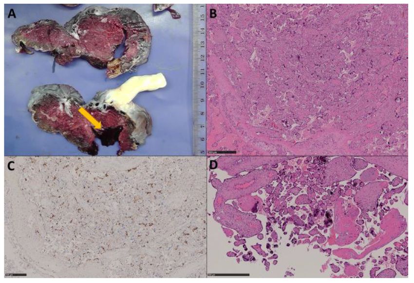
Pathological ndings of Case 3. Similarly to Cases 1 and 2, foci of villous/intervillous lymphocytic in ltration were present in
association with thrombo-hemorrhagic vasculopathy and partial trophoblast loss (A: HE, 100X; B: HE, 160X). Prevalence of
cytotoxic T cells was observed (C: anti-CD8 IHC, 50X). Villous infarction areas were also present (D: HE, 40X).
Page 14/16Figure 5
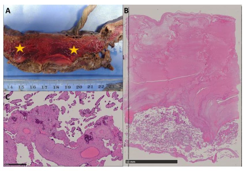
Pathological ndings of Case 4. Extensive subchorial placental abruption was noted during macroscopic examination (stars)
(A). Histological examination con rmed the presence of hemorrhagic areas located below the fetal surface (B: HE, 3X),
associated with chronic hypoxia signs (C: HE, 60X).
Page 15/16Figure 6
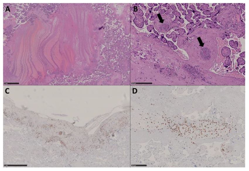
Pathological ndings of Case 5. Focal placental abruption was noted during grossing (arrow) (A). Histiocytic in ltration was
present consistently with the diagnosis of chronic histiocytic intervillositis (B: HE, 40X; C: anti-CD68 IHC, 40X). Villous
conglutination with loss of trophoblasts and microcalci cation was also observed (D: HE, 70X).
Page 16/16You can also read