Case Report Postoperative COVID-19 Pneumonia following Resection of a Large Thoracic Chondrosarcoma
←
→
Page content transcription
If your browser does not render page correctly, please read the page content below
Hindawi
Case Reports in Orthopedics
Volume 2021, Article ID 8866848, 6 pages
https://doi.org/10.1155/2021/8866848

Case Report
Postoperative COVID-19 Pneumonia following Resection of a
Large Thoracic Chondrosarcoma

          Austin C. Kaidi , Michael B. Held , Paul J. Park, and Wakenda K. Tyler
          Department of Orthopedic Surgery, Columbia University Medical Center, 622 W. 168th St, PH-11, New York, NY 10032, USA

          Correspondence should be addressed to Michael B. Held; mh3821@cumc.columbia.edu

          Received 11 September 2020; Revised 16 December 2020; Accepted 16 January 2021; Published 31 January 2021

          Academic Editor: Akio Sakamoto

          Copyright © 2021 Austin C. Kaidi et al. This is an open access article distributed under the Creative Commons Attribution License,
          which permits unrestricted use, distribution, and reproduction in any medium, provided the original work is properly cited.

          Case. A 57-year-old man presenting with two months of insidious shoulder pain was found to have a large thoracic
          chondrosarcoma invading the spinal canal. The patient’s orthopedic oncologist organized an interdisciplinary team including
          interventional radiology, thoracic surgery, neurosurgery, and plastic surgery. This allowed safe, en bloc tumor resection. The
          patient’s postoperative course was complicated by COVID-19 pneumonia, which was rapidly identified and medically managed
          with full recovery. Conclusion. Postoperative COVID-19 pneumonia can present insidiously and mimic other postoperative
          complications. Early identification and testing can promote rapid isolation, proper personal protective equipment use, and guide
          outcome-improving treatments.

1. Introduction                                                             The patient was informed that his case would be submit-
                                                                        ted for publication and provided consent.
The COVID-19 pandemic has called into question how
orthopedic surgeons should safely practice nonemergent sur-             2. Case History
geries. To date, there are few documented cases of postoper-
ative COVID-19 infection following orthopedic procedures                A 57-year-old male with hypertension presented to a nonop-
[1]. In this case, we present a thoracic chondrosarcoma com-            erative physician complaining of two months of intermittent
plicated by postoperative COVID-19 pneumonia.                           left shoulder pain localized to the scapula and lateral shoul-
     Chondrosarcomas are a group of malignant neoplasms                 der. He reported no weakness or numbness upon initial eval-
comprised of tumor cells that produce cartilaginous matrix.             uation, and the working diagnosis was scapular dyskinesis.
The only proven treatment is definitive surgical resection               The patient initially underwent one month of physical
[2]. Primary chondrosarcomas make up about 75% of all                   therapy and nonsteroidal anti-inflammatory drugs
chondrosarcomas, but they can also be seen in the setting               (NSAIDs) without improvement. Subsequent computed
of multiple enchondromatoses, such as Ollier disease or Maf-            tomography (CT) scan and magnetic resonance imagining
fucci syndrome [3]. Chest wall chondrosarcoma excisions                 (MRI) demonstrated a large exophytic mass originating at
require complex surgical approaches to avoid damaging crit-             T3 with extension cranially to C5, caudally to T5-T6, and
ical nearby structures. During the COVID-19 pandemic,                   intrathoracic extension. The mass measured approximately
postoperative recovery in a surgical intensive care unit                6:7 cm × 4:4 cm × 7:7 cm and extended into the spinal
(SICU) can increase a patients’ risk of COVID-19 exposure               canal with abutment of the spinal cord at T3-T4
from providers and other patients [4, 5]. In this case, we              (Figure 1). No metastatic chest disease was appreciated
highlight the need for swift recognition and coordinated                on either study. The patient was referred to an orthopedic
management of postoperative COVID-19 infections to allow                oncologist for further evaluation. Interval history at this
a positive outcome.                                                     time demonstrated radiating numbness and discomfort in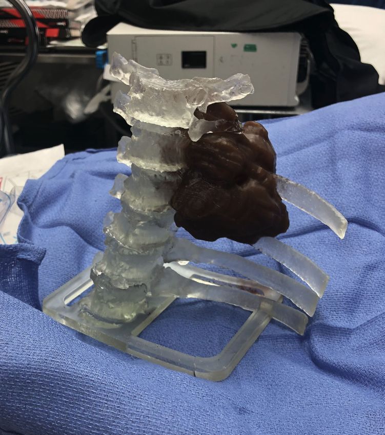
2 Case Reports in Orthopedics
(a) (b)
Figure 1: T2 MRI of patient’s thoracic spine: (a) axial view at T4 level; (b) midline sagittal with arrow at T4.
the left axilla. The patient’s orthopedic oncologist orga- racic surgery, neurosurgery, and plastic surgery were present
nized an interdisciplinary surgical care team for safe for planning and marking the incisions. This was critical to
resection. ensure the thoracotomy would not interfere with the spinal
incision and that an adequate skin bridge was left to preserve
2.1. Preoperative Planning. The size, location, and proximity coverage options for plastic surgery.
of tumor to vulnerable anatomical structures warranted a Thoracic surgery initially evaluated for intra- and extra-
multidisciplinary, coordinated approach. This care team pulmonary metastases utilizing flexible bronchoscopy and a
was organized by the patient’s orthopedic oncologist and video-assisted thoracoscopic surgery (VATS) system. Both
included interventional radiology (IR), neurosurgery, were negative. VATS was used to identify ribs 3-5 for resec-
thoracic surgery, plastic surgery, and internal medicine. tion. Thoracotomy was performed after discussion with
First, IR conducted a CT-guided biopsy of the mass. orthopedics, neurosurgery, and thoracic surgery. Osteo-
Pathology demonstrated lobulated hyaline cartilage suspi- tomies were performed along the lateral aspects of vertebral
cious for low-grade chondrosarcoma. IR also placed an infe- bodies T2-T4 using an ultrasonic bone scalpel with a margin.
rior vena cava (IVC) filter to prevent embolic sequelae. The Due to the tumor’s position, surgeons were unable to make a
patient was seen preoperatively by thoracic surgery and neu- complete cut through the vertebral body. The bone cuts were
rosurgery for multidisciplinary surgical planning. In order to taken as far as possible medially and posteriorly using the
successfully conduct an en bloc resection, it was decided that bone scalpel and curved osteotomes. At this point, the tumor
the operation would need to be sequentially staged using a was free anteriorly, a chest tube was inserted, and the thoracic
lateral and posterior approach. The lateral stage was per- wound was closed.
formed first in conjunction with thoracic surgery, the poste-
rior stage was performed next with neurosurgery, and closure 2.3. Operative Stage 2: Posterior Resection. For stage 2, the
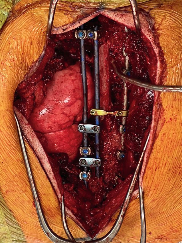
was done with plastic surgery. A 3-D printed model of the patient was positioned prone on a Jackson table with neuro-
patient’s thorax was generated to assist in preoperative monitoring. A posterior midline incision was made as previ-
planning (Figure 2). ously marked at the case’s start. Posterior laminectomies
were performed from the inferior portion of C7 to T4. The
2.2. Stage 1: Lateral Resection. On the morning of surgery, the T2-T4 nerve roots were divided, the spinal cord was mobi-
patient was afebrile without respiratory complaints, and lized to the right, and transpedicular decompression was
there was no government or hospital mandate to postpone performed from T1 to T4. Posterior osteotomies heading
elective cases. Mandatory preoperative COVID-19 testing anteriorly were connected with the osteotomies made in the
was also not standard protocol, as this was prior to wide- lateral position. This allowed for a complete en bloc resection
spread outbreak. The patient was positioned right-lateral of the tumor (Figure 3). Posterolateral spinal fusion was done
decubitus, and all teams including orthopedic oncology, tho- from C7 to T6 (Figure 4). Hydrogen peroxide irrigation was
Case Reports in Orthopedics                                                                                                     3

                                             Figure 2: 3-D model of patient’s thorax.

performed in case of local contamination, and the tumor was        CXR on POD7 also identified a new, left-sided effusion with
sent for pathological evaluation demonstrating a grade 1,          diffuse bilateral infiltrates (Figure 5).
stage pT1, and pN0 chondrosarcoma with negative bone                   On POD7, COVID-19 prevalence was low in New York
and soft tissue margins.                                           State, with fewer than 4,000 confirmed cases. However, the
     For closure, plastic surgery had initially considered a       patient’s fever and oxygen requirement raised suspicion.
latissimus dorsi flap. Given the careful planning at the case’s     Other considered diagnoses included surgical site infection,
onset however, it was possible to perform a paraspinal muscle      bacterial pneumonia, and pulmonary embolism. On POD7,
advancement flap with local tissue rearrangement to avoid           a COVID-19 nasal swab was obtained, and it returned posi-
the more time-intensive, debilitating rotational flap.              tive for COVID-19. Medical teams were immediately asked
                                                                   to help comanage. Thoracentesis was deemed unnecessary
2.4. Postoperative Course. Operative duration was greater          in the setting of a positive COVID-19 diagnosis.
than 16 hours, and estimated blood loss was 3.3 L. The                 The COVID-dedicated infectious disease (ID) team had
patient was taken to the SICU postoperatively. He was neuro-       been utilizing tests like ferritin, CRP, and D-dimer to assess
logically intact, except for expected chest wall numbness. The     infection severity and guide treatment. In a postoperative
patient was extubated on postoperative day (POD) 2 and             patient with malignancy, it was difficult to interpret these ele-
transferred to surgical step-down (SSD) on POD4 for close          vated, nonspecific values. Although the patient had mild
monitoring. Chest tubes were removed on POD5, with                 symptoms, the COVID-dedicated team recommended treat-
follow-up chest X-ray (CXR) demonstrating no pneumotho-            ment with hydroxychloroquine 600 mg twice daily for 3 days
rax or effusion. The postoperative course was unremarkable          (then 400 mg for 4 days) and azithromycin 250 mg daily for 5
until POD7, when he was febrile to 39.4°C with a new oxygen        days, after ruling out baseline QT-prolongation on EKG.
requirement (3 L nasal canula (NC) from previous room air).        This decision was determined by the patient’s risk factors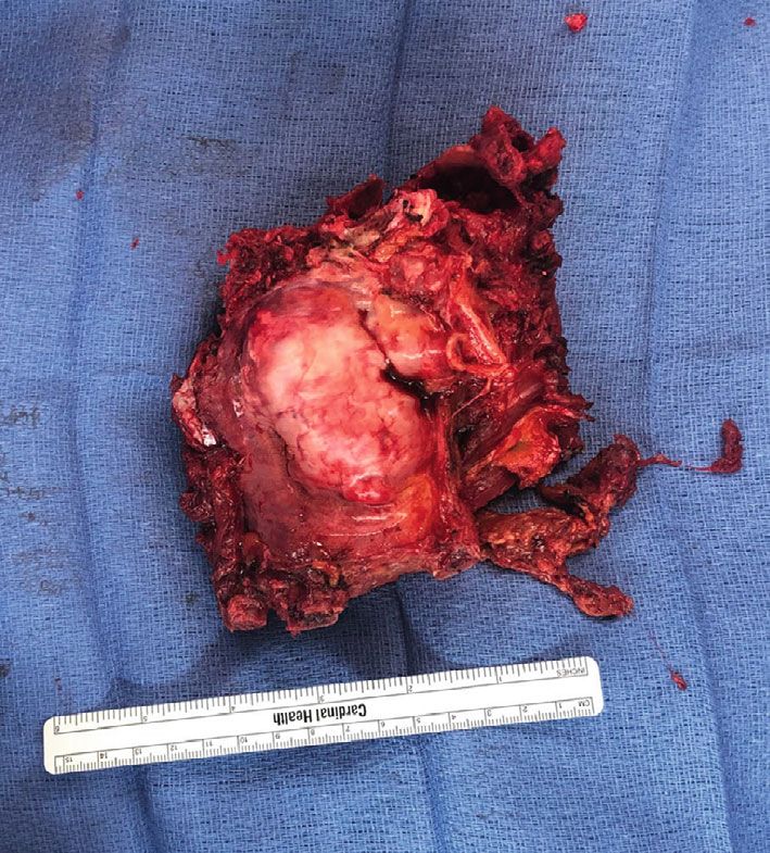
4                                                                                                       Case Reports in Orthopedics

                                        (a)                                               (b)

                                Figure 3: Removed tumor: (a) anterior surface; (b) posterior surface.

for decompensation, including intrathoracic surgery and             COVID positive on presentation. Two of the patients
malignancy history.                                                 expired; however, both had nonoperatively treated hip frac-
    The patient remained febrile through POD10 and had              tures [1]. This study is limited as it addresses neither postop-
increasing oxygen requirements to 6 L NC. Upon requiring            erative infections nor treatment. Mi et al. studied 10
6 L, he was transferred to the medicine service with surgical       orthopedic fracture patients who tested COVID positive pre-
comanagement. To minimize provider exposure, physical               operatively and evaluated whether surgical intervention was
therapy (PT) was halted and only one surgical team evaluated        safe [11]. In this study, 3 patients expired, two of which were
the patient daily. By POD19, he had completed treatment,            also nonoperatively treated hip fractures. This study did not
remained afebrile, and was breathing on room air. He                evaluate postoperative infections. Chang et al. evaluated sur-
restarted PT, began ambulating, and was transferred to acute        gical interventions on fracture patients in Singapore [12].
inpatient rehabilitation. 12-week postoperatively, the patient      They also did not address postoperative infections but did
is progressing well at home without further pulmonary               recommend utilizing inflammatory lab markers in COVID-
complications.                                                      positive patients to determine a patient’s surgical risk.
                                                                        In this report, our patient developed symptoms on
3. Discussion                                                       POD7, indicating a nosocomial or preoperative infection,
                                                                    given known 14-day incubation periods [13]. This empha-
Chondrosarcomas make up approximately 20-27% of all pri-            sizes the importance of JAAOS guidelines regarding manda-
mary malignant bone tumors [2, 6]. Even among rare chest            tory preoperative COVID-19 testing for nonemergent cases.
wall chondrosarcomas, costochondral tumors are more com-            Had this patient tested positive preoperatively, the case
mon than vertebral or costotransverse tumors. Only select           would have been postponed until infection resolution [14].
case reports have documented chondrosarcomas similar to                 Given the patient’s risk for serious complications in the
the one seen in this report [3, 7–10]. This case is unique for      setting of malignancy and major surgery, the team had a
its location, the multidisciplinary surgical approach utilized,     low threshold for COVID testing. This patient was diagnosed
and the development of postoperative COVID-19 pneumo-               with COVID-19 on the same day of symptom presentation,
nia. We believe this case provides insight regarding postoper-      even though the initial differential diagnosis was broad given
ative management of orthopedic patients during the                  his nonspecific symptoms of a mild oxygen requirement,
COVID-19 pandemic and the importance of an orthopedic               fever, and pleural effusion. A low threshold for testing
oncologist “quarterbacking” a patient’s care. Outcomes can          allowed rapid COVID team consultation and patient isola-
be improved with a dedicated COVID ID team. Shorter                 tion. Although hydroxychloroquine and azithromycin have
ICU and SSD stays may also lower nosocomial infection risk,         shown to be ineffective treatments, we believe dedicated
particularly when hospitals are inundated with COVID-19             COVID team comanagement improved this patient’s out-
patients.                                                           come [15]. Decisions to escalate care were difficult in this
     Currently, the literature on postoperative COVID-19            patient, since standard COVID-19 laboratory values were
infections following orthopedic procedures is limited. Rabie        unreliable due to his malignancy and postoperative inflam-
et al. reported 7 orthopedic trauma patients who tested             mation. A dedicated COVID team was able to more closely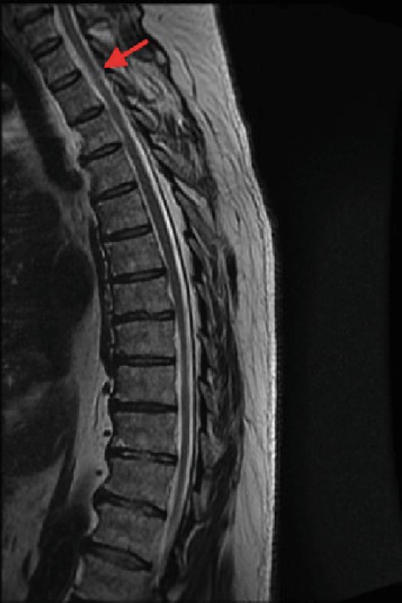
Case Reports in Orthopedics                                                                                                     5

                       Figure 4: Image of posterior midline incision after removal of tumor and spinal fusion.

                                                                    assess the patient, monitor for decompensation, and rapidly
                                                                    escalate ventilatory support when needed.
                                                                        Due to his COVID-19 pneumonia, this patient’s overall
                                                                    postoperative timeline was drastically prolonged, and he
                                                                    remained largely bedbound for three weeks. This made pre-
                                                                    operative IVC filter placement advantageous for reducing
                                                                    the risk of embolic complication [16, 17]. At our institution,
                                                                    IVC filters are routinely placed for large pelvic, sacral, and
                                                                    spinal sarcoma cases, since postoperative anticoagulation
                                                                    often must be held. If put on anticoagulation, patients’ large
                                                                    surgical spaces can form diffuse hematomas requiring multi-
                                                                    ple transfusions. Our recommended indications for preoper-
                                                                    ative IVC filter placement are as follows: (1) operations
                                                                    expected to be longer than 8 hours; (2) operations expected
  Figure 5: Chest X-ray of patient with COVID-19 pneumonia.         to require 4 or more units of blood products; (3) operations
                                                                    expected to require prolonged postoperative ICU stays; or
6 Case Reports in Orthopedics
(4) operations requiring spinal instrumentation, where post- [7] P. A. Rascoe, S. I. Reznik, and W. R. Smythe, “Chondrosar-
operative epidural bleeding could cause neural tissue coma of the thorax,” Sarcoma, vol. 2011, 7 pages, 2011.
compromise. [8] F. Bodin, C. Dissaux, J.-P. Steib, and G. Massard, “Complex
Finally, this case highlights the need for an orthopedic posterior thoracic wall reconstruction using a crossover com-
oncologist “quarterbacking” care. The orthopedic oncologist bined latissimus dorsi and serratus anterior free flap,” Euro-
coordinated care from preoperative planning to postopera- pean Journal of Cardio-Thoracic Surgery, vol. 49, no. 3,
tive management and was scrubbed for the entirety of the pp. 1008-1009, 2016.
procedure. This allowed for preoperative placement of an [9] A. Briccoli, L. Campanacci, R. Biagini, M. Rocca, C. Malaguti,
IVC filter and intraoperative closure with an advancement and M. Mercuri, “Chondrosarcoma of the ribs and sternum.
flap, sparing the patient a rotational latissimus flap, which Considerations on 20 cases treated,” Chirurgia Organi di
carries the risk of flap failure [18]. Postoperatively, the deci- Movimento, vol. 87, no. 1, pp. 17–23, 2020.
sion to COVID-test early on allowed for coordination of care [10] T. Bartalena, E. Rimondi, G. Rossi, G. Bianchi, and
with a dedicated COVID team who had more expertise in M. Alberghini, “Low grade central chondrosarcoma of the fifth
treating critically ill patients. costotransverse joint,” Australasian Radiology, vol. 51, SUPPL.
1, pp. B122–B125, 2007.
This report describes a unique primary chondrosarcoma
and provides insight on the management of postoperative [11] B. Mi, L. Chen, Y. Xiong, H. Xue, W. Zhou, and G. Liu, “Char-
COVID-19 pneumonia in orthopedic surgery. We recom- acteristics and early prognosis of COVID-19 infection in frac-
ture patients,” Journal of Bone and Joint Surgery, vol. 102,
mend universal preoperative testing, short ICU stays when
no. 9, pp. 750–758, 2020.
possible, and comanagement with a dedicated COVID team
[12] Z. Chang Liang, W. Wang, D. Murphy, and J. H. Po Hui,
for COVID-positive patients. This can optimize provider
“Novel coronavirus and orthopaedic surgery,” Journal of Bone
protection and patient outcomes.
and Joint Surgery, vol. 102, no. 9, pp. 745–749, 2020.
[13] S. A. Lauer, K. H. Grantz, Q. Bi et al., “The incubation period
Data Availability of coronavirus disease 2019 (COVID-19) from publicly
reported confirmed cases: estimation and application,” Annals
The paper describes a case report of a patient that was treated of Internal Medicine, vol. 172, no. 9, pp. 577–582, 2020.
by the authors. [14] M. E. Awad, J. C. L. Rumley, J. A. Vazquez, and J. G. Devine,
“Perioperative considerations in urgent surgical care of sus-
Consent pected and confirmed COVID-19 orthopaedic patients: oper-
ating room protocols and recommendations in the current
The patient was informed that his case would be submitted COVID-19 pandemic,” The Journal of the American Academy
for publication and provided consent. of Orthopaedic Surgeons, vol. 28, no. 11, pp. 451–463, 2020.
[15] J. Geleris, Y. Sun, J. Platt et al., “Observational study of hydro-
xychloroquine in hospitalized patients with Covid-19,” The
Conflicts of Interest New England Journal of Medicine, vol. 382, no. 25, pp. 2411–
2418, 2020.
The authors declare that they have no conflicts of interest. [16] H. Abdel-Razeq and M. Ismael-Abdulelah, “Inferior vena cava
filters in cancer patients: to filter or not to filter,” Therapeutics
References and Clinical Risk Management, vol. 7, p. 99, 2011.
[17] M. B. Pandhi, K. R. Desai, R. K. Ryu, and R. J. Lewandowski,
[1] H. Rabie, M. H. Sharafi, L. Oryadi Zanjani, and M. H. Nabian, “The role of inferior vena cava filters in cancer patients,” Sem-
“Novel coronavirus infection in orthopedic patients; report of inars in Interventional Radiology, vol. 33, no. 2, pp. 71–74,
seven cases,” Archives of Bone and Joint Surgery, vol. 8, Supple- 2016.
ment 1, pp. 302–309, 2020. [18] R. Engdahl, J. Disa, E. A. Athanasian, J. H. Healey, P. G. Cor-
[2] M. D. Murphey, E. A. Walker, A. J. Wilson, M. J. Kransdorf, deiro, and N. Fabbri, “Pedicled latissimus dorsi flap for shoul-
H. T. Temple, and F. H. Gannon, “Imaging of primary chon- der soft-tissue reconstruction after excision of a
drosarcoma: radiologic-pathologic correlation,” Radio- musculoskeletal neoplasm,” JBJS Essential Surgical Techniques,
graphics, vol. 23, no. 5, pp. 1245–1278, 2003. vol. 6, no. 2, pp. e16–1596, 2016.
[3] B. Widhe and H. C. F. Bauer, “Chest wall chondrosarcoma,”
Acta Orthopaedica, vol. 80, SUPPL. 334, pp. 82–84, 2017.
[4] J. Phua, L. Weng, L. Ling et al., “Intensive care management of
coronavirus disease 2019 (COVID-19): challenges and recom-
mendations,” The Lancet Respiratory Medicine, vol. 8, no. 5,
pp. 506–517, 2020.
[5] H. Qiu, China Critical Care Clinical Trials Group (CCCCTG),
Z. Tong et al., “Intensive care during the coronavirus epi-
demic,” Intensive Care Medicine, vol. 46, no. 4, pp. 576–578,
2020.
[6] H. V. Le, R. Wadhwa, P. Theodore, and P. Mummaneni, “Exci-
sion of thoracic chondrosarcoma: case report and review of lit-
erature,” Cureus, vol. 8, no. 7, 2016.You can also read