Volumetric Capnography - Expired volume - Hamilton Medical
←
→
Page content transcription
If your browser does not render page correctly, please read the page content below

Volumetric Capnography
Expired CO 2

               Expired volume

                                Intelligent Ventilation since 1983
Content overview – 1/2

The ventilation experts				4                     Alveolar minute ventilation – V‘alv 		    21
                                                 Dead space ventilation VDaw/VTE ratio             22
Introduction						5
                                                 What is the clinical relevance?		         23
Benefits of volumetric capnography          6    Improve ventilation quality and efficiency 24
                                                 Signs of ARDS						25
The volumetric capnogram			                 7
                                                 PEEP management 					26
The three phases					8                           Recruitment maneuver				27
Phase I – Anatomical dead space		 10             Expiratory resistance					28
Phase II – Transition phase				   11             Obstructive lung disease 				29
Phase III – Plateau phase				     12             Signs for pulmonary embolism 			           31
Slope of Phase III					13                        Hemorrhagic shock					32
Single breath CO2					14                         Optimize weaning process 			33
                                                 Monitor during patient transport		         35
Insight into the patient‘s lung condition   15
                                                 Rebreathing						36
Area X – CO2 elimination (VCO2)             17
Area Y - Alveolar dead space			             19
Area Z - Anatomical dead space			           20

Volumetric capnography - An introduction				                                                     Page 2
Content overview – 2/2

Clincial applications of trends		     37       Appendix 						49
PetCO2 versus V‘CO2 					38                    Volumetric capnography in Hamilton
Optimizing PEEP by trends				      40          Medical ventilators					50
Detecting alveolar derecruitment		 41          Loops and trends on the display			   51
                                               Volumetric capnography in monitoring 52
Test yourself 						42                         Calculation formulas					53
Multiple choice test					43                    Examples of normal values 			        54
Patient A							44                             References A – Z					55
Patient B							45                             Glossary A – Z						56
Patient C							46
Patient D							47                             Imprint							57
Solutions							48

Volumetric capnography - An introduction				                                             Page 3
The ventilation experts

Karjaghli Munir                                    Matthias Himmelstoss
Respiratory Therapist                              ICU Nurse, MSc Physics
Hamilton Medical Clinical Application Specialist   Hamilton Medical Product Manager

Volumetric capnography - An introduction				                                          Page 4
Introduction

Carbon dioxide (CO2) is the most abundant gas produced by the
human body. CO2 is the primary drive to breathe and a primary mo-
tivation for mechanically ventilating a patient. Monitoring the CO2
level during respiration (capnography) is noninvasive, easy to do,
relatively inexpensive, and has been studied extensively.

Capnography has improved over the last few decades thanks to the
developement of faster infrared sensors that can measure CO2 at
the airway opening in realtime. By knowing how CO2 behaves on
its way from the bloodstream through the alveoli to the ambient air,
physicians can obtain useful information about ventilation and per-
fusion.

There are two distinct types of capnography: Conventional,             This ebook concentrates on the use
time-based capnography allows only qualitative and semi-quan-          of volumetric capnography for
titative, and sometimes misleading, measurements, so volumetric        mechanically ventilated patients.
capnography has emerged as the preferred method to assess the
quality and quantity of ventilation.

Volumetric capnography - An introduction				                                                       Page 5
Benefits of volumetric capnography

    Improves, simplifies, and complements patient monitoring in relation to metabolism, circulation, and
    ventilation (V/Q)
    Provides information about the homogeneity or heterogeneity of the lungs
    Trend functions and reference loops allow for more comprehensive analysis of the patient condition
    Multiple clinical applications, such as detection of early signs of pulmonary emboli, COPD, ARDS, etc.
    Helps you optimize your ventilator settings
    Is easy to do and is relatively inexpensive

In short, volumetric capnography is a valuable tool to improve the
ventilation quality and efficiency for your ventilated patients.

Volumetric capnography - An introduction				                                                        Page 6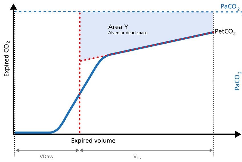
The volumetric
   capnogram

Volumetric capnography - An introduction				   Page 7
The three phases

The alveolar concentration of carbon dioxide (CO2) is the result of metabolism, cardiac output, lung per-
fusion, and ventilation. Change in the concentration of CO2 reflects perturbations in any or a combina-
tion of these factors. Volumetric capnography provides continuous monitoring of CO2 production, ven-
tilation/perfusion (V/Q) status, and airway patency, as well as function of the ventilator breathing circuit
itself.

Expired gas receives CO2 from three sequential compartments of the airways, forming three recognizable
phases on the expired capnogram. A single breath curve in volumetric capnography exhibits these three
characteristic phases of changing gas mixtures - they refer to the airway region in which they originate:

    Phase I - Anatomical dead space
    Phase II - Transition phase: gas from proximal lung areas and fast emptying lung areas
    Phase III - Plateau phase: gas from alveoli and slow emptying areas

Using features from each phase, physiologic measurements can be calculated.

Volumetric capnography - An introduction				                                                            Page 8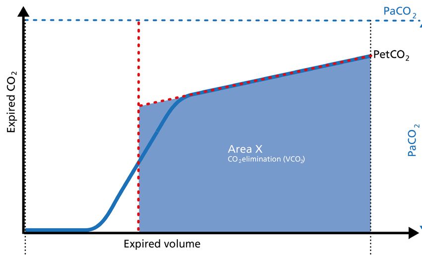
Phase I   Phase II                 Phase III
                                                              PetCO 2
Expired CO 2

                                 Expired volume

Volumetric capnography - An introduction				                      Page 9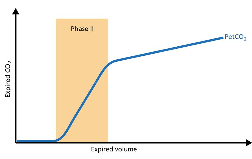
Phase I – Anatomical dead space

The first gas that passes the sensor at the onset of expiration comes from the airways and the bre-
athing circuit where no gas exchange has taken place = anatomical + artificial dead space. This gas
usually does not contain any CO2. Hence the graph shows movement along the X-axis (expired volu-
me), but no gain in CO2 on the Y-axis.

                                                                A prolonged Phase I indicates an increa-
                                                                se in anatomical dead space ventilation
                                                                (VDaw).

                                                                Presence of CO2 during Phase I indicates
                                                                rebreathing or that the sensor needs to
                                                                be recalibrated.

Volumetric capnography - An introduction				                                                      Page 10
Phase II – Transition phase
Phase II represents gas that is composed partially of distal airway volume and mixed with gas from fast
emptying alveoli. The curve slope represents transition velocity between distal airway and alveolar gas –
providing information about perfusion changes and also about airway resistances.
A prolonged Phase II can indicate an
increase in airway resistance and/or a
Ventilation/Perfusion (V/P) mismatch.
Volumetric capnography - An introduction Page 11Phase III – Plateau phase
Phase III gas is entirely from the alveoli where gas exchange takes place. This phase is representative of
gas distribution. The final CO2 value in Phase III is called end-tidal CO2 (PetCO2).
A steep slope in Phase III provides
information about lung heterogeneity
with some fast and some slow emptying
lung areas.
For example, obstructed airway results
in insufficiently ventilated alveoli,
inducing high CO2 values and increased
time constants in this region.
Volumetric capnography - An introduction Page 12Slope of Phase III
The slope of Phase III is a characteristic of the volumetric capnogram shape. This slope is measured in the
geometric center of the curve, which is defined as the middle two quarters lying between VDaw and the
end of exhalation.
Steep slope
A steep slope can be seen, for example, Normal slope
Expired CO 2
in COPD and ARDS patients.
Expired volume
Volumetric capnography - An introduction Page 13Single breath CO2 analysis Volumetric capnography - An introduction Page 14
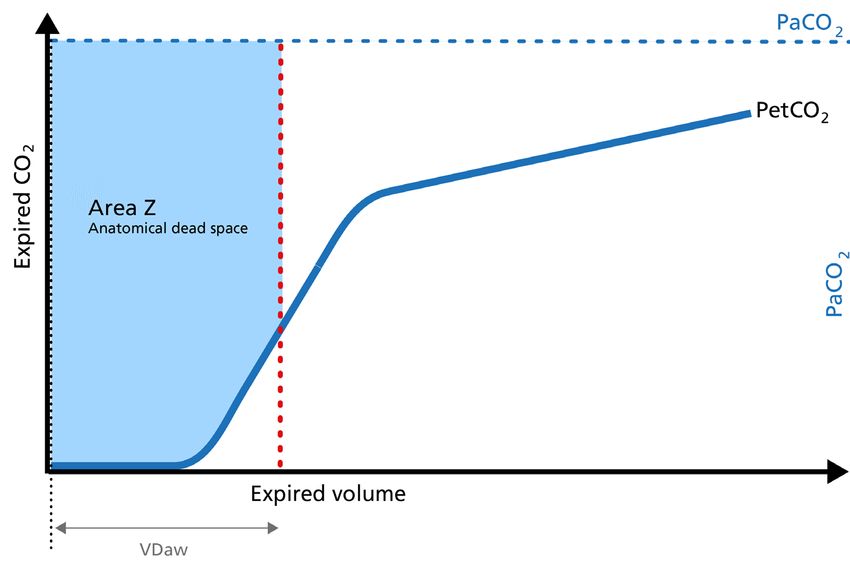
Insight into the patient‘s lung condition

The volumetric capnogram can also be divided into three areas:

    Area X - CO2 elimination
    Area Y - Alveolar dead space
    Area Z - Anatomical dead space

The size of the areas, as well as the form of the curve, can give you more insight into the patient‘s lung
condition regarding:

• Dead space fraction – VDaw /VTE
• Alveolar minute ventilation – V‘alv

Volumetric capnography - An introduction				                                                         Page 15
4
1
3
2
1. Slope of Phase III
2. Slope of Phase II
3. The intersection of lines 1 and 2 defines the limit between Phases II and III.
4. A perpendicular line is projected onto the x-axis and its position is adjusted until the areas p and q on both
sides become equal.
Volumetric capnography - An introduction Page 16Area X – CO2 elimination (V‘CO2) – 1/2 Area X represents the actual volume of CO2 exhaled in one breath (VeCO2). Adding up all of the single breaths in one minute gives you the total elimination of CO2 per minute (V‘CO2). If cardiac output, lung perfusion, and ventilation are stable, this is an assessment of the production of CO2 called V‘CO2. The V‘CO2 value displayed on the ventilator can be affected by any change in CO2 production, cardiac out- put, lung perfusion, and ventilation. It indicates instantly how the patient’s gas exchange responds to a change in ventilator settings. Monitoring trends allows for detection of sudden and rapid changes in V‘CO2. Decreasing V‘CO2 Hypothermia, deep sedation, hypothyroidism, paralysis, and brain death decrease CO2 production and induce a decrease in V‘CO2. Decreasing V‘CO2 can also be due to a decrease in cardiac output or blood loss, and may also suggest a change in blood flow to the lung areas. Pulmonary embolism, for example, exhibits V‘CO2 reduction and a slope reduction in Phase II. Volumetric capnography - An introduction Page 17
Area X – CO2 elimination (V‘CO2) – 2/2

Increase in V‘CO2
is usually due to bicarbonate infusion
or an increase in CO2 production that
can be caused by:

•   Fever
•   Sepsis
•   Seizures
•   Hyperthyroidism
•   Insulin therapy

Volumetric capnography - An introduction				   Page 18
Area Y - Alveolar dead space

Area Y represents the amount of CO2 that is not eliminated due to alveolar dead space.

Increase
Alveolar dead space is increased in cases
of lung emphysema, lung overdistension,
pulmonary embolism, pulmonary hyper-
tension, and cardiac output compromise.

Decrease
If the above mentioned conditions improve
due to successful therapy, the alveolar
dead space decreases.

Volumetric capnography - An introduction				                                             Page 19
Area Z - Anatomical dead space

Anatomical dead space measurement using a volumetric capnogram gives an effective, in-vivo measu-
re of volume lost in the conducting airway. This area represents a volume without CO2. It does not take
part in the gas exchange and consists of the airway, endotracheal tube, and artificial accessories, such as
a flextube positioned between the CO2 sensor and the patient.

An expansion of Area Z can indicate
an increase in anatomical dead space
ventilation (VDaw). Consider a reduction
of your artificial dead space volume.

A diminution of Area Z is seen when
artificial dead space volume is decreased
and when excessive PEEP is decreased.

Volumetric capnography - An introduction				                                                         Page 20
Alveolar minute ventilation – V‘alv

Phase III of the waveform represents the quantity of gas that comes from the alveoli and actively partici-
pates in gas exchange. V‘alv is calculated by subtracting the anatomical dead space (VDaw) from the tidal
volume (VTE) multiplied by the respiratory rate from the minute volume (MinVol):
V’alv =RR*Vtalv = RR*(VTE-VDaw)

                                                                                           PetCO 2
                                                                                           after recruitment
Increase

                                             Expired CO 2
An increase in V‘alv is seen after an                                                      PetCO 2
                                                                                           before recruitment
efficient recruitment maneuver and
induces a transient increase in V‘CO2.

Decrease
A decrease in V‘alv can indicate that
fewer alveoli are participating in the gas
exchange, for example, due to pulmonary
edema.                                                         Expired volume

Volumetric capnography - An introduction				                                                           Page 21
Dead space ventilation - VDaw/VTE ratio
The ratio of airway dead space (VDaw) to tidal volume (VTE) – the VDaw/VTE ratio – gives you an insight
into the effectiveness of ventilation.
PaCO2
Airway dead space (VDaw)
A rising VDaw/VTE ratio can be a sign of PetCO2
Expired CO 2
ARDS.
In a normal lung, the VDaw/VTE
ratio is between 25% and 30%.
In early ARDS, it is between 58% and
up to 83%. Tidal volume (Vt)
Expired volume
Volumetric capnography - An introduction Page 22What is the
clinical relevance?
Volumetric capnography - An introduction Page 23Improve ventilation quality and efficiency You can use the insights from the CO2 curve to improve ventilation quality and efficiency for your patients. On the following pages, you will find examples for the use of the CO2 curve in the clinical scenarios listed below: • Signs of ARDS • PEEP management • Recruitment maneuver • Expiratory resistance • Obstructive lung disease • Pulmonary embolism • Hemorrhagic shock • Optimize management of the weaning process • Monitor perfusion during patient transport • Detection of rebreathing Volumetric capnography - An introduction Page 24
Signs of ARDS - Acute respiratory distress syndrome

In ARDS, the ventilation/perfusion ratio is disturbed and changes in the slope of the volumetric capno-
gram curve can be observed.

                                                                                         Normal PetCO2

Phase I is larger due to increased                                                       ARDS

                                            Expired CO 2
anatomical dead space caused by PEEP.

The slope of Phase II is decreased due to
lung perfusion abnormalities.

The slope of Phase III is increased due
to lung heterogeneity.

                                                              Expired volume

Volumetric capnography - An introduction				                                                        Page 25
PEEP management
If PEEP is too high, the intrathoracic pressure rises, the venous return decreases, and pulmonal vascular
resistance (PVR) increases. These changes can be easily observed on the volumetric capnogram.
After PEEP
reduction
With high
An increase in Phase I shows an increase PEEP
in anatomical dead space.
Expired CO 2
A decrease in the Phase II slope indica-
tes a decrease in perfusion.
An increase in the Phase III slope depicts
a maldistribution of gas, which can be
caused by an inappropriately low PEEP
setting or an inappropriately high PEEP Expired volume
setting causing lung overdistension.
Volumetric capnography - An introduction Page 26Recruitment maneuver
The volumetric capnogram can be used to assess the effectiveness of recruitment maneuvers and might
give you an insight into the recruited lung volume.
PetCO 2
After a sucessful recruitment maneuver, after recruitment
Expired CO 2
you should see a transient increase in PetCO 2
V‘CO2. before recruitment
Phase I may decrease a little. The slope
of Phase II becomes steeper with improved
lung perfusion. The slope of Phase III im-
proves as a result of more homogeneous
lung emptying.
Expired volume
Volumetric capnography - An introduction Page 27Expiratory resistance
Concave Phase-III volumetric capnograms have been seen with obese patients and patients with increa-
sed expiratory resistance. Obese patients (Fig. 1) can have biphasic emptying and higher PetCO2 than
PaCO2. That difference suggests varying mechanical and ventilation/perfusion properties. The increase
in expiratory resistance (Fig. 2) may reflect a slow expiratory phase with a slow accumulation of alveolar
CO2. The alveoli that empty last may have more time for CO2 diffusion.
PetCO2
PaCO2
PaCO2
Expired CO 2
Expired CO 2
PetCO2
Expired volume Expired volume
Fig 1: Concave volumetric capnogram associated with obesity Fig 2: Concave volumetric capnogram associated with
increased airway resistance
Volumetric capnography - An introduction Page 28Obstructive lung disease – 1/2
When spirometry cannot be reliably performed, volumetric capnography can be used as an alternative
test to evaluate the degree of functional involvement in obstructive lung disease patients (COPD, asthma,
cystic fibrosis, etc.). Obstructive lung disease is characterized by asynchronous emptying of compartments
with different ventilation/perfusion ratios.
PetCO2 in COPD
The volumetric capnogram in COPD Normal PetCO 2
Expired CO 2
patients shows a prolonged Phase II, an
increase in PetCO2, and a continuously
ascending slope without plateau in
Phase III.
Expired volume
Volumetric capnography - An introduction Page 29Obstructive lung disease – 2/2
Patients with high airway resistance demonstrate a a decrease in the Phase II slope and a steep slope in
Phase III. The volumetric capnogram can give you insights into therapy efficiency.
PetCO2
during bronchospasm
PetCO2
A Phase II shift to the left indicates after therapy
Expired CO 2
reduced resistance.
Phase III slope shows a decrease in
steepness indicating better gas
distribution and reduced alveolar
dead space (VDalv).
Expired volume
Volumetric capnography - An introduction Page 30Signs of pulmonary embolism
Pulmonary embolism (PE) leads to an abnormal alveolar dead space that is expired in synchrony with gas
from normally perfused alveoli. This feature of PE separates it from pulmonary diseases affecting the air-
way, which are characterized by nonsynchronous emptying of compartments with an uneven ventilation/
perfusion relationship. In case of sudden pulmonary embolism, volumetric capnography has a typical
unique shape.
Normal PetCO 2
In patients with sudden pulmonary vascular
Expired CO 2
occlusion due to pulmonary embolism, Phase I
is increased due to increased anatomical dead
PetCO2
space. in PE
The slope of Phase II is decreased due to poor
lung perfusion. Phase III has a normal plateau
with low PetCO2 because the number of
functional alveoli is reduced. In this case, V‘CO2 Expired volume
drops suddenly.
Volumetric capnography - An introduction Page 31Hemorrhagic shock
Hemorrhagic shock is a condition of reduced tissue perfusion, resulting in the inadequate delivery of
oxygen and nutrients that are necessary for cellular function.
Normal PetCO 2
The expired CO2 drops drastically. Phase I
Expired CO 2
is unchanged and the slopes of Phase II
and III are unchanged, but PetCO2 is de-
creased due to the increase in alveolar
PetCO2 in
dead space. hemorrhagic shock
Expired volume
Volumetric capnography - An introduction Page 32Optimize management of the weaning process – 1/2 The volumetric capnogram and trends show the patient‘s response to the weaning trial and allow for better management of the weaning process. Indications for a successful weaning trial are: • Stable V‘alv and constant tidal volumes As ventilatory support is being weaned, the patient assumes the additional work of breathing while V‘alv remains stable and spontaneous tidal volumes remain constant. • V‘CO2 remains stable and then slightly increases The slight increase in V‘CO2 represents an increase in CO2 production as patient work of breathing in- creases in association with the decrease in ventilatory support. This suggests an increase in metabolic activity due to the additional task of breathing by the patient. Volumetric capnography - An introduction Page 33
Optimize management of the weaning process – 2/2

Indications for an unsuccessful weaning trial are:

• Dramatic increase in V‘CO2
  A more dramatic increase in V‘CO2 would suggest excessive work of breathing and the potential for
  impending respiratory decompensation. This scenario would be consistent with a visual assessment of
  increasing respiratory distress (for example, retraction, tachypnea, and agitation). The V‘CO2 will even-
  tually decrease if the patient gets exhausted.

• Decrease in V‘CO2
  As the ventilator settings are decreased, the patient is no longer able to maintain an adequate degree
  of spontaneous ventilation, and total minute ventilation falls with a decrease in CO2 elimination.

• Increased VDaw/VTE ratio
  If reducing ventilatory support is followed by a decrease in tidal volume, the VDaw/VTE ratio increases.
  This reduces ventilatory efficiency and the patient’s ability to remove CO2.

Volumetric capnography - An introduction				                                                         Page 34
Monitor perfusion during patient transport

If arterial access is not something you routinely perform when you transport a ventilated patient, PetCO2
can be used for monitoring perfusion and ventilation during transport.

A decrease in PetCO2 accompanied
by a decrease of VCO2 can signify:

•   ET tube displacement
•   Decreased cardiac output
•   Pulmonary embolism
•   Atelectasis
•   Overdistension of alveoli (for example,
    excessive PEEP)

Volumetric capnography - An introduction				                                                          Page 35
Detection of rebreathing
An elevation of the baseline during Phase I indicates rebreathing of CO2, which may be due to mechanical
problems or therapeutic use of mechanical dead space.
Normal PetCO2
Rebreathing CO2
Consider recalibration of the CO2 sensor
Expired CO 2
or reduction of the airway accessories.
Expired volume
Volumetric capnography - An introduction Page 36Clincial applications
of trends
Volumetric capnography - An introduction Page 37PetCO2 versus V‘CO2 - Opposing, asynchronous trends If the PetCO2 trend moves up while the V‘CO2 trend decreases for a while and then returns to baseline, this indicates a worsening of ventilation. If the PetCO2 trend moves down while the V‘CO2 trend increases for a while and then returns to baseline, this indicates an improvment of ventilation. Volumetric capnography - An introduction Page 38
PetCO2 versus V‘CO2 - Synchronous trends

Rising PetCO2 and V‘CO2 trends
indicate increasing CO2 production
(agitation, pain, fever).

Falling PetCO2 and V‘CO2 trends
indicate a decrease in CO2 production.

Volumetric capnography - An introduction				   Page 39
Optimizing PEEP by trends

                                                                             12                       14                         16                         18
                                                                        0 to                     2 to                       4 to                       6 to
                                                                  s   e1                   s   e1                     s   e1                     s   e1
                                                           re a                     re a                       re a                       re a
                                                  P in c                   P in c                     P in c                     P in c
                                                 E                        E                          E                          E
                                               PE                       PE                         PE                         PE
When PEEP change is associated with
an improving ventilation/perfusion
ratio, V‘CO2 shows a transient increase
for a couple of minutes and then
returns back to baseline, that is, in
equilibrium with CO2 production.

When PEEP change is associated
with a worsening of the ventilation/
perfusion ratio, V‘CO2 transiently
decreases for a few minutes and then
returns to baseline.

Volumetric capnography - An introduction				                                                                                                            Page 40
Detecting alveolar derecruitment Volumetric CO2 provides continuous monitoring to detect derecruitment and recruitment of alveoli. Alveolar ventilation and V‘CO2 will first decrease if the lung derecruits, and will then stabilize again at equilibrium. Recruitment, during, for example, a PEEP increase, can be detected by short V‘CO2 peaks before V‘CO2 returns to equilibrium. Volumetric capnography - An introduction Page 41
Test yourself

Volumetric capnography - An introduction				   Page 42
Multiple choice test

Now it is time to put your newly learned knowledge to the test. On the following pages you will find
clinical cases of intubated ICU patients, including three typical symptoms for each case. Your task is to
figure out the patient‘s condition by interpreting the volumetric capnogram.

You are presented with three possible answers of which only one is correct.
The solutions are on page 48.

Good luck!
                                                                                          A
                                                                                          B
                                                                                          C
Volumetric capnography - An introduction				                                                         Page 43
Patient A
Adult female intubated patient presents with a respiratory rate of 35 breaths/min (tachypnea) and swollen
calves. What does the volumetric capnogram indicate?
a) Pulmonary embolism
b) ARDS
Normal PetCO 2
c) Sepsis
Expired CO 2
PetCO2
in PE
Expired volume
Volumetric capnography - An introduction Page 44Patient B
Adult male intubated patient presents with a dry, nonproductive cough, crackling noises in the lungs, and a
heart rate of 110 beats/min (tachycardia). What does the volumetric capnogram indicate?
Normal PetCO 2
a) Cardiac arrest
Normal PetCO2
b) ARDS
ARDS c) Sepsis
Expired CO 2
PetCO2
in PE
Expired volume
Expired volume
Volumetric capnography - An introduction Page 45Patient C
Adult male intubated patient presents with blueness of the lips and fingernail beds (cyanosis), oxygen sa-
turation (SaO2) of 89%, and the x-ray shows overexpanded lungs. What does the volumetric capnogram
indicate?
a) PEEP is too high
PetCO2 in COPD
b) Pulmonary embolism
Normal PetCO 2 c) Severe COPD
Expired CO 2
Expired volume
Volumetric capnography - An introduction Page 46Patient D
Adult female patient, hospitalized comatose after car accident with no visible injuries, presents after
intubation with low blood pressure, hyperglycemia, and a heart rate of 118 beats/min (tachycardia).
What does the volumetric capnogram indicate?
a) Pneumothorax
b) ARDS
Normal PetCO 2
c) Hemorrhagic shock
Expired CO 2
PetCO2 in
hemorrhagic shock
Expired volume
Volumetric capnography - An introduction Page 47Solutions Patient A a) Pulmonary embolism Patient B b) ARDS Patient C c) Severe COPD Patient D c) Hemorrhagic shock Volumetric capnography - An introduction Page 48
Appendix

Volumetric capnography - An introduction				   Page 49
Volumetric capnography in Hamilton Medical ventilators

All Hamilton Medical ventilators offer volumetric capnography
either included standard or as an optional feature.

The CO2 measurement is performed using a CAPNOSTAT® 5
mainstream CO2 sensor at the patient‘s airway opening. The
CAPNOSTAT® 5 sensor provides technologically advanced mea-
surement of end-tidal carbon dioxide (PetCO2), respiratory rate,
and a clear, accurate capnogram at all respiratory rates up to
150 breaths per minute.

Volumetric capnography - An introduction				                       Page 50
Loops and trends on the display

                                      3

                2
                                  1

                                                  4

1   Current volumetric capnogram loop                 A 72-hour trend (or 96-hour with HAMILTON-S1/G5) is
                                                      available for:
2   Volumetric capnogram reference loop                              • VDaw
                                                      • PetCO2
3   Reference loop button with time and date of       • V‘CO2        • VDaw/VTE
    reference loop                                    • FetCO2       • Slope CO2
4   Most relevant CO2 values, breath by breath        • VeCO2
                                                      • ViCO2

Volumetric capnography - An introduction				                                                          Page 51
Volumetric capnography in monitoring To make your life easier, the Hamilton Medical ventilators offer an overview of all relevant CO2-related values in the monitoring window. Volumetric capnography - An introduction Page 52
Calculation formulas

Vtalv 		Alveolar tidal volume							Vtalv = Vt - VDaw

V’alv    Alveolar minute ventilation 					                   V’alv = RR*Vtalv

VCO2     Volume of CO2 eliminated/breath				VCO2 = VeCO2 - ViCO2

V‘CO2    Volume of CO2 eliminated/minute				VCO2*Number of breaths/min

FetCO2   Fractional concentration of CO2 in exhaled gas 		   FetCO2 = V’CO2/MinVol

PetCO2   Partial pressure of CO2 in exhaled gas				          PetCO2 = FetCO2*(Pb-PH2O)

VDaw/VTE Anatomical dead space fraction					                 VDaw/VTE = 1 - (PetCO2/PaCO2)

Volumetric capnography - An introduction				                                                 Page 53
Examples of normal values for ventilated patients1

Description            Unit2            Normal                                                        Reference
 VDaw                   ml               2.2 ml/kg IBW                                                 Radford 1954
 slopeCO2               %CO2/l           31324 * Vt-1.535                                              Aström 2000
                                                                                                       Weissmann 1986 /
 V’CO2                  ml/min           2.6 to 2.9 ml/min/kg
                                                                                                       Wolff 1986
 FetCO2                 %                5.1% to 6.1%                                                  Wolff 1986
 V’alv                  l/min            0.052 to 0.070 l/min/kg                                       (V‘CO2 / FetCO2)

1. These values are for illustration purposes and do not replace physician-directed treatment.
2. Bulk gas volumes, such as minute ventilation and tidal volumes, are usually measured in BTPS. Specific gas volumes are expressed in
   STPD. Conversion factors can be found in physics textbooks.

Volumetric capnography - An introduction				                                                                                      Page 54
References A – Z Anderson JT, Owings JT, Goodnight JE. Bedside noninvasive detection of acute pulmonary embolism in critically ill surgical patients. Arch Surg 1999;134(8):869–874; discussion 874–875. Aström E, Niklason L, Drefeldt B, Bajc M, Jonson B. Partitioning of dead space – a method and reference values in the awake human. Eur Respir J. 2000 Oct; 16(4):659-664. Blanch L, Romero PV, Lucangelo U. Volumetric capnography in the mechanically ventilated patient. Minerva Anestesiol. 2006 Jun;72(6):577-85. Erikson, L, Wollmer, P, Olsson, CG, et al. Diagnosis of pulmonary embolism based upon alveolar dead space analysis. Chest1989;96,357-362. Fletcher R. The single breath test for carbon dioxide [dissertation]. Lund, Sweden: University of Lund, 1980. 2nd edition revised and reprinted, Solna, Sweden: Siemens Elema, 1986. Kallet RH, Daniel BM, Garcia O, Matthay MA. Accuracy of physiologic dead space measurements in patients with acute respiratory distress syndrome using volumetric capnography: comparison with the metabolic monitor method. Respir Care. 2005 Apr;50(4):462-7. Kiiski, Ritva, and Jukka Takala. „Hypermetabolism and efficiency of CO2 removal in acute respiratory failure.“ CHEST Journal 105.4 (1994): 1198-1203. Kumar AY, Bhavani-Shankar K, Moseley HS, Delph Y. Inspiratory valve malfunction in a circle system: pitfalls in capnography. Can J Anaesth 1992;39(9):997–999. Nuckton TJ, Alonso JA, Kallet RH, Daniel BM, Pittet JF, Eisner MD, Matthay MA. Pulmonary dead-space fraction as a risk factor for death in the acute respiratory distress syndrome. N Engl J Med. 2002 Apr 25; 346(17):1281-1286. Olsson K, Jonson B, Olsson CG, Wollmer P. Diagnosis of pulmonary embolism by measurement of alveolar dead space. J Intern Med. 1998 Sep;244(3):199-207. Pyles ST, Berman LS, Modell JH. Expiratory valve dysfunction in a semiclosed circle anesthesia circuit: verification by analysis of carbon dioxide waveform. Anesth Analg 1984;63(5):536–537. Radford EP. Ventilation standards for use in artificial respiration. N Engl J Med 1954; 251:877-883. Rodger MA, Jones G, Rasuli P, Raymond F, Djunaedi H, Bredeson CN, Wells PS. Steady-state end-tidal alveolar dead space fraction and D-dimer: bedside tests to exclude pulmonary embolism. Chest 2001;120(1):115–119. Yaron M, Padyk P, Hutsinpiller M, Cairns CB. Utility of the expiratory capnogram in the assessment of bronchospasm. Ann Emerg Med. 1996 Oct;28(4):403-7. Weissman C, Kemper M, Elwyn DH, Askanazi J, Hyman AI, Kinney JM. The energy expenditure of the mechanically ventilated critically ill patient. An analysis. Chest. 1986 Feb; 89(2):254-259. Wolff G, Brunner JX, Grädel E. Gas exchange during mechanical ventilation and spontaneous breathing. Intermittent mandatory ventilation after open heart surgery. Chest. 1986 Jul; 90(1):11-17 Wolff G, Brunner JX, Weibel W, et al. Anatomical and series dead space volume: concept and measurement in clinical practice. Appl Cardiopul Pathophysiol 1989; 2:299-307. Volumetric capnography - An introduction Page 55
Glossary A – Z

f		      Frequency or resipratory rate = The number of breaths per minute
PaCO2    Partial pressure of carbon dioxide in the arterial blood; arterial carbon dioxide concentration or tension
		       It is either expressed in mmHg or in kPa
PCO2		   Partial pressure of carbon dioxide
PetCO2   End-tidal carbon dioxide
SBCO2    Single breath carbon dioxide
V‘alv		  Alveolar minute ventilation
		       The amount of minute ventilation volume that is actually participating in gas exchange
V‘CO2		  Volume of CO2 eliminated per minute
VD		     Physiological dead space
VDaw     Anatomical dead space ventilation
VDaw/VTE Anatomical dead space to tidal volume ratio
Ve		     Minute ventilation = Tidal volume multiplied by respiratory rate (Vt x f = Ve)
VeCO2    Expired CO2 volume
ViCO2		  Inspired CO2 volume
VTE		    Tidal volume is the lung volume representing the normal volume of gas displaced between inhalation
		and exhalation

Volumetric capnography - An introduction				                                                                     Page 56
Imprint

          Published by: 		   Hamilton Medical

          Authors: 			 Karjaghli Munir
          				Matthias Himmelstoss

          Release date: 		   March 2016

          Edition: 			2
More information:
www.hamilton-medical.com/volumetric-capnography
Intelligent Ventilation since 1983
Manufacturer:
Hamilton Medical AG
Via Crusch 8, 7402 Bonaduz, Switzerland
+41 58 610 10 20
info@hamilton-medical.com
www.hamilton-medical.com
ELO20151002N.01 Specifications are subject to change without notice. Some features are options. Not all features are available in all markets. For all trademarks used by Hamilton Medical AG, see www.hamilton-medical.com/trademarks. © 2020 Hamilton Medical AG.
All rights reserved.
Volumetric capnography - An introductionYou can also read