Femtosecond LASIK outcomes using the hyperopic astigmatism refractive surgery
←
→
Page content transcription
If your browser does not render page correctly, please read the page content below
EXPERIMENTAL AND THERAPEUTIC MEDICINE 21: 288, 2021

                     Femtosecond‑LASIK outcomes using the
                  VisuMax®‑MEL® 80 platform for hyperopia and
                     hyperopic astigmatism refractive surgery
 BOGDANA TĂBĂCARU1, HORIA TUDOR STANCA1, RUXANDRA ANGELA PÎRVULESCU1, SIMONA STANCA2,
    CIPRIAN DANIELESCU3, MIHNEA MUNTEANU4, COSMIN ROȘCA5 and ADRIAN COSMIN TEODORU6

    Departments of 1Ophthalmology, 2Pediatrics, ‘Carol Davila’ University of Medicine and Pharmacy, 050474 Bucharest;
             3
               Department of Ophthalmology, ‘Grigore T. Popa’ University of Medicine and Pharmacy, 700115 Iasi;
          4
            Department of Ophthalmology, ‘Victor Babeș’ University of Medicine and Pharmacy, 300041 Timisoara;
            5
              Department of Ophthalmology, Oculens Clinic, 400501 Cluj‑Napoca; 6Department of Ophthalmology,
                            Faculty of Medicine, ‘Lucian Blaga’ University, 550159 Sibiu, Romania

                                    Received November 11, 2020; Accepted December 11, 2020

                                                    DOI: 10.3892/etm.2021.9719

Abstract. The present study evaluated the efficacy, the              Introduction
safety and the predictability of the Femtosecond laser-
assisted in situ keratomileusis (Femto‑LASIK) procedure              Currently, laser‑assisted in situ keratomileusis (LASIK) is
for hyperopia and hyperopic astigmatism. We retrospectively          the default choice for refractive surgical procedures, as it can
analyzed the postoperative 12‑month evolution of 593 eyes            address a wide spectrum of high and complex ametropias (1‑5).
with hyperopia and hyperopic astigmatism that underwent              There are numerous previous reports of hyperopic LASIK
Femto‑LASIK treatment. The procedure was predictable                 showing various safety, efficiency and predictability percent-
and effective. No eye lost 2 lines of corrected distance             ages (6‑32). The new femtosecond laser technology to perform
visual acuity (CDVA), demonstrating a safety profile of the         LASIK flaps avoids many complications of the mechanical
procedure. Nine percent of the eyes gained at least one line         microkeratome (free caps, incomplete, irregular or displaced
of CDVA. The accuracy of the spherical equivalent after              flaps), being presently preferred by many surgeons (1,2,33).
12 months was 74% within ±1.0 diopter (D) of emmetropia.             In this study, we evaluated the safety, efficacy, predictability
The refractive outcomes were stable during the follow‑up             and accuracy of the refractive results of Femtosecond‑LASIK
period. There were no significant complications during the           (Femto‑LASIK) procedure using the VisuMax®‑MEL® 80
procedure. Femto‑LASIK using the VisuMax® ‑MEL ® 80                  platform for hyperopia with or without astigmatism.
platform was demonstrated to be a suitable option to correct
selected cases of hyperopia and hyperopic astigmatism.               Patients and methods
A longer follow‑up period is required to better assess the
refractive results and to detect any further regression.             Data collection. We performed a retrospective, noncom-
                                                                     parative consecutive case series study on eyes with hyperopia
                                                                     and hyperopic astigmatism that underwent Femto‑LASIK
                                                                     surgery. Patients were operated on by the same refractive
                                                                     surgeon (HTS) at the Europe Eye‑Metropolitan Hospital
                                                                     in Bucharest, Romania between June 2011 and June 2017.
                                                                     All surgeries were performed using the same femtosecond
                                                                     laser‑excimer laser platform VisuMax®‑MEL® 80 (Carl Zeiss
Correspondence to: Dr Horia Tudor Stanca, Department of
Ophthalmology, ‘Carol Davila’ University of Medicine and Pharmacy,   Meditec).
8 Eroilor Sanitari Street, 050474 Bucharest, Romania
E‑mail: hstanca@yahoo.com                                            Inclusion and exclusion criteria. The inclusion criteria for the
                                                                     surgery were: Patients of age ≥22 years, no refractive change
Dr Simona Stanca, Department of Pediatrics, ‘Carol Davila’
                                                                     for at least 2 years before surgery, stable peripheral retina
University of Medicine and Pharmacy, 8 Eroilor Sanitari Street,
050474 Bucharest, Romania                                            (normal or already treated by laser photocoagulation if at‑risk
E‑mail: simonastanca@yahoo.com                                       peripheral lesions were present), central endothelial density
                                                                     ≥2,000 cells/mm2 and good compliance (1,3,33,34).
Key words: femtosecond‑LASIK, Femto‑LASIK, hyperopia,                    The refractive inclusion criteria were: Manifest hyperopia
hyperopic astigmatism, refractive surgery                            up to 6.00 diopter (D) with or without astigmatism up to 5.00 D
                                                                     and spherical equivalent +6.00 D at most. Patients outside
                                                                     these limits were referred for intraocular surgery, either phakic
2                  TĂBĂCARU et al: HYPEROPIC Femto-LASIK OUTCOMES USING THE VisuMax®-MEL® 80 PLATFORM

intraocular lenses or refractive lens exchange, according to         draping of the eyelids, positioning of the eye to be treated
patient age and ocular biometric considerations (35‑37).             under the femtosecond laser surgical microscope, docking of
    We considered the following exclusion criteria for surgery:      the eye and proper suction, femtosecond laser assisted cutting
Eyes with corneal inadequate parameters (evidence or suspect         of the corneal flap, repositioning of the eye under the excimer
of ectasia, thinnest point on pachymetry ≤500 µm, estimated          laser surgical microscope, lifting the flap, drying the corneal
postoperative steep keratometry >50 D, insufficient corneal          bed, excimer laser ablation, lavage of the debris with saline
thickness for laser ablation‑estimated residual thickness of the     solution, repositioning of the flap, bandage contact lens appli-
stromal bed after treatment ≤300 µm) (1,3,33), eyes with ante-       cation and instillation of topical antibiotic and artificial tears.
rior segment pathology (eg. severe dry eye syndrome, ocular              The flap position, the flap regularity and the interface
inflammation or infection) (1,3,33), patients with eye‑related       clarity were examined before patient discharge.
conditions which might interfere with visual acuity (eg. cataract,
congenital or acquired macular pathology, optic nerve pathology      Postoperative care. Postoperative treatment consisted
or retinal vascular pathology) (1,38‑44), patients with previous     of topical eye drops: Antibiotic, steroid and non‑steroid
ocular trauma or any previous ocular procedures (eg. scleral,        anti‑inflammatory and artificial tears. The bandage contact
vitreo‑retinal surgery, glaucoma laser procedures or glaucoma        lens was removed on the first day of postoperative visit.
surgery) (1,45‑47) and patients taking medication with potential         The follow‑up visits were carried out at 1, 3, 6 and
ocular side effects (eg. isotretinoin, amiodarone) (1,3,33).         12 months. For each examination, a slit‑lamp examina-
    Patients with very deep‑set eyes were also excluded, as          tion of the anterior segment and several investigations were
well as patients with narrow palpebral fissures or periocular        performed which included: Manifest refraction, uncorrected
tumors (1,48‑50), as normal orbital anatomy is important in          distance visual acuity, corneal topography and tomography
order to permit the proper suction cup positioning.                  (Scheimpflug) and non‑contact tonometry. Corrected distance
    We also excluded patients with systemic diseases with            visual acuity and cycloplegic refraction were performed for
risk of postoperative low visual acuity due to possible              the eyes in which a residual refraction was determined or the
vascular complications including ischemic optic neuropathy           visual acuity was uncorrelated with the manifest refraction.
or vascular occlusion (eg. cardiovascular diseases, severe
systemic hypertension, severe dyslipidemia) (1,51‑54) and            Data analysis and statistics. Patient data were centralized
patients with systemic diseases that could interfere with the        into an Excel® (ver.14.0, Microsoft Corp.) database after being
wound‑healing process (eg. autoimmune disorders, diabetes            collected on case forms. Statistical analysis was performed
mellitus) (1,33,55).                                                 using Statistical Package for the Social Sciences (SPSS) soft-
    Pregnancy or lactation were exclusion criteria for the           ware (ver. 20, IBM® SPSS® Statistics; IBM Corp.). Normality
surgery (1,3,33).                                                    of continuous variables distribution was checked by the
    Patients unable to understand the perioperative manage-          Shapiro‑Wilk test, the statistically significance being set at a
ment, patients with unreasonable expectations or patients            P‑value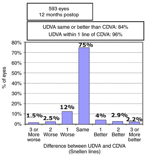
EXPERIMENTAL AND THERAPEUTIC MEDICINE 21: 288, 2021 3
Table I. Preoperative manifest and cycloplegic refraction data.
Parameter Diopters (mean ± SD, range)
Manifest sphere +3.9532±1.5282, +1.25 to +6.00
Manifest cylinder +2.6316±1.2208, +0.50 to +5.00
Manifest SEQ +4.0780±1.5723, +1.50 to +6.00
Cycloplegic sphere +5.7198±1.4351, +3.25 to +6.75
Cycloplegic cylinder +3.1320±1.4829, +0.50 to +5.00
SD, standard deviation; SEQ, spherical equivalent refraction.
Figure 2. Efficacy, shown as gained and lost Snellen lines of postoperative
uncorrected distance visual acuity (UDVA) compared to preoperative corrected
distance visual acuity (CDVA), 12‑months postoperative. Postop, postoperative.
Figure 1. Summary of the 12‑month postoperative uncorrected distance
visual acuity (UDVA) and preoperative corrected distance visual acuity
(CDVA). Refractive target was emmetropia (plano target). Postop, postopera-
tive; Preop, preoperative.
all eyes reached a UDVA of 20/63 or better 12 months after
Femto‑LASIK surgery. As shown in Fig. 2, for 84.1% of the
eyes, the postoperative 12‑month UDVA was the same or
better than the preoperative CDVA.
Safety, defined as no loss of two or more Snellen lines of
CDVA, was excellent. Twelve months after Femto‑LASIK
surgery, no eye lost 2 lines and 6 eyes (1.012%) lost one
Snellen line of CDVA (Fig. 3). Contrariwise, we found several
amblyopic eyes that gained at least one line of visual acuity, Figure 3. Safety, shown as gained and lost Snellen lines of postoperative
reflecting potential benefit of the refractive surgery, possibly corrected distance visual acuity (CDVA) compared to preoperative CDVA,
explained by reduction or elimination of optical aberrations of 12‑months postoperative. Postop, postoperative.
the hyperopic magnifying correction lenses.
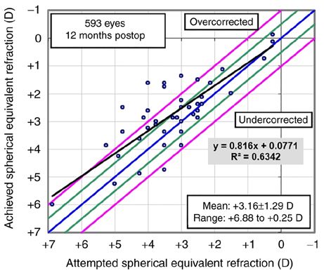
The scatterplot of the attempted spherical equivalent
refraction (SEQ) against the achieved refractive change is Fig. 7 shows the excellent evolution of SEQ after the
shown in Fig. 4. At the postoperative 12 months follow‑up surgery and its stability in time over the 12‑month follow‑up
visit, the mean sphere was +0.887±0.8746 D, the mean cylinder period. The postoperative refraction data at 1, 3, 6 and
was +0.766±0.5797 and the mean SEQ was +0.5042±0.8336. 12 months postoperative are presented in Table II. There were
The accuracy of SEQ at 12 months was 49% within ±0.50 D no intraoperative or postoperative complications during the
and respectively 74% within ±1.00 D of emmetropia (Fig. 5). follow‑up period.
Lower predictability and accuracy of SEQ was found for the
eyes with high preoperative refractive errors. Discussion
Regarding the mean refractive manifest astigmatism at the
last postoperative visit, 54 and 79% were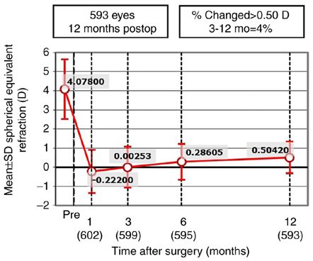
4                     TĂBĂCARU et al: HYPEROPIC Femto-LASIK OUTCOMES USING THE VisuMax®-MEL® 80 PLATFORM

                                                                               Figure 6. Summary of preoperative and 12‑months postoperative refractive
Figure 4. Scattergram of attempted vs. achieved spherical equivalent refrac-   astigmatism. Postop, postoperative; Preop, preoperative; D, diopter.
tion (SEQ), 12‑months postoperative. Postop, postoperative; D, diopter.

Figure 5. Accuracy of spherical equivalent refraction (SEQ) to intended        Figure 7. Stability of spherical equivalent refraction (SEQ) during the
target, 12‑months postoperative. Postop, postoperative; D, diopter.            follow‑up period of 12 months. Postop, postoperative.

challenging for eyes with hyperopia or hyperopic astigmatism,                  wide ablation and high correction speed has greatly improved
especially when refractive error is high (1,6‑32,58).                          prognosis (1,8) [large diameter optical areas being more resistant
    Hyperopic correction has several difficulties. Hyperopic                   to epithelial hyperplasia that is responsible for real regression (1,7)],
patients are frequently under‑corrected in regards to glass                    latent hyperopia is often the reason why refractive surgeons avoid
prescriptions prior to preoperative evaluation and have accom-                 approaching cases of hyperopia and hyperopic astigmatism or
modation reserve of different degrees or accommodation                         limit the surgeries to refractive errors below +3.0 D (1,8). Laser
spasm (1,6). Therefore, the integration of manifest and cyclo-                 refractive treatment is difficult to choose because ablation of
plegic refractions is difficult and latent hyperopia may lead to               manifest refraction in hyperopia or hyperopic astigmatism may
increasing values of manifest refraction with age (1,6). Accurate              lead postoperatively to recurrence of a degree of hyperopia, falsely
centration of the ablation is another difficult point as hyperopic             interpreted as regression of the laser procedure (1,6,7), while
patients have a wide angle Kappa (6). Hyperopic correction                     ablation of cycloplegic refraction may lead to myopic refraction
induces central corneal flattening, which may be limited by                    in the immediate postoperative period, causing an unsatisfactory
preoperative keratometry (6). Another issue concerning hyper-                  UDVA (1). In order to compensate in part for the latent hyperopia,
opic and astigmatic patients is the higher risk of de‑centration               Kanellopoulos proposed a nomogram that involves full treatment
of ablation due to the difficulty in fixation of the near target               in the case of the cylinder and the ablation of the manifest refrac-
point during laser correction (6). Considering all the above                   tion sphere with an addition of +0.25 D for the dominant eye and
listed features, the predictability and the accuracy of the refrac-            up to +0.75 D for the non‑dominant eye (1,9).
tive results in hyperopia, with or without astigmatism, are lower                  There have been several reports of hyperopic‑LASIK and
when compared to myopic refractive corrections (58).                           hyperopic astigmatism‑LASIK in the past, using different
    Although some excimer lasers have been approved for correc-                excimer laser platforms and using for the flap creation either
tions up to +6.0 D (1,7) and current technology using models with              the microkeratome or the femtosecond laser (1,6‑32). Table III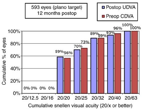
EXPERIMENTAL AND THERAPEUTIC MEDICINE 21: 288, 2021                                                                                                                             5

Table II. Postoperative refraction data.

                                                                                                            Postoperative visit
                               -----------------------------------------------------------------------------------------------------------------------------------------------------------------------------------------------
                                       1 month                                           3 months                                              6 months                                             12 months
                               -----------------------------------------------------------------------------------------------------------------------------------------------------------------------------------------------
Parameter                                                                                         Diopters (mean ± SD, range)

Manifest sphere                +0.233±1.1409,                                    +0.4393±1.1563,                                     +0.667±0.9742,                                         +0.887±0.8746,
                               -1.50 to +3.25                                    -1.25 to +3.75                                      -1.00 to +2.50                                         -1.00 to +2.75
Manifest cylinder              +0.91±0.739,                                      +0.8735±0.6021,                                     +0.7618±0.54706,                                       +0.766±0.5797,
                               +0.00 to +3.00                                    +0.25 to +2.75                                      +0.00 to +2.75                                         +0.25 to +3.25
Manifest SEQ                   -0.222±1.1320,                                    +0.00253±1.079,                                     +0.28605±0.9382,                                       +0.5042±0.8336,
                               -2.50 to +3.00                                    -1.50 to +3.375                                     -1.25 to +2.50                                         -1.00 to +3.00

SD, standard deviation; SEQ, spherical equivalent refraction.

Table III. Predictability of the SEQ between ±0.5 D and ±1.0 D of the intended target, in several published reports of LASIK
interventions for hyperopia or hyperopic astigmatism (1).

		                                                                                                                                   Follow‑up                        ±0.5 D of                         ±1.0 D of
		                                                                                                                                     period                       the intended                      the intended
Authors (Ref.) Refractive error of the treated eyes                                                                                   (months)                         target                             target

Zadok et al (20)                                 Hyperopia (SEQ
6 TĂBĂCARU et al: HYPEROPIC Femto-LASIK OUTCOMES USING THE VisuMax®-MEL® 80 PLATFORM
was emmetropia in all cases. The treatment plan was chosen accountable for all aspects of the study in ensuring that ques-
corroborating the manifest, fogging and cycloplegic refraction, tions related to the accuracy or integrity of any part of the work
considering both the accommodative reserve and patient age. are appropriately investigated and resolved.
Femtosecond‑LASIK using the VisuMax® ‑MEL ® 80
platform was demonstrated to be a suitable option to correct Ethics approval and consent to participate
selected cases of hyperopia and hyperopic astigmatism.
The postoperative results after one year demonstrated the All participants signed an informed consent in accordance
Femto‑LASIK procedure to be safe and effective. The predict- with the Declaration of Helsinki. The study was approved by
ability at 12 months was 74% within ±1 D of emmetropia. As the Ethics Committee of ‘Carol Davila’ University of Medicine
both the sphere and the cylinder plus values were reduced and and Pharmacy of Bucharest, Romania.
the need for hyperopic correction lenses was minimal, the
optical aberrations and distortion of the retinal image were Patient consent for publication
smaller, allowing us to achieve a better postoperative CDVA
with at least one line in 9% of the eyes. This manuscript does not contain case details, personal
The retrospective pattern of this report and the short period information or images that may enable an individual to be
of follow‑up of one year are limiting factors in our study. A identified.
future prospective study and a longer follow‑up period are
necessary for a better understanding of the procedure stability Competing interests
and for elaboration of nomogram ablation profiles.
The authors have no financial or proprietary interest to declare
Acknowledgements in any device presented in this article.
Professional editing, linguistic and technical assistance was References
performed by Irina Radu, Individual Service Provider.
1. Tăbăcaru B: Femtosecond Laser‑Excimer Laser Platform for
Ametropias Surgery, PhD thesis (no. TD 4697). ‘Carol Davila’
Funding University of Medicine and Pharmacy, Bucharest, Romania, 2019.
2. Shah R: History and results; indications and contraindications of
No funding was received. SMILE compared with LASIK. Asia Pac J Ophthalmol (Phila) 8:
371‑376, 2019.
3. Stanca HT, Munteanu M, Jianu DC, Motoc AG, Jecan CR,
Availability of data and materials Tăbăcaru B, Stanca S and Preda MA: Femtosecond‑LASIK
outcomes using the VisuMax®‑MEL® 80 platform for mixed
astigmatism refractive surgery. Rom J Morphol Embryol 59:
The data that support the findings of this study are available 277‑283, 2018.
from the corresponding author (HTS), upon reasonable request. 4. Tabacaru B and Stanca HT: One year refractive outcomes of
femtosecond‑LASIK in mild, moderate and high myopia. Rom J
Ophthalmol 61: 23‑31, 2017.
Authors' contributions 5. Tabacaru B and Stanca HT: Scheimpflug topographical changes
after femtosecond LASIK for mixed astigmatism‑theoretical
BT contributed to the conception and design of the study, the aspects and case study. Rom J Ophthalmol 61: 69‑75, 2017.
6. El‑Naggar MT and Hovaghimian DG: Assessment of refractive
acquisition, analysis and interpretation of the data of the study. outcome of femtosecond‑assisted LASIK for hyperopia correc-
She also contributed to the drafting of the work and its critical tion. Electron Physician 9: 3958‑3965, 2017.
revision for important intellectual content. HTS contributed to 7. Motwani M and Pei R: Treatment of moderate‑to‑high hyperopia
with the WaveLight Allegretto 400 and EX500 excimer laser
the conception and design of the study, the acquisition, analysis systems. Clin Ophthalmol 11: 999‑1007, 2017.
and interpretation of the data of the study, contributed to the 8. Motwani M: Topographic‑guided treatment of hyperopic correc-
drafting of the work and its critical revision for important intel- tions with a combination of higher order aberration removal with
WaveLight® Contoura and wavefront‑optimized hyperopic treat-
lectual content. RAP contributed to the acquisition, analysis and ment. Clin Ophthalmol 12: 1021‑1029, 2018.
interpretation of data of the study, contributed to the drafting 9. Kanellopoulos AJ: Topography‑guided hyperopic and hyperopic
of the work and its critical revision for important intellectual astigmatism femtosecond laser‑assisted LASIK: Long‑term
experience with the 400 Hz eye‑Q excimer platform. Clin
content. SS contributed to the conception and design of the study, Ophthalmol 6: 895‑901, 2012.
contributed to the drafting of the work and its critical revision for 10. Suarez E, Torres F and Duplessie M: LASIK for correction of
important intellectual content. CD contributed to the conception hyperopia and hyperopia with astigmatism. Int Ophthalmol
Clin 36: 65‑72, 1996.
and design of the study, contributed to the drafting of the work 11. Esquenazi S: Five‑year follow‑up of laser in situ keratomileusis
and its critical revision for important intellectual content. MM for hyperopia using the Technolas Keracor 117C excimer laser.
contributed to the acquisition, the analysis and interpretation of J Refract Surg 20: 356‑363, 2004.
12. Zadok D, Raifkup F, Landau D and Frucht‑Pery J: Long‑term
data of the study, to the drafting of the work and its critical revi- evaluation of hyperopic laser in situ keratomileusis. J Cataract
sion for important intellectual content. CR contributed to the Refract Surg 29: 2181‑2188, 2003.
analysis and interpretation of data of the study, to the drafting 13. Jaycock PD, O'Brart DP, Rajan MS and Marshall J: 5‑year follow‑up
of LASIK for hyperopia. Ophthalmology 112: 191‑199, 2005.
of the work and its critical revision for important intellectual 14. Varley GA, Huang D, Rapuano CJ, Schallhorn S, Boxer Wachler BS
content. ACT contributed to the analysis and interpretation of and Sugar A; Ophthalmic Technology Assessment Committee
data of the study, to the drafting of the work and its critical Refractive Surgery Panel, Ameican Academy of Ophthalmology:
LASIK for hyperopia, hyperopic astigmatism, and mixed astig-
revision for important intellectual content. All authors read and matism: A report by the American academy of ophthalmology.
approved the final version of the manuscript and agreed to be Ophthalmology 111: 1604‑1617, 2004.EXPERIMENTAL AND THERAPEUTIC MEDICINE 21: 288, 2021 7
15. Spades L, Sabetti L, D'Alessandri L and Balestrazzi E: 38. Stanca HT, Stanca S, Tabacaru B, Boruga M and Balta F:
Photorefractive keratectomy and LASIK for the correction of Bevacizumab in Wet AMD treatment: A tribute to the thirteen
hyperopia: 2‑year follow‑up. J Refract Surg 22: 131‑136, 2006. years of experience from the beginning of the anti‑VEGF era in
16. Alió J, Galal A, Ayala MJ and Artola A: Hyperopic LASIK with Romania. Exp Ther Med 18: 4993‑5000, 2019.
esiris/schwind technology. J Refract Surg 22: 772‑781, 2006. 39. Goldberg I, Jay Katz L, Mansouri K, Pakravan M and Yazdani S:
17. Antonios R, Arba Mosquera S and Awwad ST: Hyperopic laser A refractive surgery candidate with optic nerve head cupping.
in situ keratomileusis: Comparison of femtosecond laser and J Ophthalmic Vis Res 7: 248‑256, 2012.
mechanical microkeratome flap creation. J Cataract Refract 40. Stanca HT, Suvac E, Munteanu M, Jianu DC, Motoc AGM,
Surg 41: 1602‑1609, 2015. Roşca GC and Boruga O: Giant cell arteritis with arteritic
18. Desai RU, Jain A and Manche EE: Long‑term follow‑up of anterior ischemic optic neuropathy. Rom J Morphol Embryol 58:
hyperopic laser in situ keratomileusis correction using the Star 281‑285, 2017.
S2 excimer laser. J Cataract Refract Surg 34: 232‑237, 2008. 41. Moshirfar M, D Wagner W, H Linn S, W Brown T, L Goldberg J,
19. el‑Agha MS, Johnston EW, Bowman RW, Cavanagh HD and T Gomez A, C Ronquillo Y and C Hoopes P: Corneal refractive
McCulley JP: Excimer laser treatment of spherical hyperopia: surgery in patients with history of optic neuritis. J Ophthalmic
PRK or LASIK? Trans Am Ophthalmol Soc 98: 59‑66, 2000. Vis Res 14: 436‑441, 2019.
20. Zadok D, Maskaleris G, Montes M, Shah S, Garcia V and 42. Munteanu M, Rosca C and Stanca H: Sub‑inner limiting
Chayet A: Hyperopic laser in situ keratomileusis with the Nidek membrane hemorrhage in a patient with Terson syndrome. Int
EC‑5000 excimer laser. Ophthalmology 107: 1132‑1137, 2000. Ophthalmol 39: 461‑464, 2019.
21. Davidorf JM, Eghbali F, Onclinx T and Maloney RK: Effect of 43. Arevalo JF: Managing retinal detachment after refractive
varying the optical zone diameter on the results of hyperopic laser surgery: With current knowledge, we cannot determine whether
in situ keratomileusis. Ophthalmology 108: 1261‑1265, 2001. prophylactic treatment is indicated in candidates for refractive
22. Rashad KM: Laser in situ keratomileusis for the correction surgery. Retina Today 2017: 36‑40, 2017.
of hyperopia from +1.25 to +5.00 diopters with the Technolas 44. Vatsa S and Sood S: Macular dystrophy in a post LASIK patient.
Keracor 117C laser. J Refract Surg 17: 113‑122, 2001. DJO 30: 60‑62, 2020.
23. Llovet F, Galal A, Benitez‑del‑Castillo JM, Ortega J, Martin C 45. Stanca HT, Munteanu M, Jianu DC, Motoc AGM, Tăbăcaru B,
and Baviera J: One‑year results of excimer laser in situ keratomi- Stanca S, Ungureanu E, Boruga VM and Preda MA: New
leusis for hyperopia. J Cataract Refract Surg 35: 1156‑1165, 2009. perspectives in the use of laser diode transscleral cyclophoto-
24. Alió JL, El Aswad A, Vega‑Estrada A and Javaloy J: Laser in situ coagulation. A prospective single center observational cohort
keratomileusis for high hyperopia (>5.0 diopters) using optimized study. Rom J Morphol Embryol 59: 869‑872, 2018.
aspheric profiles: Efficacy and safety. J Cataract Refract Surg 39: 46. Donnenfeld ED: Correcting astigmatism after glaucoma surgery.
519‑527, 2013. Glaucoma Today 2011: 29‑32, 2011.
25. Kanellopoulos AJ, Conway J and Pe LH: LASIK for hyperopia 47. Preda MA, Karancsi OL, Munteanu M and Stanca HT: Clinical
with the WaveLight excimer laser. J Refract Surg 22: 43‑47, 2006. outcomes of micropulse transscleral cyclophotocoagulation in
26. Gil‑Cazorla R, Teus MA, de Benito‑Llopis L and Mikropoulos DG: refractory glaucoma‑18 months follow‑up. Lasers Med Sci 35:
Femtosecond laser vs. mechanical microkeratome for hyperopic 1487‑1491, 2020.
laser in situ keratomileusis. Am J Ophthalmol 152: 16‑21.e2, 48. Chang JSM, Law AKP, Ng JCM and Cheng MSY: Femtosecond
2011. laser in situ keratomileusis flap creation in narrow palpebral
27. Ditzen K, Fiedler J and Pieger S: Laser in situ keratomileusis for fissure eyes without suction. Case Rep Ophthalmol 8: 341‑348,
hyperopia and hyperopic astigmatism using the Meditec MEL 70 2017.
spot scanner. J Refract Surg 18: 430‑434, 2002. 49. Boruga O, Bălăşoiu AT, Giuri S, Munteanu M, Stanca HT,
28. Lian J, Ye W, Zhou D and Wang K: Laser in situ keratomileusis Iovănescu G and Preda MA: Caruncular late‑onset junctional
for correction of hyperopia and hyperopic astigmatism with the nevus: Apropos of an anatomo‑clinical observation. Rom J
Technolas 117C. J Refract Surg 18: 435‑438, 2002. Morphol Embryol 58: 1461‑1464, 2017.
29. El‑Agha MS, Bowman RW, Cavanagh D and McCulley JP: 50. Moshirfar M, Shah TJ, Skanchy DF, Linn SH, Kang P and
Comparison of photorefractive keratectomy and laser in situ Durrie DS: Comparison and analysis of FDA reported visual
keratomileusis for the treatment of compound hyperopic astig- outcomes of the three latest platforms for LASIK: Wavefront
matism. J Cataract Refract Surg 29: 900‑907, 2003. guided Visx iDesign, topography guided WaveLight allegro
30. Pineda‑Fernández A, Rueda L, Huang D, Nur J and Jaramillo J: contoura and topography guided Nidek EC‑5000 CATz. Clin
Laser in situ keratomileusis for hyperopia and hyperopic astig- Ophthalmol 11: 135‑147, 2017.
matism with the Nidek EC‑5000 excimer laser. J Refract Surg 17: 51. J Shah T, Moshirfar M and C Hoopes P: Safety of the excimer
670‑675, 2001. laser in LASIK and PRK for patients with implantable cardiac
31. Roesler C and Kohnen T: Changes of functional optical zone devices: Our clinical experience in the past two decades.
after LASIK for hyperopia and hyperopic astigmatism. J Refract J Ophthalmic Vis Res 14: 530‑531, 2019.
Surg 34: 476‑481, 2018. 52. Stanca HT, Petrović Z and Munteanu M: Transluminal Nd:YAG
32. Salz JJ and Stevens CA; LADARVision LASIK Hyperopia Study laser embolysis‑a reasonable method to reperfuse occluded
Group: LASIK correction of spherical hyperopia, hyperopic branch retinal arteries. Vojnosanit Pregl 71: 1072‑1077, 2014.
astigmatism, and mixed astigmatism with the LADARVision 53. Cobo‑Soriano R, Beltrán J and Baviera J: LASIK outcomes in
excimer laser system. Ophthalmology 109: 1647‑1658, 2002. patients with underlying systemic contraindications: A prelimi-
33. Tăbăcaru B, Stanca S, Mocanu V, Zemba M, Stanca HT and nary study. Ophthalmology 113: 1118.e1‑e8, 2006.
Munteanu M: Intraoperative flap‑related complications in 54. Savoiu Balint G, Iovanescu G, Stanca H, Popoiu CM, Boia E,
FemtoLASIK surgeries performed with Visumax® femtosecond Popovici RA and Bolintineanu SL: The protective effect of
laser: A ten‑year Romanian experience. Exp Ther Med 20: HDL‑cholesterol in patients with essential hypertension. Rev
2529‑2535, 2020. Chim 68: 949‑952, 2017.
34. Stanca TH, Tabacaru B and Celea C: Correlations between 55. Spadea L and Paroli MP: Laser refractive surgery in diabetic
confocal microscopy and histological aspects of normal cornea. patients: A review of the literature. Clin Ophthalmol 6: 1775‑1783,
Rom J Ophthalmol 59: 19‑23, 2015. 2012.
35. Schallhorn SC, Schallhorn JM, Pelouskova M, Venter JA, 56. Reinstein DZ, Archer TJ and Randleman JB: JRS standard for
Hettinger KA, Hannan SJ and Teenan D: Refractive lens exchange reporting astigmatism outcomes of refractive surgery. J Refract
in younger and older presbyopes: Comparison of complication Surg 30: 654‑659, 2014.
rates, 3 months clinical and patient‑reported outcomes. Clin 57. Waring GO III: Standard graphs for reporting refractive surgery.
Ophthalmol 11: 1569‑1581, 2017. J Refract Surg 16: 459‑466, 2000.
36. Mălăescu M, Stanca HT, Tăbăcaru B, Stănilă A, Stanca S and 58. Spierer O, Mimouni M, Nemet A, Rabina G and Kaiserman I:
Danielescu C: Accuracy of five intraocular lens formulas in eyes Hyperopic laser keratorefractive surgery: Do steep corneas have
with trifocal lens implant. Exp Ther Med 20: 2536‑2543, 2020. worse outcomes? Int Ophthalmol 40: 1885‑1895, 2020.
37. Alió JL, Grzybowski A and Romaniuk D: Refractive lens
exchange in modern practice: When and when not to do it? Eye This work is licensed under a Creative Commons
Vis (Lond) 1: 10, 2014. Attribution-NonCommercial-NoDerivatives 4.0
International (CC BY-NC-ND 4.0) License.You can also read