Artificial intelligence using convolutional neural networks for real-time detection of early esophageal neoplasia in Barrett's esophagus (with video)
←
→
Page content transcription
If your browser does not render page correctly, please read the page content below
ORIGINAL ARTICLE: Clinical Endoscopy

     Artificial intelligence using convolutional neural networks for
     real-time detection of early esophageal neoplasia in Barrett’s
     esophagus (with video)
            Rintaro Hashimoto, MD, PhD,1 James Requa,2 Tyler Dao,2 Andrew Ninh,2 Elise Tran,1 Daniel Mai,1 Michael Lugo,1
            Nabil El-Hage Chehade, MD,1 Kenneth J. Chang, MD,1 Williams E. Karnes, MD,1 Jason B. Samarasena, MD1

            Orange, Irvine, California, USA

     Background and Aims: The visual detection of early esophageal neoplasia (high-grade dysplasia and T1 cancer)
     in Barrett’s esophagus (BE) with white-light and virtual chromoendoscopy still remains challenging. The aim of
     this study was to assess whether a convolutional neural artificial intelligence network can aid in the recognition
     of early esophageal neoplasia in BE.
     Methods: Nine hundred sixteen images from 65 patients of histology-proven early esophageal neoplasia in BE
     containing high-grade dysplasia or T1 cancer were collected. The area of neoplasia was masked using image anno-
     tation software. Nine hundred nineteen control images were collected of BE without high-grade dysplasia. A con-
     volutional neural network (CNN) algorithm was pretrained on ImageNet and then fine-tuned with the goal of
     providing the correct binary classification of “dysplastic” or “nondysplastic.” We developed an object detection
     algorithm that drew localization boxes around regions classified as dysplasia.
     Results: The CNN analyzed 458 test images (225 dysplasia and 233 nondysplasia) and correctly detected early
     neoplasia with sensitivity of 96.4%, specificity of 94.2%, and accuracy of 95.4%. With regard to the object detection
     algorithm for all images in the validation set, the system was able to achieve a mean average precision of .7533 at
     an intersection over union of .3
     Conclusions: In this pilot study, our artificial intelligence model was able to detect early esophageal neoplasia in
     BE images with high accuracy. In addition, the object detection algorithm was able to draw a localization box
     around the areas of dysplasia with high precision and at a speed that allows for real-time implementation. (Gastro-
     intest Endosc 2020;91:1264-71.)

Abbreviations: AI, artificial intelligence; BE, Barrett’s esophagus; CNN,   Copyright ª 2020 by the American Society for Gastrointestinal Endoscopy
convolutional neural network; DL, deep learning; mAP, mean average          0016-5107/$36.00
precision; NBI, narrow-band imaging; WLI, white-light imaging.              https://doi.org/10.1016/j.gie.2019.12.049
DISCLOSURE: The following authors disclosed financial relationships: J.     Received October 16, 2019. Accepted December 30, 2019.
Requa: Employee of Docbot Inc. D. Tyler, A. Ninh, W. E. Karnes: Co-
                                                                            Current affiliations: H. H. Chao Comprehensive Digestive Disease Center,
founder and equity holder of Docbot Inc. K. J. Chang: Consultant for
                                                                            Division of Gastroenterology & Hepatology, Department of Medicine,
Olympus, Cook Medical, Medtronics, Endogastric Solution, Erbe,
                                                                            University of California, Irvine, Orange, California, USA (1), Docbot Inc,
Apollo, Mederi, Ovesco, Mauna Kea, and Pentax. J. Samarasena: Co-
                                                                            Irvine, California, USA (2).
founder and equity holder of Docbot Inc; consultant for Medtronic,
Olympus, US Endoscopy, Mauna Kea, Motus, and Pentax. All other              Reprint requests: Jason Samarasena, MD, Division of Gastroenterology,
authors disclosed no financial relationships.                               University of California Irvine, 333 City Blvd West, Suite 400, Orange,
                                                                            CA 92868.

                       This video can be viewed directly                    If you would like to chat with an author of this article, you may contact Dr
                                                                            Samarasena at jsamaras@hs.uci.edu.
                       from the GIE website or by using
                       the QR code and your mobile de-
                       vice. Download a free QR code
                       scanner by searching “QR Scanner”
                       in your mobile device’s app store.

1264 GASTROINTESTINAL ENDOSCOPY Volume 91, No. 6 : 2020                                                                          www.giejournal.org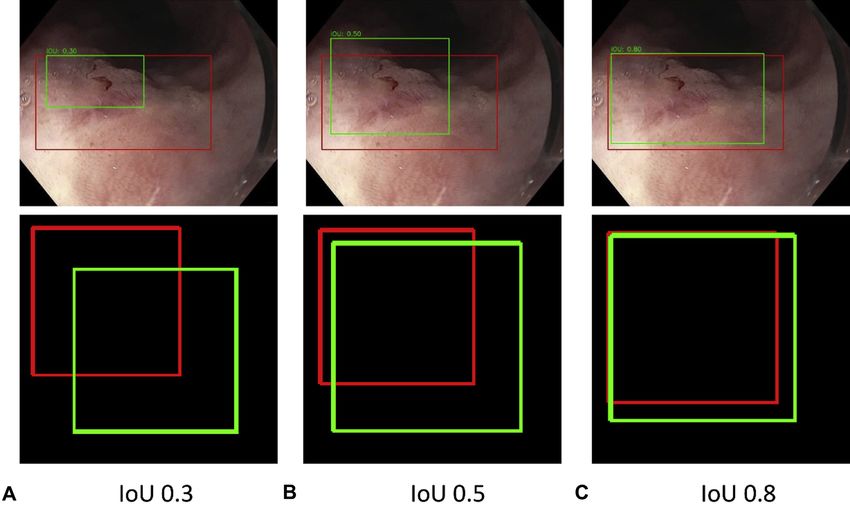
Hashimoto et al                                                                                             Using AI to detect early BE

   Esophageal cancer is the eighth most common cancer
and the sixth leading cause of cancer death worldwide
with an estimated incidence of 52,000 cases in 2012.1
Barrett’s esophagus (BE) is a known risk factor for the
development of esophageal adenocarcinoma.2 Prognosis
is strongly related to stage at diagnosis.2 However, more
than 40% of patients with esophageal adenocarcinoma
are diagnosed after the disease has metastasized, and the
5-year survival rate is less than 20%.2,3 The risk of
esophageal adenocarcinoma rises when BE is associated
with dysplasia.4,5 Most GI societies recommend
endoscopic surveillance for BE subjects to enable early
detection of dysplasia or carcinoma.6-8 However, the effect
of endoscopic surveillance for BE has been disappointing.9
   Current guidelines recommend endoscopic surveillance
in patients with BE with random 4-quadrant biopsy speci-
mens obtained every 1 to 2 cm to detect dysplasia,10 but
this method is invasive, time-consuming, and difficult to
comply with.11 As a result, the American Society for           Figure 1. White-light endoscopic image of a dysplastic area of Barrett’s
Gastrointestinal Endoscopy set a performance threshold         esophagus. Red box is expert annotation. Green box is prediction by arti-
for optical diagnosis. That is, if an imaging technology is    ficial intelligence algorithm.
able to achieve a per-patient sensitivity of 90%, a negative
predictive value of 98%, and a specificity of 80% for detect-   endoscopy database. The endoscope used over this time
ing early esophageal neoplasm,8 then random 4-quadrant bi-     period was the Olympus 190 series upper endoscope
opsy sampling could be avoided. Because this requirement is    (190 HQ and 190 H; Olympus, Center Valley, Pa, USA).
believed to be difficult to achieve for nonexperts,12 a great   The images were categorized by image quality (excellent,
number of technologies and imaging enhancements have           adequate, or poor) and by the following imaging tech-
been studied and developed to help improve detection.          niques: white-light imaging (WLI), narrow-band imaging
Among these are chromoendoscopy, endocytoscopy, probe-         (NBI), standard focus, and Near Focus (Olympus). Near
based confocal laser–induced endomicroscopy, image-            focus is a proprietary imaging technique to Olympus
enhanced endoscopy, volumetric laser endomicroscopy,           endoscopes that enables a change to the focal length of
magnification endoscopy, and wide-area transepithelial          the scope lens such that objects within 2 mm of the
sampling with computer-assisted 3-dimensional analysis.13      endoscope tip are maintained in clear focus. Only excellent
Many of these have disadvantages with respect to learning      and adequate images were used.
curve, cost, and time issues.                                     Nine hundred sixteen images from 70 patients were retro-
   A simple real-time diagnosis support system would be of     spectively collected of histology-proven dysplasia (high-grade
great help for endoscopists in the detection of Barrett’s      dysplasia and T1 adenocarcinoma) in BE, and 916 control im-
dysplasia. Recently, artificial intelligence (AI) using deep    ages from 30 patients were collected of either histology-
learning (DL) with convolutional neural networks (CNNs)        proven or confocal laser endomicroscopy–proven BE without
has emerged and showed great results in the diagnosis          dysplasia. We did not include cases with low-grade dysplasia
and detection of lesions in the esophagus,14-17 stom-          for this pilot study. A large diversity of dysplastic lesions was
ach,18-21 small bowel,22 and colon.23-26 However, no study     chosen for training in this study to optimize the algorithm.
has been reported on an application of DL for detection        Neoplastic lesions in the image dataset ranged from 3 to
of early neoplasia within BE. We conducted a pilot study       20 mm in size, and most lesions were deemed subtle, such
on the endoscopic detection of early esophageal neoplasia      that a lesser-trained endoscopist could miss them.
on BE using a DL system showing promising results.
                                                               Annotation of images
                                                                   All neoplastic lesions in the selected images were anno-
METHODS                                                        tated independently by 2 expert endoscopists (R.H. and
                                                               J.S.) who were not blinded to endoscopic findings and pa-
Upper endoscopy and dysplasia image dataset                    thology. Images were annotated with a single rectangular
   We retrospectively reviewed all endoscopic images of        box using image annotation software (Fig. 1, red box).
patients with early esophageal neoplasia in BE, defined in      When multiple neoplastic lesions were noted in an
this case as high-grade dysplasia or T1 stage adenocarci-      image, multiple rectangular boxes were used. This
noma, proven by histology at our institution between           annotation was used as the ground truth for the training
January 2016 and November 2018 using the electronic            of the automated detection algorithm.

www.giejournal.org                                             Volume 91, No. 6 : 2020 GASTROINTESTINAL ENDOSCOPY 1265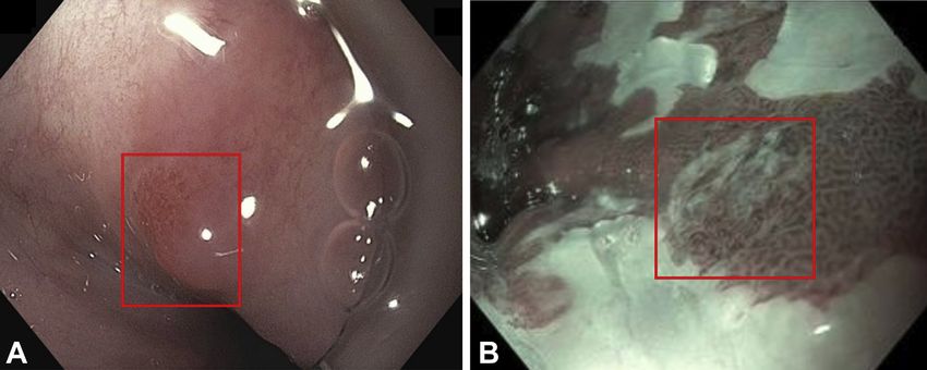
Using AI to detect early BE Hashimoto et al
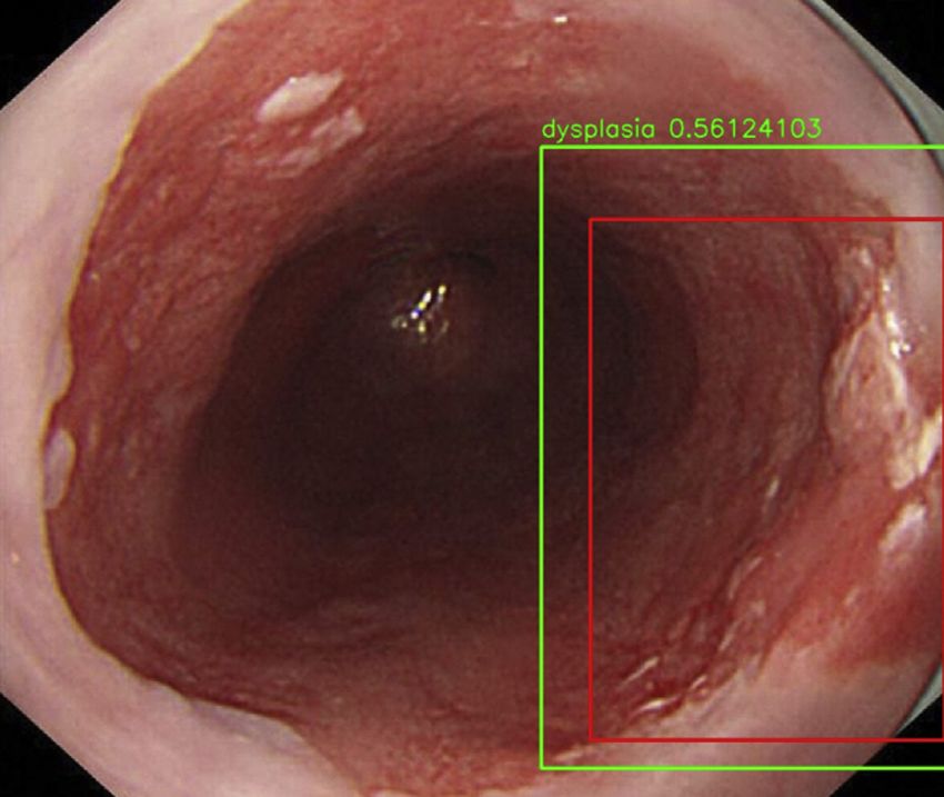
13x13 Class Probability
32px x 32px Map
Input Image Feature Maps
(real-time endoscopy feed) grid
Final
Detection
Bounding
Resized to Boxes
416px x 416px
CNN layers
x K anchors x C classes
Conv Conv 3 Conv 3 Conv 5 Conv class
MaxPool MaxPool MaxPool MaxPool MaxPool
5 Conv 3 Conv Conv (x, y, w, h, obj score)
probability NMS
13x13xKx(5+C)
Post-Processing
Non-Maximum Suppression
Figure 2. Illustration of the deep learning system. CNN, Convolutional neural network; NMS, non-maximum suppression.
CNN architecture leakage, we carefully split the training and validation sets
We developed and designed a CNN model architecture such that each set only contained unique images from
from 2 primary modules: feature extraction and classifica- different patients. This ensured the algorithm metrics
tion. The base module of our algorithm is responsible for were validated on patients who were not part of the training
automated feature extraction and borrows from the set. In the inference stage, the data produced by the training
Inception-ResNet-v2 algorithm developed by Google AI were used to generate test set predictions. Various augmen-
(Mountain View, Calif, USA) that achieves state-of-the-art ac- tations to the test set generated an additional layer of pre-
curacy on popular image classification benchmarks. The dictions. Predictions were then averaged to create a
head module of our algorithm is designed for transforming composite probability and then fed into an Adam optimizer
extracted features from the base layers (50 mm þ parame- to produce a binary result between 0 to .5 and .5 to 1 for
ters) into a graded scale that allows for pathologic classifica- final classification data. Any value .5 was classified as the
tion. The sigmoid activation function maps the model’s presence of dysplasia and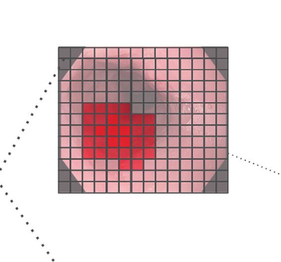
Hashimoto et al                                                                                                                        Using AI to detect early BE

Figure 3. Endoscopic images with varying intersections of union (IoUs): A, IoU .3. B, IoU .5. C, IoU .8. In this study, we set the IoU threshold as .3 for
true positive on object detection.

   For localization accuracy, we used mean average preci-                                                    Barrett’s - Dysplasia vs Non-Dysplasia
sion (mAP), a standard metric used in AI. Average precision
serves to determine the area under the precision recall                                                                                                      200
curve and carries a value between 0 and 1. To calculate                                                             220                       13             175
                                                                                             non-dysplasia
mAP, we need to use the intersection of union, which mea-
                                                                                                                                                             150
sures the overlap between prediction and ground truth.
                                                                                True label

We predefined an intersection over union threshold at .3                                                                                                      125
to classify whether the box prediction by the algorithm                                                                                                      100
was a true positive or a false positive. An intersection
                                                                                                                                                             75
over union threshold of .3 was chosen because it was                                             dysplasia              8                 217
believed that in real-time clinical endoscopy this level of                                                                                                  50
lesion targeting was more than sufficient to aid in the clin-                                                                                                 25
ical identification of a region of neoplasia during a
screening or surveillance BE examination (Fig. 3).                                                                                                 ia
                                                                                                                            ia

                                                                                                                                               as
                                                                                                                        as

                                                                                                                                              l
                                                                                                                                           sp
                                                                                                                        pl

                                                                                                                                         dy
                                                                                                                    ys

Statistical analysis
                                                                                                                    d
                                                                                                                 n-
                                                                                                               no

   All statistical analyses were performed using R software
                                                                                                                            Predicted label
(version 3.3.3; The R Foundation for Statistical Computing,
Vienna, Austria). Categorical data were compared with the                       Figure 4. A 2  2 table showing results of the artificial intelligence algo-
c2 and t tests and continuous variables with the Mann-                          rithm performance of binary classification of dysplasia versus no dysplasia
Whitney U test. Differences between variables with                              per image.
P < .05 were considered significant.
                                                                                RESULTS
Ethics
  This study was approved by the Institutional Review                           Binary classification per image
Board of the University of California Irvine Medical Center                        A total of 458 images, 225 images of dysplastic lesions
(UCI IRB HS no. 2008-6258).                                                     from 26 patients and 233 images of nondysplastic BE

www.giejournal.org                                                             Volume 91, No. 6 : 2020 GASTROINTESTINAL ENDOSCOPY 1267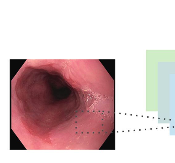
Using AI to detect early BE                                                                                                                               Hashimoto et al

TABLE 1. Results of AI binary diagnosis: per-image analysis

                                                               Sensitivity (%)                     P value                    Specificity (%)                     P value

  AI diagnosis by white-light imaging                           98.6 (144/146)                       .023                      88.8 (95/107)                       .0007
  AI diagnosis by narrow-band imaging                            92.4 (73/79)                                                 99.2 (125/126)
  AI diagnosis by standard focus                                96.6 (141/146)                        .89                      89.9 (98/109)                        .005
  AI diagnosis by near focus                                     96.2 (76/79)                                                 98.4 (122/124)
  Comprehensive AI diagnosis                                    96.4 (217/225)                                                94.2 (220/233)
Values in parentheses are n/N. A total of 458 images (dysplastic 225, nondysplastic 233) were used for validation. The number of dysplastic images of white-light imaging,
narrow-band imaging, standard focus, and near focus was 146, 79, 146, and 79 and nondysplastic images 107, 126, 109, and 124, respectively.
AI, Artificial intelligence.

TABLE 2. Results of AI binary diagnosis: per-patient analysis

                                                                                              Sensitivity (%)                                                     P value

  AI diagnosis by white-light imaging                                                           94.7 (18/19)                                                        .74
  AI diagnosis by narrow-band imaging                                                           91.7 (11/12)
  AI diagnosis by standard focus                                                                95.2 (20/21)                                                        .68
  AI diagnosis by near focus                                                                    91.7 (11/12)
  Comprehensive AI diagnosis                                                                    92.3 (24/26)
Values in parentheses are n/N. Validation set includes images of dysplasia from 26 patients in total. Images of white-light imaging, narrow-band imaging, standard focus, and
near focus came from 19, 12, 21, and 12 patients, respectively.
AI, Artificial intelligence.

from 13 patients, were used for validation. For binary clas-                             Object detection
sification (dysplasia vs nondysplasia) sensitivity, specificity,                              In our validation set with an intersection of union at .3,
and accuracy per image were 96.4%, 94.2%, and 95.4%,                                     overall mAP was .7533. mAP for NBI only was higher at
respectively (Fig. 4, Table 1). The sensitivity per image                                .802, and mAP for near focus images was .819. The overall
for WLI only was 98.6% and for NBI was 92.4%. The                                        sensitivity (ie, recall) was 95.6%. The sensitivity for WLI was
sensitivity per image for standard focus images only was                                 94.1%, NBI 98.2%, standard focus 96.8%, and near focus
96.6% and for near focus was 96.2%. NBI specificity                                       97.5%. The overall positive predictive value (ie, precision)
(99.2%) was higher than WLI specificity (88.8%) (P Z                                      was 89.2%. The positive predictive value for WLI was
.0007). Near focus specificity (98.4%) was higher than                                    87.8%, NBI 92.1%, standard focus 86.3%, and near focus
standard focus (89.9%, P Z .005).                                                        96.3% (Table 3).

Binary classification per patient                                                         Speed
   The CNN correctly diagnosed 24 of 26 patients (92.3%)                                    On GPU gtx1070 setting, the binary classifier runs at
as having dysplasia. The sensitivities of dysplasia diagnosis                            around 72 frames per s (299x299 pixel image size). The
per patient by WLI, NBI, false standard focus, and near                                  YOLO v2 runs at around 45 frames per s (416 x 416p x im-
focus were 94.7%, 91.7%, 95.2%, and 91.7%, respectively                                  age size). Video 1 (available online at www.giejournal.org)
(Table 2).                                                                               demonstrates how this algorithm could work in real time.

False-negative and -positive lesions on binary
classification                                                                            DISCUSSION
    Eight of 225 dysplastic images (3.6%) with 4 patients
showed a false negative in the binary classification. Six of                                 Our pilot study for detection of early esophageal
8 false-negative images were from 2 patients but had                                     neoplasm in BE using CNN demonstrated a high sensitivity
another true-positive image and therefore did not affect                                 (96.4%), specificity (94.2%), and accuracy (95.4%) per im-
per-patient sensitivity. Two of 8 false-negative images                                  age for binary classification (dysplasia vs no dysplasia) on
from 2 patients had no true-positive images. All false neg-                              an internal validation study. Our localization algorithm
atives were small subtle lesions less than 5 mm in size                                  also detected most lesions correctly with a mAP of .7533,
(Fig. 5). For most false-positive lesions, it appears that                               sensitivity of 95.6%, and positive predictive value of 89.2%.
the algorithm detected an area of nodularity and/or raised                                  DL for the detection of lesions in the GI tract is rapidly
tissue as dysplastic.                                                                    emerging.14-31 However, there still remains limited

1268 GASTROINTESTINAL ENDOSCOPY Volume 91, No. 6 : 2020                                                                                             www.giejournal.org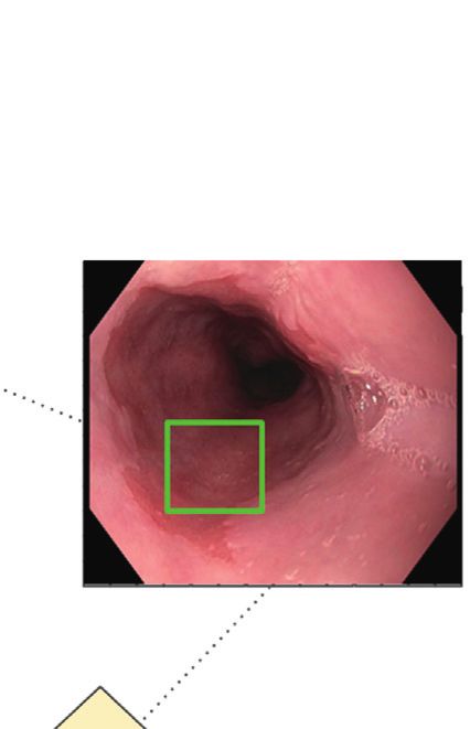
Hashimoto et al                                                                                                             Using AI to detect early BE

Figure 5. Two cases with negative binary result. Both lesions showed high-grade dysplasia in histopathologic examination. A, A 4-mm nodular lesion from
buried Barrett’s esophagus. B, A 4-mm slightly depressed lesion.

TABLE 3. Results of AI object detection for binary positive image: per-image analysis

                                                   Sensitivity (%)            P value             Positive predictive value (%)              P value

 AI diagnosis by white-light imaging                94.1 (318/338)              .018                       87.8 (318/362)                      .14
 AI diagnosis by narrow-band imaging                98.2 (164/166)                                         92.1 (164/178)
 AI diagnosis by standard focus                     96.8 (328/346)               .24                       86.3 (328/380)                     .0003
 AI diagnosis by near focus                         97.5 (154/158)                                         96.3 (154/160)
 Comprehensive AI diagnosis                         95.6 (482/504)                                         89.2 (482/540)
Values in parentheses are n/N.
AI, Artificial intelligence.

literature that focuses on early esophageal neoplasia in                          To improve the impact of endoscopic surveillance of
BE. de Groof et al29 reported a study of automated                            BE, a real-time AI video overlay assisting an endoscopist
detection of early esophageal neoplasia in BE using                           detect neoplastic lesions is certainly desirable. Our
machine learning with a hand-crafted algorithm. They                          group has already achieved a real-time video overlay
used 100 endoscopic images from 44 patients with a sensi-                     with colonoscopy algorithms including polyp detection
tivity of 83% and a specificity of 83%.28 They also published                  and optical pathology and believe this is certainly
a prospective pilot study of this algorithm with promising                    feasible.23 The advantage of real-time AI over other tech-
data. However, this algorithm was not fast enough to be                       nologies aimed to improve dysplasia detection is that the
used in real time. Also reported was the application of AI                    learning curve is negligible. Ideally, the system would
for volumetric laser endomicroscopy, resulting in a                           simply flag areas of concern for the endoscopist to inter-
sensitivity and specificity of 90% and 93%.30,31 However,                      rogate further with more detailed imaging or a biopsy
volumetric laser endomicroscopy requires time and                             sampling. An algorithm, well trained by expert endoscop-
equipment. A Japanese group recently reported a pilot                         ists, has the potential to elevate the image interpretation
study using DL with CNN on detection mainly of early                          skill set of an endoscopist to a level near that of an im-
squamous cell carcinoma with a sensitivity of 98%.14                          aging expert. If an AI algorithm were able to achieve
Ebigbo et al16 used computer-aided diagnosis in the evalua-                   American Society for Gastrointestinal Endoscopy Preser-
tion of early esophageal adenocarcinoma in BE. This system                    vation and Incorporation of Valuable Endoscopic Innova-
showed sensitivity and specificity of 97% and 88% and was                      tions threshold for the detection of early esophageal
trained on only 148 images, significantly less than our                        neoplasia, it could decrease the number of unnecessary
training set. In a follow-up study piloting this system in                    biopsy samplings performed during surveillance
real time, the same group was able to assess the system in                    examinations.
14 patients with promising results, and their system was                          There were performance differences on binary classifi-
trained on 129 images.17 The speed of the algorithm                           cation with specific imaging modalities. NBI showed better
prediction required pausing the live video display to make                    sensitivity and specificity than WLI, and near focus showed
a prediction. Our current prototype as seen in Video 1                        better specificity than standard focus. Although the effect
does not require pausing and also does not require a                          of NBI on detection of neoplasia is controversial,32,33 our
second monitor to display the user interface as in the                        study suggests that NBI and near focus may alter tissue
system shown in their publication.                                            imaging characteristics and detail in such a way that

www.giejournal.org                                                           Volume 91, No. 6 : 2020 GASTROINTESTINAL ENDOSCOPY 1269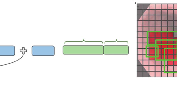
Using AI to detect early BE                                                                                                                  Hashimoto et al

subtle imaging differences between dysplastic and                                    grade dysplasia: a meta-analysis. Gastrointest Endosc 2008;67:
nondysplastic tissue are enhanced and are better                                     394-8.
                                                                                6.   Shaheen NJ, Falk GW, Iyer PG, et al. ACG clinical guideline: diagnosis
differentiated by the algorithm.                                                     and management of Barrett’s esophagus. Am J Gastroenterol
   Because overfitting and spectrum bias can cause overes-                            2016;111:30-50.
timation of accuracy, we were extremely careful in select-                      7.   Fitzgerald RC, di Pietro M, Ragunath K, et al. British Society of Gastro-
ing a highly diverse group of images from each patient.                              enterology guidelines on the diagnosis and management of Barrett’s
Furthermore, images of specific patients used in the                                  oesophagus. Gut 2014;63:7-42.
                                                                                8.   Evans JA, Early DS, Fukami N, et al. The role of endoscopy in Barrett’s
training set were not used in the validation set. Rectangular                        esophagus and other premalignant conditions of the esophagus. Gas-
polygons for annotation and prediction were chosen to                                trointest Endosc 2012;76:1087-94.
minimize processing power such that the algorithm can                           9.   Codipilly DC, Chandar AK, Singh S, et al. The effect of endoscopic sur-
make predications at >40 frames per s and real-time                                  veillance in patients with Barrett’s esophagus: a systematic review and
evaluation is possible (as evidenced in Video 1). With                               meta-analysis. Gastroenterology 2018;154:2068-86.
                                                                               10.   Sharma P, Savides TJ, Canto MI, et al. The American Society for Gastro-
more complex polygons with independent corners and/                                  intestinal Endoscopy PIVI (Preservation and Incorporation of Valuable
or vertices there becomes a significantly increased level                             Endoscopic Innovations) on imaging in Barrett’s Esophagus. Gastroint-
of processing power needed, thereby slowing down the                                 est Endosc 2012;76:252-4.
frame rate for prediction and diminishing the utility in                       11.   Tavakkoli A, Appelman HD, Beer DG, et al. Use of appropriate surveil-
real time.                                                                           lance for patients with nondysplastic Barrett’s esophagus. Clin Gastro-
                                                                                     enterol Hepatol 2018;16:862-9.
   Performance detection depends on the neural network                         12.   ASGE Technology Committee; Thosani N, Abu Dayyeh BK, et al. ASGE
architecture used. Ghatwary et al34 compared several DL                              Technology Committee systematic review and meta-analysis assessing
object detection methods to identify esophageal                                      the ASGE Preservation and Incorporation of Valuable Endoscopic Inno-
adenocarcinoma using 100 WL images from 39 patients                                  vations thresholds for adopting real-time imaging-assisted endoscopic
and concluded that single-shot multibox detector methods                             targeted biopsy during endoscopic surveillance of Barrett’s esophagus.
                                                                                     Gastrointest Endosc 2016;83:684-98.
outperformed regional-based CNN, fast regional-based                           13.   Sami SS, Iyer PG. Recent advances in screening for Barrett’s esophagus.
CNN, and faster regional-based CNN with a sensitivity of                             Curr Treat Options Gastroenterol 2018;16:1-14.
.90, specificity of .88, precision of .70, and recall of .79.34                 14.   Horie Y, Yoshio T, Aoyama K, et al. Diagnostic outcomes of esophageal
We chose to use YOLO v2, which is similar to the single-                             cancer by artificial intelligence using convolutional neural networks.
shot multibox detector but has a faster frames per second                            Gastrointest Endosc 2019;89:25-32.
                                                                               15.   Cai SL, Li B, Tan WM, et al. Using deep learning system in endoscopy
prediction to use this algorithm in real time.                                       for screening of early esophageal squamous cell carcinoma (with
   Our study has limitations. First, this study was a single-                        video). Gastrointest Endosc 2019;90:745-53.
center retrospective study and lacked external validation.                     16.   Ebigbo A, Mendel R, Probst A, et al. Computer-aided diagnosis using
That is, although this algorithm worked very well on our                             deep learning in the evaluation of early oesophageal adenocarcinoma.
validation set of images, it was not tested on endoscopic                            Gut 2019;68:1143-5.
                                                                               17.   Ebigbo A, Mendel R, Probst A, et al. Real-time use of artificial intel-
images from other centers or other scope manufacturers,                              ligence in the evaluation of cancer in Barrett’s oesophagus. Gut
and this is a future goal for this project. Second, the num-                         2020;69:615-6.
ber of early esophageal neoplasia in BE was not large.                         18.   Hirasawa T, Aoyama K, Tanimoto T, et al. Application of artificial intel-
   In summary, this is among the first studies in which                               ligence using a convolutional neural network for detecting gastric can-
AI using CNNs was examined for detection of early                                    cer in endoscopic images. Gastric Cancer 2018;21:653-60.
                                                                               19.   Kanesaka T, Lee TC, Uedo N, et al. Computer-aided diagnosis for iden-
esophageal neoplasm in BE. Although this is a pilot                                  tifying and delineating early gastric cancers in magnifying narrow-
study, the results were very promising. The speed of                                 band imaging. Gastrointest Endosc 2018;87:1339-44.
the algorithm is fast enough to enable true real-time                          20.   Zhang X, Chen F, Yu T, et al. Real-time gastric polyp detection using
detection, and our results suggest a real-time AI detec-                             convolutional neural networks. PLoS One 2019;14:e0214133.
tion system for dysplasia in BE can likely be achieved                         21.   Li L, Chen Y, Shen Z, et al. Convolutional neural network for the diag-
                                                                                     nosis of early gastric cancer based on magnifying narrow band imag-
in the near future.                                                                  ing. Gastric Cancer 2020;23:126-32.
                                                                               22.   Aoki T, Yamada A, Aoyama K, et al. Automatic detection of erosions and
                                                                                     ulcerations in wireless capsule endoscopy images based on a deep con-
REFERENCES                                                                           volutional neural network. Gastrointest Endosc 2019;89:357-63.
                                                                               23.   Urban G, Tripathi P, Alkayali T, et al. Deep learning localizes and iden-
 1. Arnold M, Soerjomataram I, Ferlay J, et al. Global incidence of oesopha-         tifies polyps in real time with 96% accuracy in screening colonoscopy.
    geal cancer by histological subtype in 2012. Gut 2015;64:381-7.                  Gastroenterology 2018;155:1069-78.
 2. Hur C, Miller M, Kong CY, et al. Trends in esophageal adenocarcinoma       24.   Misawa M, Kudo SE, Mori Y, et al. Artificial intelligence-assisted polyp
    incidence and mortality. Cancer 2013;119:1149-58.                                detection for colonoscopy: initial experience. Gastroenterology
 3. Thrift AP. The epidemic of oesophageal carcinoma: Where are we                   2018;154:2027-9.
    now? Cancer Epidemiol 2016;41:88-95.                                       25.   Klare P, Sander C, Prinzen M, et al. Automated polyp detection in the
 4. Curvers WL, Ten Kate FJ, Krishnadath KK, et al. Low-grade dysplasia in           colorectum: a prospective study (with videos). Gastrointest Endosc
    Barrett’s esophagus: overdiagnosed and underestimated. Am J Gastro-              2019;89:576-82.
    enterol 2010;105:1523-30.                                                  26.   Wang P, Xiao X, Brown JR, et al. Development and validation of a deep-
 5. Rastogi A, Puli S, El-Serag HB, et al. Incidence of esophageal                   learning algorithm for the detection of polyps during colonoscopy. Nat
    adenocarcinoma in patients with Barrett’s esophagus and high-                    Biomed Eng 2018;2:741.

1270 GASTROINTESTINAL ENDOSCOPY Volume 91, No. 6 : 2020                                                                                www.giejournal.org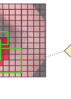
Hashimoto et al                                                                                                              Using AI to detect early BE

27. Redmon J, Farhadi A. YOLO9000: better, faster, stronger. The IEEE        31. Fonollà R, Scheeve T, Struyvenberg MR, et al. Ensemble of deep con-
    Conference on Computer Vision and Pattern Recognition 2017:                  volutional neural networks for classification of early barrett’s
    7263-71. Available at: https://arxiv.org/abs/1612.08242. Accessed            neoplasia using volumetric laser endomicroscopy. Appl Sci 2019;9:
    November 22, 2019.                                                           2183.
28. van der Sommen F, Zinger S, Curvers WL, et al. Computer-aided detec-     32. Beg S, Mensa M, Fullard M, et al. Impact of advanced endoscopic im-
    tion of early neoplastic lesions in Barrett’s esophagus. Endoscopy           aging on Barrett’s esophagus in daily clinical practice. Gastrointest En-
    2016;48:617-24.                                                              dosc 2018;87:1189-94.
29. de Groof J, van der Sommen F, van der Putten J, et al. The Argos proj-   33. Xiong YQ, Ma SJ, Hu HY, et al. Comparison of narrow-band imaging
    ect: the development of a computer-aided detection system to                 and confocal laser endomicroscopy for the detection of neoplasia in
    improve detection of Barrett’s neoplasia on white light endoscopy.           Barrett’s esophagus: a meta-analysis. Clin Res Hepatol Gastroenterol
    United Eur Gastroenterol J 2019;7:538-47.                                    2018;42:31-9.
30. Swager AF, van der Sommen F, Klomp SR, et al. Computer-aided detec-      34. Ghatwary N, Zolgharni M, Ye X. Early esophageal adenocarcinoma
    tion of early Barrett’s neoplasia using volumetric laser endomicro-          detection using deep learning methods. Int J Comput Assist Radiol
    scopy. Gastrointest Endosc 2017;86:839-46.                                   Surg 2019;14:611-21.

                                                Submit to GIE's sister journal, VideoGIE
                                                   Now indexed in PubMed Central!

                     VideoGIE is an Open Access, online-only journal, indexed in PubMed Central. Submit
                     video cases of endoscopic procedures used in the study, diagnosis, and treatment of
                     digestive diseases.
                     VideoGIE publishes the following article types:
                     • Case Reports: Reports of the diagnosis and management of digestive diseases using
                               a single case.
                     • Case Series: Reports of the diagnosis and management of digestive diseases using 3
                               or more cases.
                     • Tools and Techniques: Educational videos demonstrating the use of a particular
                               endoscopic tool or technique. The goal of this section is to help trainees,
                               endoscopy nurses, and technicians learn how best to use the tools of endos-
                               copy for high-quality care.
                     All manuscripts must be submitted online at http://www.editorialmanager.com/vgie

www.giejournal.org                                                           Volume 91, No. 6 : 2020 GASTROINTESTINAL ENDOSCOPY 1271
Using AI to detect early BE                                                    Hashimoto et al

    916 images of dysplasia             916 images of non-dysplasia
       from 70 patients                       from 30 patients

                 1832 images of Barrett’s esophagus

        1374 images for training
   (691 dysplasia/683 non-dysplasia)

   Algorithm developed using CNN
                                          Validation
                                            study

                                              458 images for test
                                       (225 dysplasia/233 non-dysplasia)

Supplementary Figure 1. Study flow of this study. CNN, Convolutional
neural network.

1271.e1 GASTROINTESTINAL ENDOSCOPY Volume 91, No. 6 : 2020                 www.giejournal.org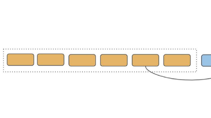
You can also read