Towards Understanding the Whiplash Condition at 3 Tesla
←
→
Page content transcription
If your browser does not render page correctly, please read the page content below
Clinical Neurology
Towards Understanding the Whiplash
Condition at 3 Tesla
Elliott JM, PT, Ph.D.1,2; McMahon K, Ph.D.3; Cowin G Ph.D.3; Parrish TB, Ph.D.4
1
Department of Physical Therapy and Human Movement Sciences, Feinberg School of Medicine, Chicago, IL, USA
2
School of Health and Rehabilitation Sciences, Division of Physiotherapy, Centre for Clinical Research Excellence
in Spinal Pain, Injury and Health. The University of Queensland, Brisbane, Australia
3
Centre for Advanced Imaging, The University of Queensland, Brisbane, Australia
4
Department of Biomedical Engineering and Radiology, Feinberg School of Medicine, Chicago, IL, USA
The accurate and consistent radiological jects with whiplash injury, it is clinically taken to determine fracture prevalence
observation of head and neck soft-tissue important that objective and quantifi- in this population. More importantly,
damage in patients with whiplash inju- able measures to characterize the whip- CT is unable to assess soft tissue damage
ries has been largely inconsistent and lash condition be made available. This is in the cervical spine and surrounding
highly variable [6, 33, 34, 39, 40, 44]. especially important for the exploration muscles.
As a result there exists the proposition and development of more informed treat- Conventional magnetic resonance imag-
that tissue damage does not, or cannot ment strategies aimed at retarding if not ing (MRI) has largely failed to consistently
occur, as a result of a low-speed motor preventing the expression of persistent reveal soft-tissue damage in patients
vehicle collision [9]. Engineering appli- pain for some patients. with whiplash, but this may relate to the
cations [26–28, 37, 38, 49] and con- For many of the laboratory-created use of generic clinical protocols (typi-
trolled animal studies [48, 51-53] have lesions shown to occur in animal and cally 1.5T and lower) and limitations in
informed us of what can happen to a bioengineering models, there are cur- the resolution. The advent of higher-field
number of vulnerable tissues in the cer- rently very few, if any, clinical means for systems (3T and greater) has provided
vical region following whiplash, includ- their diagnosis available to practicing a foundation for measuring physiologic
ing the facet capsule, dorsal root gan- clinicians. Plain films lack sensitivity for processes that could be associated with
glion and nerve roots [36, 46, 48, 51–53] ruling out bony lesions and the images tissue damage. Preliminary MRI evidence
At the forefront of clinical enquiry how- lack the detail to quantify strained facet identifying the unique expression of neck
ever, is how to best determine what has joint capsules and/or tears in discs. muscle degeneration (fatty infiltrates)
happened to these tissues in patients Computed tomography (CT) can identify at 4-weeks post injury in those who tran-
with whiplash injury. Due to the persis- some cervical spine fractures but rigorous sit from acute to chronic pain suggests
tent nature of symptoms in some sub- longitudinal studies have not been under- that this may be so [12]. Muscle changes
1A 1B 1C
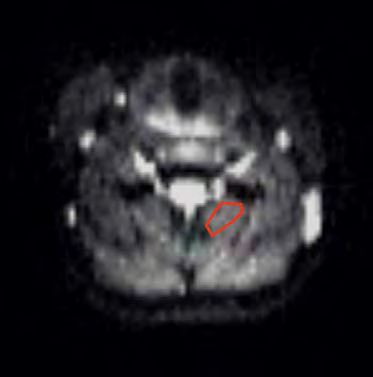
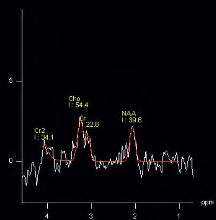
1 (1A) Localization of SVS for the cervical cord at the C2/3 segmental level with (1B) corresponding metabolic peaks for NAA, Cr, and Cho in
healthy control and (1C) subject with chronic whiplash (adapted from Elliott et al., Spinal Cord [16]).
44 MAGNETOM Flash · 1/2012 · www.siemens.com/magnetom-world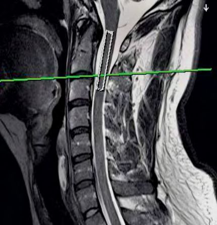
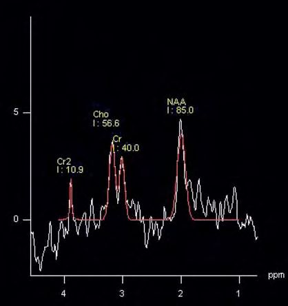
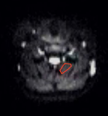
2A

                                                                                                                                  Neurology Clinical

                 b-value = 0 s/mm2                                   b-value = 50 s/mm2                             b-value = 250 s/mm2

     2A DWI scans of a ROI over the left cervical multifidus muscle (b-values of 0 – 250 s/mm2).

did not occur in patients with lower                 which may be contributing to their pain         ADVANCED shim* WIP that provides a
levels of initial pain or in patients with           and disability.                                 robust and rapid shim map. The typical
chronic non-traumatic neck pain [13]                                                                 shim result is a FWHM of ~15 Hz at 3T,
suggesting traumatic factors play a role             MR Spectroscopy –                               which translates into a metabolite line
in altering the make-up of the neck                  metabolite scale                                width of 6 Hz. The optimized shim is
muscles.                                             Alterations in MR visible metabolites,          achieved by manually setting the shim
We have developed a comprehensive                    such as Lactate (Lac), N-Acetylaspartate        volume to be slightly larger (5 mm in
advanced MR imaging protocol that                    (NAA), Creatine (Cr) and Choline (Cho)          each direction) than the actual acquisi-
assesses the cervical spine at the meta-             have been demonstrated in neurological          tion voxel. Following this acquisition,
bolic, microscopic and macroscopic                   disorders [5, 31] traumatic brain injury        a quick (8 average) acquisition is obtained
level. Furthermore, by applying this pro-            [7], and cervical myelopathy [24] and may       without water suppression to use as a
tocol, in tandem with clinical signs and             have predictive capacity [7]. Our previ-        standard over time, which is helpful with
symptoms, over several weeks, it may be              ous work has reported the presence of           repeated measures. These 30-second
possible to classify which patients are              altered cord biochemistry in a small            spectra assess the total amount of water
at risk for transiting to a persistent pain          sample of patients with chronic whiplash        present and can be used for control over
state. This non-invasive methodology to              related pain and disability [16]. How-          placement of the voxel over time and
quantify several physiologic processes               ever, this study did not resolve the tem-       calculate actual concentration of metab-
may afford clinicians the ability to triage          poral development of these changes or           olites.
their patients with confidence. Further-             if they are unique to those with poor           *Work in progress. This information about this product
more, it may be possible to determine if             functional recovery. Such work is well           is preliminary. The product is under development and
                                                                                                      not commercially available in the U.S., and its future
a person has suffered a traumatic event,             underway.
                                                                                                      availability cannot be ensured.
                                                     The current protocol uses a single voxel
                                                     spectroscopic (SVS) technique to investi-
                                                                                                     Diffusion-weighted imaging
      Table 1: SVS_SE Sequence                       gate the spinal cord at the C2-C3 level.
                                                     Using a high resolution T2 TSE sagittal
                                                                                                     of muscle – microscopic scale
                                                     scan along with the axial and coronal local-    Diffusion-weighted imaging (DWI) of the
                    TR 2000 ms                       izer scans, a long rectangular voxel            muscle system has the potential advan-
                                                     (5 mm x 7 mm x 40 mm) is placed in the          tage over conventional sequences to
                   TE 135 ms                         middle of the cord (Fig. 1). The para-          help uncover the early physiological
                                                     meters are listed in Table 1. The acquisi-      mechanisms underlying the manifesta-
                                                     tion is not cardiac triggered but this is       tion of intra- or inter-muscular fatty
           160 averages with HEP
                                                     possible to reduce movement artifacts           infiltrates. Diffusion properties of water
                                                     induced by CSF flow. We currently are           have been quantified with DWI in many
            coil elements neck 1,2
                                                     using a long TE (135 ms) SVS PRESS              different organ systems (e.g. brain and
                                                     acquisition to reduce the contamination         spinal cord, kidneys, heart, lumbar
             coil elements spine 1
                                                     of short T2 metabolite species as well          intervertebral disc and the prostate) [1,
                                                     as have lactate out of phase with the           2, 29, 22, 25, 30, 35, 43, 17, 18, 47, 3,
                                                     nearby lipid signal. This acquisition is        4, 50]. Normal water diffusion is affected
 Table 1: Parameters for the SVS_SE
 Sequence.                                           5:28 minutes after the voxel has been           by the presence and orientation of phys-
                                                     shimmed properly. We are using the              ical tissue barriers (e.g. cell membranes,

                                                                                  MAGNETOM Flash · 1/2012 · www.siemens.com/magnetom-world               45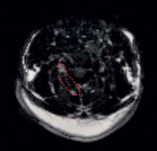
Clinical Neurology
2B proteins, myelin sheaths and/or lipids)
ADC_WAD [45]. The measure quantifies the micro-
50 100 150 200 250 300
scopic movements of water diffusion
0 via the mean apparent diffusion coeffi-
- 0,05 cient (ADC) [20, 45, 39]. Passive enlarge-
- 0,1 ment of the muscle cell following tissue
damage may be associated with an
- 0,15
increase in ADC [23]. We have shown
- 0,2
altered ADC maps for the cervical multi-
- 0,25
fidus in a small sample of subjects with
- 0,3 chronic whiplash when compared to
- 0,35 healthy controls [16], suggesting a pas-
b-values (s/mm2)
- 0,4 sive enlargement of the muscle cell (e.g.
atrophy) (Figs. 2A, B). However, the tem-
poral development of such changes and
ADC_Control whether they are unique to those at risk
50 100 150 200 250 300 for chronicity is unknown at this stage.
0
Such evidence could provide for a sensi-
- 0,05
tive indicator of early tissue injury at a
stage when muscle degeneration remains
- 0,1 potentially salvageable [23, 32]. Thus,
the potential diagnostic and prognostic
- 0,15 value of in-vivo DWI sequences for
quantifying the temporal degeneration
- 0,2 of muscle tissue, at a cellular level, in
b-values (s/mm2) whiplash is clear.
- 0,25
Diffusion-weighted
2B ADC maps (at 1.5T) for the left cervical multifidus muscle in patients with chronic
whiplash associated disorders (WAD) and healthy controls [16]. imaging parameters:
The DWI scan uses a spin-echo, echo-
planar acquisition with an in-plane reso-
lution of 1.6 mm, a thickness of 5 mm,
3
TR 4000 ms and TE 65 ms to reduce arti-
facts. The acquisition is taken in the
axial plane using an inversion pulse to
reduce the signal from fat with a TI of
160 ms. The diffusion scan is the simple
3-scan trace acquisition but the number
of diffusion-weightings is increased to 5.
The b-values used at 3T are 0, 50, 100,
200, and 300 s/mm2. This is quite differ-
ent from brain DWI where a typical
b-value is 1000 s/mm2. Due to signal-to-
noise constraints and distortions of the
signal, the b-values are small. From all of
the b-value data, trace and ADC images
are generated for each of the 14 slices in
7:08 minutes.
Fat/water imaging –
macroscopic scale
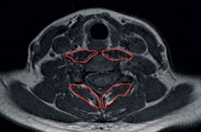
3 T1-weighted axial MR image at the C6 vertebral level demonstrating outlined ROIs for
The demonstration of neck muscle fatty
the right and left longus colli and the right and left posterior cervical multifidus. Increased
infiltrates on T1-weighted imaging in
signal, indicative of fatty infiltration, is noted in both sets of muscles in a subject with chronic
whiplash associated disorders. chronic whiplash (Fig. 3) [11, 14, 15] is
interesting as such findings were not
46 MAGNETOM Flash · 1/2012 · www.siemens.com/magnetom-world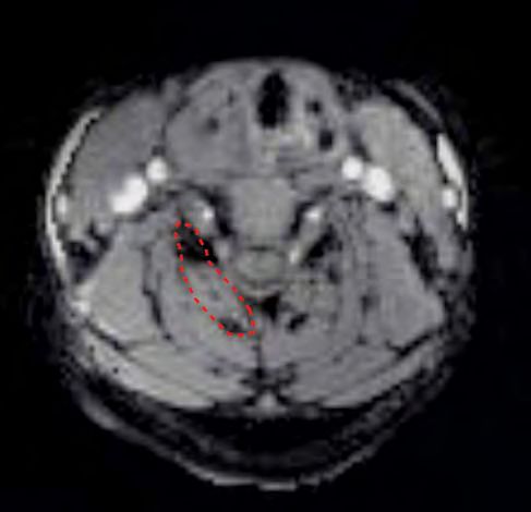
4A

featured in those with chronic non-trau-        This method has been applied success-
matic neck pain [13] and their expression       fully in the liver and musculoskeletal
(between 4 weeks and 3 months post-             application using an iterative least squares
injury event) on standard T1-weighted           solution called IDEAL [41, 42]. The
images appears unique to only those who         method we have used in the study of
transit [12]. It is postulated that these       whiplash subjects collects 8 different
muscle changes represent one neuro-             echo times sufficiently spaced on the unit
physiologic basis for the transition to         circle to provide adequate phase infor-
chronic pain in this population. While          mation for the variable projection
the mechanisms underlying their tempo-          (VARPRO) algorithm*, generating a glob-
ral development and contribution                ally optimal solution for the water/fat
towards the transition remain unclear,          decomposition [21].                                       4B
it is possible that newer MRI techniques        Saurabh Shah implemented the VARPRO
(MRS and DWI) could help quantify ear-          algorithm and acquisition sequence in
lier physiologic changes at the spinal cord     the Cardiovascular R&D team located at
and muscle cell that may precede observ-        Northwestern University, Chicago, IL,
able muscle changes on T1-weighted              USA. Currently this feature is a WIP at
sequences. An earlier detection of such         syngo MR B17 software (Fig. 4A, B).
mechanisms could prove crucial for              A three-dimension 230 mm field-of-view
identifying the early presence of select        (FOV) axial gradient echo acquisition
biochemical changes in spinal cord metab-       was used to collect the data required for
olism with the attendant later changes          the VARPRO algorithm. The sequence
in muscle physiology and the develop-           parameters are TR 23.81 ms, 8 echo times
ment of chronic pain and disability.            with a spacing of 1.78 ms starting at
                                                1.36 ms. A single slab is placed over the
Fat/water separation                            cervical spine with 36 partitions and a
Several approaches are possible to              partition thickness of 3 mm and slab
measure the water and fat composition           oversampling of 22% to prevent aliasing                     4 ROIs in (4A) fat (VARPRO* based) and (4B)
of a voxel. These include a dual acquisi-       in the 3D direction. The in-plane resolu-                  water for the right cervical multifidus muscle.
tion method, where one image is fat             tion is 1.4 mm using a rectangular                         The ROIs are automatically copied to the same
suppressed [19] (water image) and a stan-       FOV of 75% resulting in an acquisition                     location for both images and relative differences
                                                                                                           (signal loss) between the fat and water images
dard image is collected (fat and water          time of 2:06 minutes.
                                                                                                           can be calculated.
image). The difficulty of this type of acqui-
                                                *Work in progress. This information about this product
sition is that it relies on the uniform fre-
                                                 is preliminary. The product is under development and
quency difference between water and              not commercially available in the U.S., and its future     References
fat across the whole volume of excitation,       availability cannot be ensured.                          1 Basser PJ, Mattiello J, LeBihan D. MR diffusion
which is often difficult to obtain espe-                                                                    tensor spectroscopy and imaging. Biophys J
                                                                                                            1994;66:259-67.
cially at high magnetic fields. A fat sup-
pressed acquisition using a short-tau           Conclusions                                               2 Basser PJ, Pierpaoli C. Microstructural and physio-
                                                                                                            logical features of tissues elucidated by quantita-
inversion recovery (STIR) sequence is pos-      The observed alterations in spinal cord                     tive-diffusion-tensor MRI. J Magn Reson B 1996;
sible, but the T1 of fat has to be assumed,     biochemistry, muscle water diffusion,                       111:209-19.
which may vary depending on the sub-            and fatty infiltration in chronic whiplash                3 Beattie P. Diffusion-Weighted Magnetic Resonance
                                                                                                            Imaging of the Musculoskeletal System: An Emerg-
ject or the evolution of the infiltration of    [16] provides preliminary evidence for
                                                                                                            ing Technology with Potential to Impact Clinical
the muscle [8]. An alternative is the           the early detection and classification of                   Decision Making. J Orthop Sports Phys Ther 2011.
Dixon method [10], where one collects           the patient with a whiplash injury. Cur-                  4 Beattie PF, Arnot CF, Donley JW, et al. The imme-
data at an echo time when water and             rent studies indicate that the physiologic                  diate reduction in low back pain intensity following
fat are in-phase and at an echo time            measures assessed with the multi-dimen-                     lumbar joint mobilization and prone press-ups is
                                                                                                            associated with increased diffusion of water in the
when water and fat are out of phase. The        sional imaging protocol outlined above
                                                                                                            L5-S1 intervertebral disc. JOSPT 2010;40:256-64.
data can be combined in such a way              show promise for detecting which patients                 5 Blamire AM, Cader S, Lee M, et al. Axonal damage
that they generate a fat and water image.       may be at risk for transitioning from                       in the spinal cord of multiple sclerosis patients
This method works well when there are           acute to persistent pain following a low-                   detected by magnetic resonance spectroscopy.
                                                                                                            Magnetic Resonance in Medicine 2007;58:880-5.
no field inhomogeneities, which is often        velocity traumatic event involving the
                                                                                                          6 Borchgrevink G, Smevik O, Haave I, et al. MRI of
not the case. Current methods collect           head and neck.                                              cerebrum and cervical columna within two days
multiple echo time data to improve the                                                                      after whiplash neck sprain injury. Injury 1997;
estimation of the fat and water images.                                                                     28:331-5.

                                                                                 MAGNETOM Flash · 1/2012 · www.siemens.com/magnetom-world                   47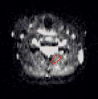
Clinical Neurology

 7 Brenner T, Freier MC, Holshouser BA, et al. Pre-          24 Holly LT, Freitas B, McArthur DL, et al. Proton        41 Reeder SB, Pineda AR, Wen Z, et al. Iterative de-
   dicting neuropsychologic outcome after traumat-              magnetic resonance spectroscopy to evaluate               composition of water and fatwith echo asymmetry
   ic brain injury in children. Pediatr Neurol 2003;            spinal cord axonal injury in cervical spondylotic         and least-squares estimation (IDEAL): Application
   28:104-14.                                                   myelopathy. J Neurosurg Spine 2009;10:194-200.            with fast spin-echo imaging. Magn Reson Med
 8 Bydder GM, Steiner RE, LH. B. MR imaging of the           25 Hsieh TJ, Chang JM, Chuang HY, et al. End-stage           2005;54:636–44.
   liver using short TI inversion recovery. J Comput            renal disease: in vivo diffusion-tensor imaging        42 Reeder SB, Wen Z, Yu H, et al. Multicoil Dixon
   Assist Tomogr 1985;9(6):1084-90.                             of silent white matter damage. Radiology 2009;            chemical species separation with an iterative least
 9 Curatolo M, Bogduk N, Ivancic PC, et al. The Role            252:518-25.                                               squares estimation method. Magn Reson Med
   of Tissue Damage in Whiplash Associated Disor-            26 Ito S, PM, Ivancic PC, Pearson AM. Spinal canal           2004 2004;51:35–45.
   ders: Discussion Paper 1. Spine 2011; 36(25 Sup-             narrowing during simulated whiplash. spine             43 Ries M, Jones RA, Basseau F, et al. Diffusion tensor
   pl): S309-15.                                                2004;29:1330-9.                                           MRI of the human kidney. J Magn Reson Imaging
10 Dixon W. Simple proton spectroscopic imaging.             27 Ivancic PC, Sha D, Lawrence BD, et al. Effect of          2001;14:42-9.
   Radiology. 1984;153:189-94.                                  Active Head Restraint on Residucal Neck Instabili-     44 Ronnen HR, de Korte PJ, Brink PR, et al. Acute
11 Elliott JM, Jull G, Noteboom JT, et al. Fatty infiltra-      ty due to Rear Impact. Spine 2010;35:2071-78.             whiplash injury: is there a role for MR imaging?
   tion in the cervical extensor muscles in persistent       28 Ivancic PC, Pearson AM, Panjabi MM, et al. Injury         – a prospective study of 100 patients. Radiology
   whiplash-associated disorders: a magnetic reso-              of the anterior longitudinal ligament during              1996;Oct;201:93-6.
   nance imaging analysis. Spine (Phila Pa 1976)                whiplash simulation. Eur Spine J 2004;13:61-8.         45 Sehy JV, Ackerman JJH, Neill JJ. Evidence that both
   2006;31:E847-55.                                          29 Jones DK, Simmons A, Williams SC, et al. Non-             fast and slow ADC components arise from intra-
12 Elliott JM, Pedler A, Kenardy J, et al. The temporal         invasive assessment of axonal fiber connectivity          cellular space. Magn Res Med 2002;48:765-70.
   development of Fatty infiltrates in the neck mus-            in the human brain via diffusion tensor MRI.           46 Siegmund GP, Winkelstein BA, Ivancic PC, et al.
   cles following whiplash injury: an association with          Magn Reson Med 1999;42:37-41.                             The anatomy and biomechanics of acute and
   pain and posttraumatic stress. PLoS One 2011;             30 Kataoka M, Kido A, Yamamoto A, et al. Diffusion           chronic whiplash injury. Traffic Inj Prev 2009;
   6:e21194.                                                    tensor imaging of kidneys with respiratory trig-          10:101-12.
13 Elliott JM, Sterling M, Noteboom JT, et al. Fatty            gering: optimization of parameters to demonstrate      47 Sosnovik DE, Wang R, Dai G, et al. Diffusion MR
   infiltrate in the cervical extensor muscles is not a         anisotropic structures on fraction anisotropy             tractography of the heart. J Cardiovasc Magn
   feature of chronic, insidious-onset neck pain.               maps. J Magn Reson Imaging 2009;29:736-44.                Reson 2009;11:47.
   Clin Radiol 2008;63:681-7.                                31 Kendi AT, Tan FU, Kendi M, et al. MR spectroscopy      48 Svensson MY, Aldman B, Bostrom O, et al. [Nerve
14 Elliott JM, Sterling M, Noteboom JT, et al. The              of cervical spinal cord in patients with multiple         cell damages in whiplash injuries. Animal experi-
   clinical presentation of chronic whiplash and the            sclerosis. Neuroradiology 2004;46:764-9.                  mental studies]. Orthopade 1998;27:820-6.
   relationship to findings of MRI fatty infiltrates in      32 Lindenberg R, Renga V, Zhu LL, et al. Structural       49 Tominaga Y, Maak TG, Ivancic PC, et al. Head-
   the cervical extensor musculature: a preliminary             integrity of corticospinal motor fibers predicts          turned rear impact causing dynamic cervical inter-
   investigation. Eur Spine J 2009;18:1371-8.                   motor impairment in chronic stroke. Neurology;            vertebral foramen narrowing: implications for
15 Elliott JM, O’Leary S, Sterling M, et al. Magnetic           74:280-7.                                                 ganglion and nerve root injury. J Neurosurg Spine
   resonance imaging findings of fatty infiltrate            33 Myran R, Kvistad KA, Nygaard OP, et al. Magnetic          2006;4:380-7.
   in the cervical flexors in chronic whiplash. Spine           resonance imaging assessment of the alar liga-         50 Virta A, Barnett A, Pierpaoli C. Visualizing and
   (Phila Pa 1976) 2010;35:948-54.                              ments in whiplash injuries: a case-control study.         characterizing white matter fiber structure and
16 Elliott JM, Pedler AR, Cowin G, et al. Spinal                Spine 2008;33:2012-6.                                     architecture in the human pyramidal tract using
   cord metabolism and muscle water diffusion in             34 Myran R, Zwart JA, Kvistad KA, et al. Clinical char-      diffusion tensor MRI Magn Reson Imaging
   whiplash. Spinal Cord 2011; PMID: 21383759.                  acteristics, pain and disability in relation to alar      1999;17:1121-33.
17 Frindel C, Robini M, Croisille P, et al. Comparison          ligament MRI findings. Spine (Phila Pa 1976) 2011.     51 Winkelstein BA, McLendon RE, Barbir A, et al. An
   of regularization methods for human cardiac diffu-        35 Notohamiprodjo M, Glaser C, Herrmann KA, et al.           anatomical investigation of the human cervical
   sion tensor MRI. Med Image Anal 2009;13:405-18.              Diffusion tensor imaging of the kidney with parallel      facet capsule, quantifying muscle insertion area.
18 Frindel C, Robini M, Rapacchi S, et al. Towards in           imaging: initial clinical experience. Invest Radiol       J Anat 2001 Ap;198:455-61.
   vivo diffusion tensor MRI on human heart using               2008;43:677-85.                                        52 Winkelstein BA, NR, Richardson WJ, et al. The cer-
   edge-preserving regularization. Conf Proc IEEE            36 Ortengren T, Hansson HA, Lovsund P, et al.                vical facet capsule and its role in whiplash injury:
   Eng Med Biol Soc 2007;2007:6008-11.                          Membrane leakage in spinal ganglion nerve cells           a biomechanical investigation. Spine 2000 May
19 Haase A, Frahm J, Hänicke W, et al. 1H NMR                   induced by experimental whiplash extension                15;25:1238-46.
   chemical shift selective (CHESS) imaging. Phys               motion: a study in pigs. J Neurotrauma                 53 Winkelstein BA, Rutkowski MD, Sweitzer SM, et al.
   Med Biol 1985;30.                                            1996;13:171-80.                                           Nerve injury proximal or distal to the DRG induces
20 Hagmann P, Jonasson L, Maeder P, et al. Under-            37 Panjabi MM, Ito S, Pearson AM, et al. Injury mech-        similar spinal glial activation and selective cytokine
   standing diffusion MR imaging techniques: from               anisms of the cervical intervertebral disc during         expression but differential behavioral responses
   scalar diffusion-weighted imaging to diffusion               simulated whiplash. Spine 2004;29:1217-25.                to pharmacologic treatment. J Comp Neurol 2001;
   tensor imaging and beyond. Radiographics                  38 Panjabi MM, Maak TG, Ivancic PC, et al. Dynamic           439:127-39.
   2006;26 Suppl 1:S205-23.                                     intervertebral foramen narrowing during simulated
21 Hernando D, Kellman P, Haldar JP, et al. Estima-             rear impact. Spine 2006;31:E128-34.
   tion of water/fat images, B0 field map and T2*            39 Pettersson K, Hildingsson C, Toolanen G, et al.           Contact
   map using VARPRO. In Proceedings of the 16th                 Disc Pathology after whiplash injury: a prospec-          James M. Elliott PT, Ph.D.
   Annual Meeting of ISMRM, Toronto, Canada.                    tive magnetic resonance imaging and clinical              Department of Physical Therapy
   2008:1517.                                                   investigation. Spine 1997;22:283-8.                       and Human Movement Sciences
22 Hoischen A, Landwehr C, Kabisch S, et al.                 40 Pettersson K, Hildingsson C, Toolanen G, Fager-           Feinberg School of Medicine
   Array-CGH in unclear syndromic nephropathies                 lund M, Bjornebrink J. MRI and neurology in acute         Northwestern University
   identifies a microdeletion in Xq22.3-q23. Pediatr            whiplash trauma. No correlation in prospective            645 N. Michigan Ave
   Nephrol 2009;24:1673-81.                                     examination of 39 cases. Acta Orthopaed Scand             Suite 1100
23 Holl N, Echaniz-Laguna A, Bierry G, et al.                   1994;65:525-8.                                            Chicago, IL, 60611
   Diffusion-weighted MRI of denervated muscle:                                                                           USA
   a clinical and experimental study. Skeletal Radiol                                                                     j-elliott@northwestern.edu
   2008;37:1111-7.

48 MAGNETOM Flash · 1/2012 · www.siemens.com/magnetom-world
You can also read