Detection of unknown primary neuroendocrine tumours (CUP-NET) using 68Ga-DOTA-NOC receptor PET/CT
←
→
Page content transcription
If your browser does not render page correctly, please read the page content below
Eur J Nucl Med Mol Imaging (2010) 37:67–77
DOI 10.1007/s00259-009-1205-y

 ORIGINAL ARTICLE

Detection of unknown primary neuroendocrine tumours
(CUP-NET) using 68Ga-DOTA-NOC receptor PET/CT
Vikas Prasad & Valentina Ambrosini &
Merten Hommann & Dieter Hoersch & Stefano Fanti &
Richard P. Baum

Received: 30 November 2008 / Accepted: 12 June 2009 / Published online: 18 July 2009
# Springer-Verlag 2009

Abstract                                                              pancreas (16), rectum/colon (2), lungs (2) and paragan-
Purpose This bi-centric study aimed to determine the role             glioma (1). CT alone (on retrospective analyses) confirmed
of receptor PET/CT using 68Ga-DOTA-NOC in the detection               the findings in 12 of 59 patients (20%). The mean SUVmax
of undiagnosed primary sites of neuroendocrine tumours                of identified previously unknown pNET and SI-NET were
(NETs) and to understand the molecular behaviour of the               18.6±9.8 (range: 7.8–34.8) and 9.1±6.0 (range: 4.2–27.8),
primarily undiagnosed tumours.                                        respectively. SUVmax in patients with previously known
Methods Overall 59 patients (33 men and 26 women, age:                pNET and SI-NET were 26.1±14.5 (range: 8.7–42.4) and
65±9 years) with documented NET and unknown primary were              11.3±3.7 (range: 5.6–17.9). The SUVmax of the unknown
enrolled. PET/CT was performed after injection of approxi-            pNET and SI-NET were significantly lower (p
68                                                                                  Eur J Nucl Med Mol Imaging (2010) 37:67–77

                                                                 68
imaging tests (MRI, CT and US). The site of the occult                Ga-DOTA-NOC labelling
primary tumour often remains unidentified after conven-
tional imaging investigations (chest X-ray, abdominal and        The elution of 68Ga from 68Ge/68Ga generator (obtained
pelvic CT, mammography in women) (20–70% of cases) [1,           from Eckert and Ziegler, Berlin, Germany) and its labelling
2] or autopsy (30–82%) [2–4] . Overall, in approximately         and associated quality control were performed as described
3% of the patients, the site of origin of a histologically       in a previous publication [21]. Shortly, preconcentration
documented carcinoma is not identified clinically [5–9].         and purification of the initial generator eluate were
Early identification of the primary tumour is a fundamental      performed using a miniaturised column with organic
prerequisite for changing a patient’s prognosis and prolonging   cation-exchanger resin and hydrochloric acid/acetone eluent.
the survival [10].                                               The purified fraction was used for the labelling of nanomolar
   Whole-body PET/CT using 18F-FDG has been successfully         amounts of octreotide derivatives either in pure aqueous
used for the detection of CUP of the most common                 solution or in buffers. Using the generator post-eluate
malignancies, namely adenocarcinoma, squamous cell carci-        processing system, >97% of the initially eluted 68Ga activity
noma and poorly differentiated carcinoma, identifying the        was obtained within 4 min as a 0.4-ml volume of a
primary lesion in 24–40% of patients [11–16]. However, the       hydrochloric acid/acetone fraction. The initial amount of
role of 18F-FDG PET is limited in slowly growing tumours,        68
                                                                   Ge (IV) was decreased by a factor of 104, whereas initial
such as well-differentiated neuroendocrine tumours (NET)         amounts of Zn(II), Ti(IV) and Fe(III) were reduced by
[17]. Among the novel tracers that may provide a more            factors of 105, 102 and 10, respectively. The processed 68Ga
accurate localisation of NET, 18F-DOPA [18] and recently         fraction was directly transferred to solutions containing 30–
68
  Ga-DOTA-NOC [19, 20] have been suggested.                      50 µg of DOTA-NOC. Labelling yields of >95% were
   The aim of this bi-centric study was to evaluate the          achieved within 10 min. Overall yields reached 70% at
clinical usefulness of 68Ga-DOTA-NOC receptor PET/CT             20 min after generator elution relative to the eluted 68Ga
in the identification of the primary tumour site in NET          activity, not corrected for decay. 68Ga labelling of DOTA-
patients and to understand the molecular behaviour of            NOC was performed following the procedure described by
primarily undiagnosed carcinomas.                                Meyer et al. [22].
                                                                    At the Zentralklinik Bad Berka, all patients were
                                                                 examined on a dual-modality PET/CT tomograph (biograph
Materials and methods                                            duo, Siemens Medical Solutions). The CT component of
                                                                 the biograph BGO duo corresponds to a Somatom Emotion
Fifty-nine patients with histologically proven NET and           Duo (Siemens Medical Solutions), a 2-row spiral CT
unknown primary tumour were enrolled at the Department           system with a maximum continuous scan time of 100 s
of Nuclear Medicine/PET Centre, Bad Berka, Germany and           and a maximum rotation speed of 75 rpm. The PET
at the Unit of Nuclear Medicine, S. Orsola-Malpighi              components of the combined PET/CT tomograph are based
Hospital, Bologna, Italy, between July 2004 and February         on a full-ring lutetium orthosilicate (LSO) PET system.
2007. Six patients were enrolled at the University Hospital      Although the CT images constructed out of the raw data
in Bologna and the remaining 53 at the Zentralklinik in Bad      from the PET/CT data were inferior to the diagnostic CT,
Berka. This study was performed in accordance with               the use of contrast agents and acquisition of CT images
German regulations (as published by the Federal Office           during the venous phase of the contrast agent assured that
for Radiation Protection, BfS) and approved by the Ethics        image quality was suitable for diagnostic purposes. At the
Committee of the S. Orsola-Malpighi Hospital of Bologna.         S. Orsola-Malpighi Hospital of Bologna, patients were
Each patient was extensively informed about the PET/CT           examined using a dedicated hybrid PET/CT tomograph
procedure and possible adverse effects. Written informed         (Discovery LS scanner, GE Medical Systems, Waukesha,
consent was obtained from all patients.                          WI, USA). The patients fasted 6 h before the scans were
   The inclusion criteria were (1) biopsy-proven NET and         carried out (intravenous injection of 185 MBq 68Ga-DOTA-
(2) an unidentified primary tumour (negative physical            NOC, uptake time 60 min). PET scan emission images were
examination and conventional imaging).                           recorded for 4 min per bed position; for non-uniform
   All patients underwent multislice CT, magnetic reso-          attenuation correction, CT images were used (acquisition
nance imaging (MRI 1.5 T) and ultrasonography (USG).             parameters: 140 kV, 90 mA, 0.8 s, tube rotation, 5 mm
In patients with suspicion/evidence of primary in the            thickness). PET images were acquired from the skull base
pancreas, endosonography (EUS) was performed. The                to the middle part of the thigh.
maximum time interval between last morphological imag-              At the Zentralklinik Bad Berka, acquisition started 60–
ing study (chest X-ray, CT and/or MRI or USG) and PET/           90 min after injection of approximately 100 MBq (46–
CT was 3 weeks.                                                  260 MBq) 68Ga-DOTA-NOC. Depending upon the severity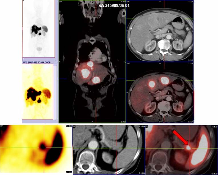
Eur J Nucl Med Mol Imaging (2010) 37:67–77                                                                                    69

of symptoms in functionally active tumours (e.g. carcinoid             At the Zentralklinik Bad Berka, image analysis of the
syndrome), patients were either advised to stop octreotide         CT was performed on a syngo viewing station by an
therapy (if on treatment) 6 weeks prior to 68Ga-DOTA-              experienced radiologist (more than 5 years of CT experi-
NOC PET/CT study when using a long-acting formulation              ence). The PET/CT images were assessed using the e.soft
[e.g. sandostatin long-acting repeatable (LAR) or somatuline]      workstation (Siemens Medical Solutions) by two experi-
or in cases with severe symptoms, convert from LAR to s.c.         enced nuclear medicine physicians. In Bologna, the PET/CT
application. All patients at Bad Berka were asked to drink 1.5 l   images were assessed using the GE Xeleris workstation (GE)
of a water-equivalent oral contrast dispersion (Gastrografin).     by two experienced nuclear medicine physicians.
This dispersion is used routinely in PET/CT without known              First, the maximum intensity projection (MIP) images
adverse side effects to the accumulation of 68Ga-DOTA-NOC.         were visually inspected in varying scales. Thereafter, each
Immediately before the PET/CT examination, patients were           single transversal slice was viewed from head to mid-thigh
asked to void the bladder. Patients were positioned head first     in combination with the corresponding CT image and the
supine on the common patient handling system with the              fused image slice and each focal, abnormal tracer uptake
arms raised in accordance with standard CT practice.               was recorded by slice number and anatomical localisation.
   First, a topogram was acquired over 1,024 mm axially.           Finally, manually selected regions of interest (ROIs) were
Coaxial whole-body imaging ranges were defined on the              automatically drawn on a single slice of the reconstructed
topogram, covering an area from the skull to the upper             PET images using a 50% standardised uptake value (SUV)
thighs (7–8 PET bed positions, or 90–100 cm, depending             threshold and the software provided by e.soft. Any area
on the size of the patient). After 100 ml of intravenous           with intensity greater than background that could not be
contrast (by an automated injection pump) administration,          explained by physiological activity was considered to be
contrast-enhanced CT was acquired in the craniocaudal              indicative of tumour tissue.
direction with a 30-s delay. CT was performed in spiral                For semi-quantitative analysis SUVmax was calculated.
mode using a continuous acquisition at 130 kVp, 115 mAs,           However, because of the dependency of SUVmax on
4-mm collimation, 5-mm slice width, a table feed of 8 mm           different PET scanners used at two centres, only the
per rotation at 0.8-s rotation time and 2.4-mm slice spacing.      patients enrolled at the Zentralklinik Bad Berka were taken
During the CT acquisition a limited breath-hold protocol           into consideration for comparison of the SUVmax of
was followed, which required the patients to hold their            primary pancreatic NET and ilium/jejunum/duodenum with
breath in normal expiration. After completion of the CT,           the SUVmax of known pancreatic NETs in 49 patients and
patients were moved automatically to the PET toward the            that of known ileum/jejunum/duodenum NETs in 11
rear of the gantry, where two-dimensional PET emission             patients. The results of PET/CT were also correlated with
scanning subsequently started in the caudocranial direction        CT alone. Histopathological confirmation was not possible in
with the bladder/pelvis region being scanned first. An             all of the cases. Hence, the lesions detected on 68Ga-DOTA-
emission scan time of 1–2min per bed position was used             NOC PET/CT were considered to be positive by follow-up
for all patients (depending upon the patient’s weight and          for a minimum duration of 6 months with 68Ga-DOTA-NOC
height), which resulted in a total emission scan time of no        PET/CT, ultrasound, biochemical markers or in the case of
more than 24 min and a total PET/CT examination time of            pancreatic lesions with EUS.
about 30 min (including patient positioning, CT and PET
imaging). In Bologna, the emission scan time was 2–4 min           Statistical analyses
per bed position according to the scanner used (GE
Discovery STE, GE Discovery LS), which resulted in a               Statistical analysis was performed using dedicated statistical
total emission scan time of no more than 12–24 min and a           software. Correlation and significance levels were calculated
total PET/CT examination of about 30 min (including the            using SPSS 13. Values were tested for significance applying
PET/CT scan and patient positioning).                              the nonparametric Mann Whitney test for SUV. Because of
                                                                   the lack of normally distributed data, Spearman’s rank
Data reconstruction and image analysis                             correlation was used.

The CT transmission images were used for attenuation
correction of the PET emission data. After scatter and             Results
attenuation correction, PET emission data were recon-
                                                                   68
structed using an attenuation-weighted ordered subsets                Ga-DOTA-NOC PET/CT was performed in 59 patients
expectation maximisation approach with 2 iterations and            with documented NET (confirmed on biopsy from meta-
8 subsets on 128×128 matrices and with a 5-mm Gaussian             static lesions) and unknown primary tumour (Table 1).
                                                                   68
post-reconstruction filtering.                                        Ga-DOTA-NOC PET/CT could localise the site of the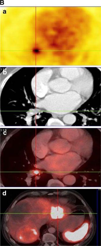
70                                                                              Eur J Nucl Med Mol Imaging (2010) 37:67–77

Table 1 Patients’ characteristics
                                    Age                                          65±9years

                                    Male to female ratio                         33:26
                                    Primary tumour                               Total detected: 35/59
                                                                                 Pancreas, n=16
                                                                                 Ileum/jejunum/duodenum, n=14
                                                                                 Rectum/colon, n=2
                                                                                 Lungs, n=2
                                                                                 Paraganglioma, n=1
                                    WHO classification                           Well-differentiated, n=45
                                                                                 Moderately differentiated, n=2
                                                                                 Poorly differentiated, n=2
                                                                                 Undifferentiated, n=2
                                                                                 NA, n=8
                                    pNET SUVmax                                  Previously known tumours: 26.1±14.5
                                                                                 Previously unknown: 18.6±9.8
                                    SI-NET SUVmax                                Previously known: 11.3±3.7
                                                                                 Previously unknown: 9.1±6.0
NA not available, pNET pancre-
atic neuroendocrine tumour,         Ki-67 (n=22)                                 High-grade (Ki-67>15%): 4/22 (18%)
SI-NET small intestinal                                                          Low-grade (Ki-67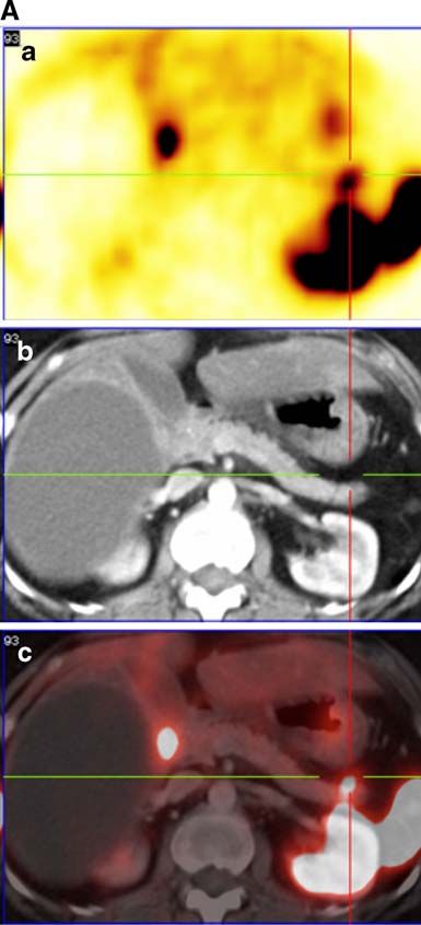
Eur J Nucl Med Mol Imaging (2010) 37:67–77                                                                                        71

Fig. 2 A Multifocal primary pNET(in head and tail) detected on
receptor PET/CT. EUS later confirmed the lesion in the head but
missed the one in the pancreatic tail. B Patient with large liver
metastases from a neuroendocrine tumour (unknown primary) referred
for PRRT. Receptor PET/CT detected the primary in the right
                                                                        Fig. 2 (continued)
bronchus confirmed later on by bronchoscopy. a) 68Ga-DOTA-NOC
PET image, b) CT image, c) PET/CT fusion image, d) PET/CT fusion
image of the liver lesions. C Neuroendocrine tumour patient with CUP
referred for PRRT. 68Ga-DOTA-NOC PET/CT revealed the primary in
                                                                        findings in 12 of 59 patients (20%). According to the WHO
the rectum (I). Additionally, pararectal lymph node metastases were     classification, 45 patients had well-differentiated endocrine
clearly detectable (II). The patient was referred for surgery. a) PET   carcinoma, 2 had moderately differentiated endocrine
image, b) CT image, c) PET/CT fusion image. D Primary ileum             carcinoma, 2 had poorly differentiated endocrine carcinoma
neuroendocrine carcinoma without detection on receptor PET/CT. The
patient was later on operated and the primary was resected
                                                                        and 2 had undifferentiated endocrine carcinoma. In the
                                                                        remaining eight cases the WHO classification was not
                                                                        available (Fig. 3, Table 3).
which was CT negative; persistent elevated gastrin level in                The average time interval between the first biopsy-proven
follow-up confirmed the findings of PET/CT. In one patient              diagnosis of NET and the first evidence of primary on 68Ga-
there was indirect evidence of tumour in the pancreatic                 DOTA-NOC receptor PET/CT was found to be approximately
head/processus uncinatus region; one patient had EUS                    29 months. CT alone (with knowledge of the PET results,
confirmation, while the fourth patient had CT correlation.              Table 4) showed abnormal findings in 12 of 59 patients
Overall 80% (4/5) of the PET/CT findings of tumour in the               (20%). In 24 of 59 patients (41%), the primary remained
pancreatic head were confirmed. CT alone confirmed the                  undiagnosed, even after 68Ga-DOTA-NOC receptor PET/CT.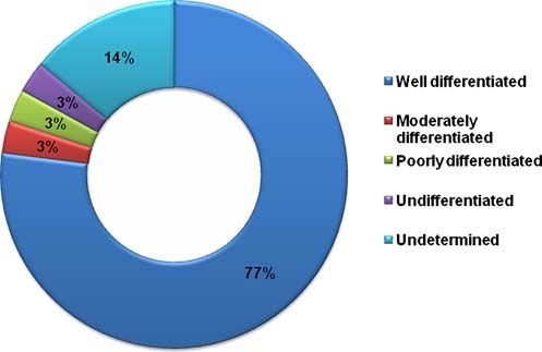
72                                                                                 Eur J Nucl Med Mol Imaging (2010) 37:67–77

Fig. 2 (continued)

    PET/CT with 68Ga-DOTA-NOC identified lymph node             patient was diagnosed 11 years after the first histopatho-
metastases in 30 patients (only 16 of 30 were detected by       logical correlation from liver lesions.
CT), liver metastases in 46 cases (38 of 46 evident also on        The Ki-67 values (proliferation index) were available in 22
CT scan) and bone metastases in 17 cases (8 of 17               of 59 patients: 4 of 22 patients (18%) had high-grade NET
identified by CT). For the identification of the primary,       (Ki-67 value>15%), 16 of 22 (73%) low-grade NET (Ki-67<
68
   Ga-DOTA-NOC PET alone in many instances was not              5%) and 2 of 22 (9%) had Ki-67 in between 5 and 15%.
sufficient in giving the exact anatomical location; a PET/CT       Based on 68Ga-DOTA-NOC receptor PET/CT, 6 of 59
fusion image was required for the correct diagnosis. Also,      patients were operated and the primary was removed
some of the metastatic lesions in liver were picked up only     (4 pancreatic, 1 ileal and 1 rectal tumour) resulting in a
on CT; however, no additional information regarding the         management change in approximately 10% of the patients.
site of the primary could be ascertained only on the basis of   In the remaining 29 patients, because of the far advanced
the CT scan.                                                    stage of the disease (due to distant metastases), the primary
    In the patients enrolled at the PET Centre in Bad Berka,    tumours were not operated. Additional histopathological
the SUVmax of the patients with CUP were compared with          sampling was available from one patient with one bronchial
the SUVmax obtained in the other 49 patients with known         carcinoid (through bronchoscopy).
pancreatic NET and in 11 patients with known ileum/
jejunum/duodenum NET. The mean SUVmax of identified
previously unknown primary pancreatic NET was 18.6±9.8          Discussion
(range: 7.8–34.8) and 9.1±6.0 (range: 4.2–27.8) of SI-NET.
SUVmax in patients with previously known pNET and SI-           The majority of CUP are adenocarcinomas or undifferentiated
NET were 26.1±14.5 (range: 8.7–42.4) and 11.3±3.6               tumours and less commonly, squamous cell carcinoma, mela-
(range: 5.6–17.9). The SUVmax of the unknown pNET               noma or sarcoma; neuroendocrine tumours frequently present
and SI-NET was significantly lower (p
Eur J Nucl Med Mol Imaging (2010) 37:67–77 73
Fig. 2 (continued)
significant prognostic importance identified by multivariate and 5 years, respectively [23, 25–27]. A literature survey
analysis. Lymph node involvement and neuroendocrine shows that the detection of the primary tumour site prolongs
histology were found to be associated with longer survival; survival (for about 1–2 years) [10]. FDG PET has been
male sex, increasing number of involved organ sites, reported to be useful for the detection of the primary tumour
adenocarcinoma histology and hepatic involvement were in patients with a negative diagnostic work-up. However,
unfavourable prognostic factors [24]. until now, no systematic study has been performed to
The prognosis of patients with CUP is poor. As a group,
the median survival is reported to be approximately 3–
4 months with less than 25 and 10% of patients alive at 1
Table 2 Comparison of the sensitivity of 68Ga-DOTA-NOC PET/CT
and CT alone for detection of the primary tumour and sites of
metastases of neuroendocrine tumours
68
Site Ga-DOTA-NOC CT
PET/CT
Primary tumour 35/59 (59%) 12/59 (20%)
Liver metastases 46/59 (78%) 38/59 (64%)
Lymph node metastases 30/59 (51%) 16/69 (27%)
Bone metastases 17/59 (29%) 8/59 (14%)
Lung metastases 3/59 (5%) 3/59 (5%) Fig. 3 WHO classification of the previously unknown primary
neuroendocrine tumours detected on 68Ga-DOTA-NOC PET/CT74 Eur J Nucl Med Mol Imaging (2010) 37:67–77 Table 3 Different primary tumours detected and their Ki-67, WHO classification and positivity on CT Patient number Diagnosis/site WHO classification Ki-67 CT Zentralklinik Bad Berka 1 Pancreas tail Undifferentiated 15% 2 Unknown Unknown 3 Unknown Well-differentiated 4 Unknown Well-differentiated 5 Unknown Unknown
Eur J Nucl Med Mol Imaging (2010) 37:67–77                                                                                       75

Table 3 (continued)

Patient number    Diagnosis/site                                                 WHO classification        Ki-67    CT

 49               Paraganglioma                                                  Well-differentiated                +ve
 50               Small intestine                                                Well-differentiated
 51               Ileum                                                          Well-differentiated       5%
 52               Unknown                                                        Well-differentiated
 53               Unknown                                                        Unknown                   5%
University of Bologna
 54               Pancreas head                                                  Poorly differentiated
 55               Duodenum                                                       Well-differentiated
 56               Unknown                                                        Well-differentiated
 57               Pancreas body                                                  Well-differentiated       2%
 58               Pancreas head and body                                         Well-differentiated
 59               Unknown                                                        Well-differentiated       2%

determine the incidence and clinical course of unknown                decided to remove the primary. One patient was primarily
primary in patients with neuroendocrine tumours [28, 29].             referred for peptide receptor radionuclide therapy (PRRT)
    Numerous factors are known to have an influence on the            after chemotherapy. 68Ga-DOTA-NOC PET/CT revealed
survival and prognosis of patients with neuroendocrine                only faint somatostatin receptor expression in the liver
tumours, among which the presence of liver metastases is              metastases (negative on CT) along with multiple necrotic
the single most important factor. A correlation has been              lesions; the primary was detected in the rectum, along with
found between the size of the primary tumour and the                  presacral lymph nodes. Based upon these findings, the
probability of metastases for small intestinal carcinoids.            patient was referred for surgery. Another patient who had
Metastases to the liver can be found in 15–25% of tumours             peritoneal and liver metastases was diagnosed with a primary
if the tumour diameter is less than 1 cm, 58–80% if it is 1–          tumour in the ileum. The primary was removed and the liver
2 cm and more than 75% if the tumour size is more than                metastases were subsequently treated with PRRT. Somato-
2 cm [28]. This makes it even more important to detect the            statin receptor PET/CT has been shown to be superior to
                                                                      111
primary tumour at the earliest stage. Surgery remains one of             In-OctreoScan, the currently accepted gold standard in the
the treatment options with palliative as well curative intent         diagnosis of NET [30]. However, 111In-OctreoScan SPECT
(if performed at a stage when the primary is completely               alone often fails in localising the exact site of the primary
resectable or if there are no or few metastases) [28]. In the         tumour (if detected) because of lack of morphological/
absence of any concrete evidence of primary on conven-                anatomical information. 111In-OctreoScan SPECT/CT over-
tional imaging, exploratory laparotomy (for unknown                   comes some of these challenges; however, the sensitivity and
gastroenteropancreatic neuroendocrine tumours) becomes                resolution of gamma camera imaging is inferior to PET.
more challenging. In this study, the detection of primaries               Affinity for a broader spectrum of somatostatin receptor
could lead to the surgical removal in 10% of the cases. Of            subtypes (SSTR 2, 3 and 5) makes 68Ga-DOTA-NOC an
the four pancreatic NETs resected, one had no evidence of             interesting radiotracer for the detection of neuroendocrine
any lymph node, bone or liver metastases. Two patients had            tumours [31–33]. Our results, for the first time, have clearly
only peripancreatic lymph nodes. The fourth patient had               shown that 68Ga-DOTA-NOC receptor PET/CT fusion
both lymph node and liver metastases but still the surgeons           imaging is not only sensitive, but also highly specific in
                                                                      picking up the unknown primary. The sensitivity of
Table 4 Primary tumours found on CT after retrospective analyses of
                                                                      receptor PET/CT was roughly three times higher as
PET results                                                           compared to CT alone. When compared to the literature
                                                                      [34], the detection rate of 68Ga-DOTA-NOC was found to
Primary site                                          CT positive     be much higher than for 111In-OctreoScan (35% as
Small intestine (duodenum, ilium and jejunum)         2/14 (14.3%)
                                                                      compared to 59% for 68Ga-DOTA-NOC PET). One of the
Pancreas                                              8/16 (50%)
                                                                      probable explanations for the inability of 68Ga-DOTA-NOC
Lungs (bronchus)                                      1/2 (50%)
                                                                      receptor PET/CT to pick up the primary—even when
                                                                      metastatic lesions are detected—is the sometimes extremely
Paraganglioma                                         1/1 (100%)
                                                                      small size of the primary tumour or receptor down-regulation
Overall                                               12/35 (34%)
                                                                      due to prolonged somatostatin therapy (e.g. using LAR
76 Eur J Nucl Med Mol Imaging (2010) 37:67–77
octreotide). The average time interval between the first in the processus uncinatus of the pancreas often poses a
biopsy-proven diagnosis of NET and the first evidence of diagnostic challenge. In cases of a high degree of suspicion
primary on 68Ga-DOTA-NOC receptor PET/CT was these patients should undergo EUS to confirm or rule out
~29 months. It may be hypothesised that the metastatic the primary tumour in the pancreatic head [39]. CT
lesions, being comparatively new as compared to the morphological information is also helpful in such cases.
primary, are subject to less intense down-regulation of One of the patients in this study showed focal uptake of
68
receptors [35]. Also, the different degree of differentiation Ga-DOTA-NOC in the processus uncinatus of the
(different biological behaviour, e.g. growth rate) of the pancreas (SUVmax 11.7). CT in this patient although it did
primary tumour and the metastatic lesions may account for not show any definite evidence of mass lesion in the head
the absence of somatostatin receptor positivity of the primary showed evidence of atrophy of the pancreas cauda and tail
on PET/CT. with dilated ductus pancreaticus indirectly suggestive of a
Our data also clearly suggest that for proper staging of lesion in the pancreas head. In another patient, there was
NET and also for the follow-up, it is more appropriate to evidence of increased uptake in the head (primary tumour
use 68Ga-DOTA-NOC receptor PET/CT rather than CT or 9.1) and processus uncinatus (physiological SUVmax 4.7) of
68
Ga-DOTA-NOC PET alone. The addition of morphological the pancreas. The results were confirmed by the EUS. CT
information from CT was found to be absolutely essential in findings did not show any mass lesion.
pinpointing the exact site of the primary tumour, particularly Another limitation of this study is that the SUVmax were
in the abdominal region. not corrected for the size of the lesion to rule out partial
Most of the patients had low-grade NET (Ki-67Eur J Nucl Med Mol Imaging (2010) 37:67–77 77
12. Alberini JL, Belhocine T, Hustinx R, Daenen F, Rigo P. Whole- 27. Ringenberg QS. Tumors of unknown origin. Med Pediatr Oncol
body positron emission tomography using fluorodeoxyglucose in 1985;13:301–6.
patients with metastases of unknown primary tumours (CUP 28. Jensen RT. Endocrine tumors of the gastrointestinal tract and
syndrome). Nucl Med Commun 2003;24:1081–6. pancreas. In: Kasper DL, Fauci AS, Longo DL, et al., editors.
13. Bohuslavizki KH, Klutmann S, Kröger S, Sonnemann U, Buchert Harrison’s principles of internal medicine. 16th ed. New York:
R, Werner JA, et al. FDG PET detection of unknown primary McGraw-Hill; 2005
tumors. J Nucl Med 2000;41:816–22. 29. Rufini V, Calcagni ML, Baum RP. Imaging of neuroendocrine
14. Freudenberg LS, Fischer M, Antoch G, Jentzen W, Gutzeit A, tumors. Semin Nucl Med 2006;36:228–7.
Rosenbaum SJ, et al. Dual modality of 18F-fluorodeoxyglucose- 30. Gabriel M, Decristoforo C, Kendler D, Dobrozemsky G, Heute D,
positron emission tomography/computed tomography in patients Uprimny C, et al. 68Ga-DOTA-Tyr3-octreotide PET in neuroendo-
with cervical carcinoma of unknown primary. Med Princ Pract crine tumors: comparison with somatostatin receptor scintigraphy
2005;14:155–60. and CT. J Nucl Med 2007;48:508–18.
15. Gutzeit A, Antoch G, Kühl H, Egelhof T, Fischer M, Hauth 31. Wild D, Mäcke HR, Waser B, Reubi JC, Ginj M, Rasch H, et al.
E, et al. Unknown primary tumors: detection with dual- 68Ga-DOTANOC: a first compound for PET imaging with high
modality PET/CT—initial experience. Radiology 2005; affinity for somatostatin receptor subtypes 2 and 5. Eur J Nucl
234:227–34. Med Mol Imaging 2005;32:724.
16. Kole AC, Nieweg OE, Pruim J, Hoekstra HJ, Koops HS, 32. Wild D, Schmitt JS, Ginj M, Mäcke HR, Bernard BF, Krenning E,
Roodenburg JL, et al. Detection of unknown occult primary tumors et al. DOTA-NOC, a high-affinity ligand of somatostatin receptor
using positron emission tomography. Cancer 1998;82:1160–6. subtypes 2, 3 and 5 for labelling with various radiometals. Eur J
17. Adams S, Baum R, Rink T, Schumm-Dräger PM, Usadel KH, Hör Nucl Med Mol Imaging 2003;30:1338–47.
G. Limited value of fluorine-18 fluorodeoxyglucose positron 33. Hofmann M, Maecke H, Börner R, Weckesser E, Schöffski P, Oei
emission tomography for the imaging of neuroendocrine tumours. L, et al. Biokinetics and imaging with the somatostatin receptor
Eur J Nucl Med 1998;25:79–83. PET radioligand (68)Ga-DOTATOC: preliminary data. Eur J Nucl
18. Ambrosini V, Tomassetti P, Rubello D, Campana D, Nanni C, Med 2001;28:1751–7.
Castellucci P, et al. Role of 18F-dopa PET/CT imaging in the 34. Savelli G, Lucignani G, Seregni E, Marchianò A, Serafini G,
management of patients with 111In-pentetreotide negative GEP Aliberti G, et al. Feasibility of somatostatin receptor scintigraphy
tumours. Nucl Med Commun 2007;28:473–7. in the detection of occult primary gastro-entero-pancreatic (GEP)
19. Baum RP, Prasad V, Hommann M, Hörsch D. Receptor PET/CT neuroendocrine tumours. Nucl Med Commun 2004;25:445–9.
imaging of neuroendocrine tumors. Recent Results Cancer Res 35. Hofland LJ, Lamberts SW. The pathophysiological consequences
2008;170:225–42. of somatostatin receptor internalization and resistance. Endocr
20. Ambrosini V, Tomassetti P, Castellucci P, Campana D, Montini G, Rev 2003;24:28–47.
Rubello D, et al. Comparison between 68Ga-DOTA-NOC and 36. Eisenhofer G, Siegert G, Kotzerke J, Bornstein SR, Pacak K.
18F-DOPA PET for the detection of gastro-entero-pancreatic and Current progress and future challenges in the biochemical
lung neuro-endocrine tumours. Eur J Nucl Med Mol Imaging diagnosis and treatment of pheochromocytomas and paraganglio-
2008;35:1431–8. mas. Horm Metab Res 2008;40:329–37.
21. Zhernosekov KP, Filosofov DV, Baum RP, Aschoff P, Bihl H, 37. Mundschenk J, Unger N, Schulz S, Höllt V, Schulz S, Steinke R,
Razbash AA, et al. Processing of generator-produced 68Ga for et al. Somatostatin receptor subtypes in human pheochromocytoma:
medical application. J Nucl Med 2007;48:1741–8. subcellular expression pattern and functional relevance for octreo-
22. Meyer GJ, Macke H, Schuhmacher J, Knapp WH, Hofmann M. tide scintigraphy. J Clin Endocrinol Metab 2003;88:5150–7.
68Ga-labelled DOTA-derivatised peptide ligands. Eur J Nucl Med 38. Miederer M, Seidl S, Buck A, Scheidhauer K, Wester HJ,
Mol Imaging 2004;31:1097–104. Schwaiger M, et al. Correlation of immunohistopathological
23. Neumann KH, Nystrom JS. Metastatic cancer of unknown origin: expression of somatostatin receptor 2 with standardised uptake
nonsquamous cell type. Semin Oncol 1982;9:427–34. values in 68Ga-DOTATOC PET/CT. Eur J Nucl Med Mol
24. Abbruzzese JL, Abbruzzese MC, Hess KR, Raber MN, Lenzi R, Imaging 2009;36:48–52.
Frost P. Unknown primary carcinoma: natural history and 39. Zimmer T, Scherübl H, Faiss S, Stölzel U, Riecken EO,
prognostic factors in 657 consecutive patients. J Clin Oncol Wiedenmann B. Endoscopic ultrasonography of neuroendocrine
1994;12:1272–80. tumours. Digestion 2000;62(Suppl 1):45–50.
25. Altman E, Cadman E. An analysis of 1539 patients with cancer of 40. Wieder H, Beer AJ, Poethko T, Meisetschlaeger G, Wester HJ,
unknown primary site. Cancer 1986;57:120–4. Rummeny E, et al. PET/CT with Gluc-Lys-([(18)F]FP)-TOCA:
26. Moertel CG, Reitemeier RJ, Schutt AJ, Hahn RG. Treatment of correlation between uptake, size and arterial perfusion in somatostatin
the patient with adenocarcinoma of unknown origin. Cancer receptor positive lesions. Eur J Nucl Med Mol Imaging 2008;35:264–
1972;30:1469–72. 271.You can also read