2018 AHA/ACC Guideline for the Management of Adults With Congenital Heart Disease (ACHD) - American College of Cardiology Foundation and ...
←
→
Page content transcription
If your browser does not render page correctly, please read the page content below
2018 AHA/ACC Guideline for the
Management of Adults With Congenital
       Heart Disease (ACHD)

© American College of Cardiology Foundation and American Heart Association, Inc.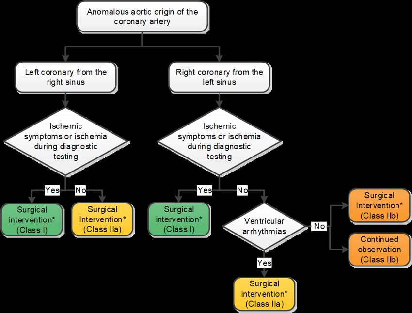
Publication Information
This slide set is adapted from the 2018 AHA/ACC Guideline for
  the Management of Adults With Congenital Heart Disease

 Published on August 16, 2018, available at: Circulation and
       Journal of the American College of Cardiology.

  The full-text guidelines are also available on the following
websites: AHA (professional.heart.org) and ACC (www.acc.org)
2018 Adults With Congenital Heart Disease Guideline
Writing Committee
Karen K. Stout, MD, FACC, Chair†
Curt J. Daniels, MD, Vice Chair*†‡
Jamil A. Aboulhosn, MD, FACC, FSCAI*§ Stephanie Fuller, MD, MS, FACC#
Biykem Bozkurt, MD, PhD, FACC, FAHA║ Michelle Gurvitz, MD, FACC**
Craig S. Broberg, MD, FACC*† Paul Khairy, MD, PhD*†
Jack M. Colman, MD, FACC† Michael J. Landzberg, MD, FACC*†
Stephen R. Crumb, DNP, AACC† Arwa Saidi, MB, BCH, FACC*†
Joseph A. Dearani, MD, FACC¶ Anne Marie Valente, MD, FACC, FAHA, FASE††
George F. Van Hare, MD‡‡
†ACC/AHA Representative. ‡International Society for Adult
Congenital Heart Disease Representative. §Society for
Cardiovascular Angiography and Interventions. ║ACC/AHA Task
Force on Clinical Practice Guidelines Liaison. ¶Society of
Thoracic Surgeons Representative. #American Association for
Thoracic Surgery Representative. **ACC/AHA Task Force on
Performance Measures Liaison. ††American Society of
Echocardiography Representative. ‡‡Heart Rhythm Society
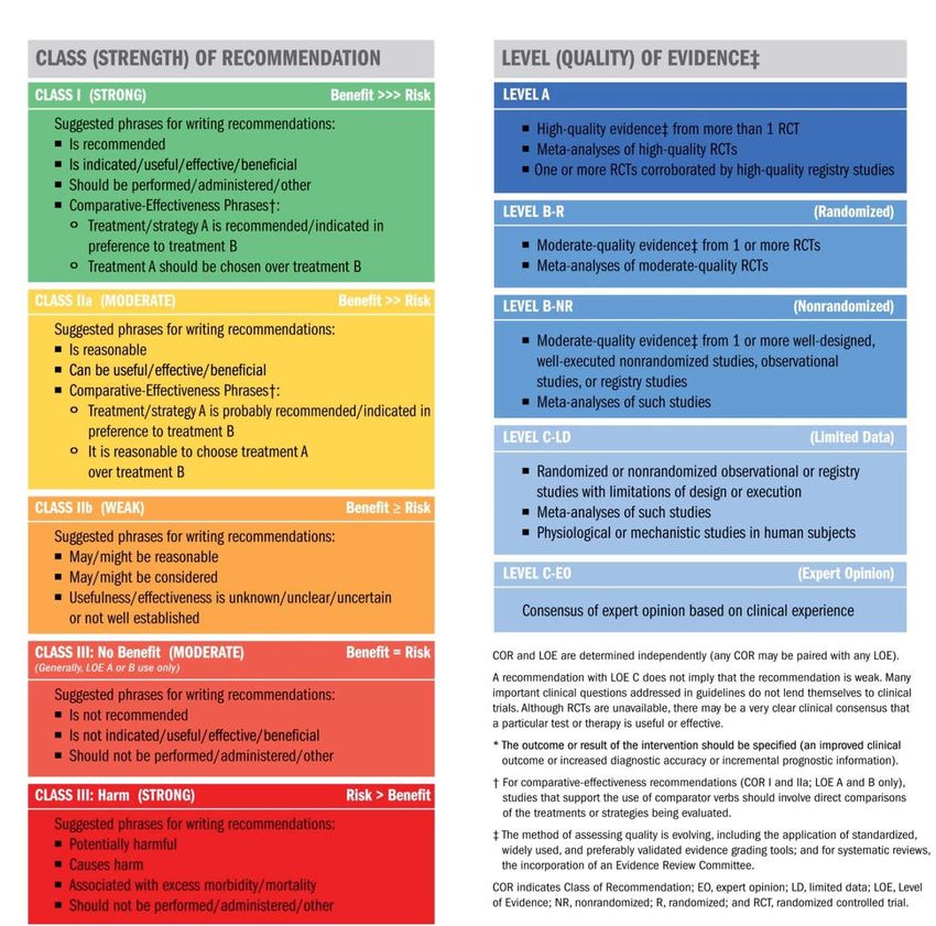
Representative.Applying Class of
Recommendation and
Level of Evidence to
Clinical Strategies,
Interventions,
Treatments, or
Diagnostic Testing
in Patient Care*
(Updated August 2015)Systematic Reviews on ACHD • “Medical Therapy for Systemic Right Ventricles: A Systematic Review (Part 1) • “Interventional Therapy Versus Medical Therapy for Secundum Atrial Septal Defect: A Systematic Review (Part 2)
ACHD AP CLASSIFICATION
                (CHD Anatomy + Physiological Stage = ACHD AP Classification)

• CHD Anatomy
(This list is not meant to be comprehensive; other conditions may be important in individual patients. ASD, atrial septal defect; AVSD,
atrioventricular septal defect; CCTGA, congenitally corrected transposition of the great arteries; CHD, congenital heart disease; d-TGA, dextro-
transposition of the great arteries; FC, functional class; HCM, hypertrophic cardiomyopathy; l-TGA, levo-transposition of the great arteries;
NYHA, New York Heart Association; TGA, transposition of the great arteries; and VSD, ventricular septal defect. )

                                                                   I: Simple
 Native disease
 • Isolated small ASD
 • Isolated small VSD
 • Mild isolated pulmonic stenosis
 Repaired conditions
 • Previously ligated or occluded ductus arteriosus
 • Repaired secundum ASD or sinus venosus defect without significant residual shunt or chamber
     enlargement
 • Repaired VSD without significant residual shunt or chamber enlargement

                                                                                                     (Con’t.)
ACHD AP CLASSIFICATION
• CHD Anatomy
II: Moderate Complexity
Repaired or unrepaired conditions
• Aorto-left ventricular fistula
• Anomalous pulmonary venous connection, partial or total
• Anomalous coronary artery arising from the pulmonary artery
• Anomalous aortic origin of a coronary artery from the opposite sinus
• AVSD (partial or complete, including primum ASD)
• Congenital aortic valve disease
• Congenital mitral valve disease
• Coarctation of the aorta
• Ebstein anomaly (disease spectrum includes mild, moderate, and severe variations)
• Infundibular right ventricular outflow obstruction
• Ostium primum ASD
• Moderate and large unrepaired secundum ASD
• Moderate and large persistently patent ductus arteriosus
• Pulmonary valve regurgitation (moderate or greater)
• Pulmonary valve stenosis (moderate or greater)
• Peripheral pulmonary stenosis
• Sinus of Valsalva fistula/aneurysm
• Sinus venosus defect
• Subvalvar aortic stenosis (excluding HCM; HCM not addressed in these guidelines)
• Supravalvar aortic stenosis
• Straddling atrioventricular valve
• Repaired tetralogy of Fallot
• VSD with associated abnormality and/or moderate or greater shunt
(Con’t.)ACHD AP CLASSIFICATION
• CHD Anatomy
III: Great Complexity (or Complex)
• Cyanotic congenital heart defect (unrepaired or palliated, all forms)
• Double-outlet ventricle
• Fontan procedure
• Interrupted aortic arch
• Mitral atresia
• Single ventricle (including double inlet left ventricle, tricuspid atresia, hypoplastic left
heart, any other anatomic abnormality with a functionally single ventricle)
• Pulmonary atresia (all forms)
• TGA (classic or d-TGA; CCTGA or l-TGA)
• Truncus arteriosus
• Other abnormalities of atrioventricular and ventriculoarterial connection (i.e.,
crisscross heart, isomerism, heterotaxy syndromes, ventricular inversion)
(Con’t.)ACHD AP CLASSIFICATION
• Physiological State
A
• NYHA FC I symptoms
• No hemodynamic or anatomic sequelae
• No arrhythmias
• Normal exercise capacity
• Normal renal/hepatic/pulmonary function
B
• NYHA FC II symptoms
• Mild hemodynamic sequelae (mild aortic enlargement, mild ventricular
enlargement, mild ventricular dysfunction)
• Mild valvular disease
• Trivial or small shunt (not hemodynamically significant)
• Arrhythmia not requiring treatment
• Abnormal objective cardiac limitation to exercise
(Con’t.)ACHD AP CLASSIFICATION
• Physiological Stage
C
• NYHA FC III symptoms
• Significant (moderate or greater) valvular disease; moderate or greater ventricular
dysfunction (systemic, pulmonic, or both)
• Moderate aortic enlargement
• Venous or arterial stenosis
• Mild or moderate hypoxemia/cyanosis
• Hemodynamically significant shunt
• Arrhythmias controlled with treatment
• Pulmonary hypertension (less than severe)
• End-organ dysfunction responsive to therapy
D
• NYHA FC IV symptoms
• Severe aortic enlargement
• Arrhythmias refractory to treatment
• Severe hypoxemia (almost always associated with cyanosis)
• Severe pulmonary hypertension
• Eisenmenger syndrome
• Refractory end-organ dysfunction2018 ACHD Clinical Practice Guidelines
Access to CareAccess to Care
Recommendation for Access to Care
COR LOE Recommendation
Physicians caring for patients with ACHD should support access to
B-NR care by 1) assuring smooth transitions for adolescents and young
adults from pediatric to adult providers (Level of Evidence: B-NR);
I and 2) promoting awareness of the need for lifelong specialized
C-EO care through outreach and educational programs (Level of
Evidence: C-EO).2018 ACHD Guideline Delivery of Care
Delivery of Care
                        Recommendations for Delivery of Care
COR    LOE                          Recommendations
               Patients with ACHD AP classification IB-D, IIA-D, and IIIA-D*
 I     B-NR    should be managed in collaboration with an ACHD cardiologist .

               Cardiac    surgery,    catheter-based   interventional cardiac
               procedures, and electrophysiological procedures involving
 I             congenital heart lesions in patients with ACHD should be
       C-LD
               performed by operators with expertise in CHD procedures and in
               collaboration with an ACHD cardiologist.

*See details on the ACHD Anatomic and Physiological classification system.
2018 ACHD Guideline Evaluation of Suspected and Known CHD
Electrocardiogram

                        Recommendations for Electrocardiogram
COR   LOE                              Recommendations
             A standard 12-lead electrocardiogram (ECG) is recommended in adults
             with CHD with serial assessment depending on the specific ACHD AP
 I    C-EO   classification or when symptoms develop or worsen.

             Ambulatory electrocardiographic monitoring should be performed in
             patients with CHD who are at risk of tachyarrhythmia, bradyarrhythmia or
 I    C-EO   heart block, or when symptoms possibly of arrhythmic origin develop.

                                                            (Con’t.)
Ionizing Radiation principles
Recommendation for Ionizing Radiation Principles
COR LOE Recommendation
Strategies to limit and monitor radiation exposure are recommended
I B-NR during imaging of patients with ACHD, with studies not involving
ionizing radiation chosen whenever appropriate.
(Con’t.)Echocardiography
Recommendations for Echocardiography
COR LOE Recommendations
Intraoperative TEE is recommended to guide surgical repair of CHD in
I B-NR adults.
Patients with ACHD should undergo transthoracic echocardiography
(TTE) for initial assessment, with timing of serial assessment based on
I C-EO
anatomic and physiological severity and clinical status.
(Con’t.)CMR Imaging
Recommendations for CMR Imaging
COR LOE Recommendations
In patients with ACHD who have or who are at risk of developing RV
I B-NR enlargement and dysfunction, serial CMR is recommended for quantitative
assessment of RV size and function.
CMR can be useful in the initial evaluation and serial assessment of
IIa C-LD selected patients with CHD based on anatomic complexity and clinical
status.Cardiac Computed Tomography
Recommendation for Cardiac Computed Tomography
COR LOE Recommendation
CCT imaging can be useful in patients with ACHD when information
that cannot be obtained by other diagnostic modalities is important
IIa C-LD enough to justify the exposure to ionizing radiation.Cardiac Catheterization
Recommendations for Cardiac Catheterization
COR LOE Recommendations
Cardiac catheterization (hemodynamic and/or angiographic) in patients
with ACHD AP classification II and III, or interventional cardiac
I C-LD catheterization in patients with ACHD AP classification I to III should be
performed by, or in collaboration with, cardiologists with expertise in
ACHD.
In patients with a low or intermediate pretest probability of coronary
artery disease (CAD), use of CT coronary angiography is reasonable to
IIa B-NR exclude significant obstructive CAD when cardiac catheterization has
significant risk or because of patient preference.Exercise Testing
Recommendations for Exercise Testing
COR LOE Recommendations
In patients with ACHD, cardiopulmonary exercise testing (CPET) can be
IIa B-NR useful for baseline functional assessment and serial testing.
In symptomatic patients with ACHD, a 6-minute walk test can be useful
IIa C-LD to objectively assess symptom severity, functional capacity, and
response to therapy.2018 ACHD Guideline Transition Education
Transition Education

                         Recommendation for Transition Education
COR   LOE                                 Recommendation
             Clinicians caring for patients with CHD should deliver developmentally
             appropriate transition education to adolescent and young patients with CHD,
 I    B-NR   and to their families/support network.
2018 ACHD Guideline Exercise and Sports
Exercise and Sports

                     Recommendations for Exercise and Sports

COR   LOE                             Recommendations
             Clinicians should assess activity levels at regular intervals and
 I    C-LD   counsel patients with ACHD about the types and intensity of
             exercise appropriate to their clinical status.
             CPET can be useful to guide activity recommendations for patients
IIa   C-LD
             with ACHD.
             Cardiac rehabilitation can be useful to increase exercise capacity
IIa   B-NR   in patients with ACHD.
2018 ACHD Guideline Mental Health and Neurodevelopmental Issues
Mental Health and Neurodevelopmental Issues
               Recommendations for Mental Health and Neurodevelopmental Issues
COR      LOE                               Recommendations
 I      B-NR     Patients with ACHD should be evaluated for depression and anxiety.

                 Referral for mental health evaluation and treatment is reasonable in
IIa     B-NR     patients with ACHD.
                 Neurodevelopmental or neuropsychological testing may be considered
                 in some patients with ACHD to guide therapies that enhance academic,
IIb     B-NR
                 behavioral, psychosocial, and adaptive functioning.
2018 ACHD Guideline Concomitant Syndromes
Concomitant Syndromes
                   Recommendation for Concomitant Syndromes
COR   LOE                            Recommendation
             Genetic testing for 22q11 deletions is reasonable for patients with
IIa   B-NR   conotruncal cardiac defects.
2018 ACHD Guideline Noncardiac Medical Issues
Noncardiac Medical Issues
                   Recommendation for Noncardiac Medical Issues
COR   LOE                            Recommendation
             Patients with ACHD at risk for hepatitis C should be screened and
 I    C-LD   vaccinated for viral hepatitis and treated as appropriate.
2018 ACHD Guideline Noncardiac Surgery
Noncardiac Surgery
                            Recommendations for Noncardiac Surgery
COR      LOE                                  Recommendations
                 Optimization before and close surveillance after invasive procedures,
 I       C-LD    regardless of the complexity of the anatomic defect or type of procedure
                 is beneficial for patients with ACHD.
                 In patients with ACHD AP classification IB-D, IIA-D, and IIIA-D*
 I      B-NR     noncardiac surgical and interventional procedures should be performed
                 in a hospital with or in consultation with experts in ACHD when possible.

 *See Table on the ACHD AP classification system
2018 ACHD Guideline Pregnancy, Reproduction, and Sexual Health
Pregnancy
                                          Recommendations for Pregnancy
 COR      LOE                                       Recommendations
                  Women with CHD should receive prepregnancy counseling with input from an ACHD
  I      C-LD     cardiologist to determine maternal cardiac, obstetrical and fetal risks, and potential
                  long-term risks to the mother.
                  An individualized plan of care that addresses expectations and contingencies should
  I      C-LD     be developed for and with women with CHD who are pregnant or who may become
                  pregnant and shared with the patient and all caregivers.
              Women with CHD receiving chronic anticoagulation should be counseled, ideally
  I      B-NR before conception, on the risks and benefits of specific anticoagulants during
              pregnancy.
              Women with ACHD AP classification IB-D, IIA-D, and IIIA-D* should be managed
  I      B-NR collaboratively during pregnancy by ACHD cardiologists, obstetricians, and
              anesthesiologists experienced in ACHD.

*See Table on the ACHD AP classification system                                        (Con’t.)
Pregnancy
In collaboration with an ACHD cardiologist to ensure accurate assessment of
pregnancy risk, patients at high risk of maternal morbidity or mortality, including
women with pulmonary arterial hypertension (PAH), Eisenmenger syndrome, severe
I C-EO
systemic ventricular dysfunction, severe left-sided obstructive lesions, and/or ACHD
AP classification ID, IID, and IIID* should be counseled against becoming pregnant or
be given the option of terminating pregnancy.
Men and women of childbearing age with CHD should be counseled on the risk of CHD
I B-NR
recurrence in offspring.
Exercise testing can be useful for risk assessment in women with ACHD AP
IIa B-NR classification IC-D, IIA-D, and IIIA-D* who are considering pregnancy.
IIa B-NR When either parent has CHD, it is reasonable to perform fetal echocardiography.
*See Table on the ACHD AP classification system
(Con’t.)Contraception
Recommendations for Contraception
COR LOE Recommendations
Women of childbearing potential with CHD should be counseled about the
I C-LD risks associated with pregnancy and appropriate contraceptive options .
Estrogen-containing contraceptives are potentially harmful for women with
CHD at high risk of thromboembolic events (e.g., cyanosis, Fontan
III: Harm B-NR
physiology, mechanical valves, prior thrombotic events, PAH) .
(Con’t.)2018 ACHD Guideline Heart Failure and Transplant
Heart Failure
                        Recommendation for Heart Failure
COR   LOE                          Recommendation
             Consultation with ACHD and HF specialists is recommended for
 I    C-LD   patients with ACHD and HF or severe ventricular dysfunction.
2018 ACHD Guideline Palliative Care
Palliative Care
                       Recommendation for Palliative Care

COR   LOE                            Recommendation
             Discussion of end-of-life issues and advance directives can be
IIa   B-NR   beneficial for patients with ACHD or their surrogates.
2018 ACHD Guideline Shunt Lesions
Atrial Septal Defect
                       Recommendations for Atrial Septal Defect

COR   LOE                                Recommendations
                                  Diagnostic
             Pulse oximetry at rest and during exercise is recommended for
 I    C-EO   evaluation of adults with unrepaired or repaired ASD with residual shunt
             to determine the direction and magnitude of the shunt.
             CMR, CCT, and/or TEE are useful to evaluate pulmonary venous
 I    B-NR   connections in adults with ASD .
             Echocardiographic imaging is recommended to guide percutaneous ASD
 I    B-NR   closure .

                                                                         (Con’t.)
Atrial Septal Defect
Therapeutic
In adults with isolated secundum ASD causing impaired functional capacity,
right atrial and/or RV enlargement, and net left-to-right shunt sufficiently large
to cause physiological sequelae (e.g., pulmonary–systemic blood flow ratio
[Qp:Qs] ≥1.5:1) without cyanosis at rest or during exercise, transcatheter or
I B-NRSR
surgical closure to reduce RV volume and improve exercise tolerance is
recommended, provided that systolic PA pressure is less than 50% of systolic
systemic pressure and pulmonary vascular resistance is less than one third of
the systemic vascular resistance.
Adults with primum ASD, sinus venosus defect or coronary sinus defect
causing impaired functional capacity, right atrial and/or RV enlargement and
net left-to-right shunt sufficiently large to cause physiological sequelae (e.g.,
I B-NR Qp:Qs ≥1.5:1) without cyanosis at rest or during exercise, should be surgically
repaired unless precluded by comorbidities, provided that systolic PA
pressure is less than 50% of systemic pressure and pulmonary vascular
resistance is less than one third of the systemic vascular resistance.
In asymptomatic adults with isolated secundum ASD, right atrial and RV
enlargement, and net left-to-right shunt sufficiently large to cause
physiological sequelae (e.g., Qp:Qs 1.5:1 or greater), without cyanosis at rest
IIa C-LDSR or during exercise, transcatheter or surgical closure is reasonable to reduce
RV volume and/or improve functional capacity, provided that systolic PA
pressure is less than 50% of systemic pressure and pulmonary vascular
resistance is less than one third systemic resistance.
(Con’t.)Atrial Septal Defect
Surgical closure of a secundum ASD in adults is reasonable when a
concomitant surgical procedure is being performed and there is a net left-
IIa C-LD to-right shunt sufficiently large to cause physiological sequelae (e.g., Qp:Qs
1.5:1 or greater) and right atrial and RV enlargement without cyanosis at
rest or during exercise.
Percutaneous or surgical closure may be considered for adults with ASD
when net left-to-right shunt (Qp:Qs) is 1.5:1 or greater, PA systolic pressure
IIb B-NR is 50% or more of systemic arterial systolic pressure, and/or pulmonary
vascular resistance is greater than one third of the systemic resistance.
ASD closure should not be performed in adults with PA systolic pressure
greater than two thirds systemic, pulmonary vascular resistance greater
III: Harm C-LD than two thirds systemic, and/or a net right-to-left shunt.
(Con’t.)Secundum ASD
Secundum ASD
Shunt
direction
Right-to-left
Left-to-right (e.g., Eisenmenger Bosentan
syndrome) (Class I)
PDE-5 inhibitors
Confirm PAH diagnosis (Class IIa)
Hemodynamic
(often requiring invasive
assessment
hemodynamic Yes Combination
assessment) therapy*
Pulmonary vascular (Class I)
Pulmonary vascular (Class IIa)
resistanceAnomalous Pulmonary Venous Connections
Recommendations for Anomalous Pulmonary Venous Connections
COR LOE Recommendations
Diagnostic
CMR or CTA is recommended for evaluation of partial anomalous
I B-NR
pulmonary venous connection.
Cardiac catheterization can be useful in adults with partial anomalous
IIa B-NR
pulmonary venous connection to further define hemodynamics.
(Con’t.)Anomalous Pulmonary Venous Connections
Therapeutic
Surgical repair is recommended for patients with partial anomalous pulmonary
venous connection when functional capacity is impaired and RV enlargement is
present, there is a net left-to-right shunt sufficiently large to cause physiological
I B-NR
sequelae (e.g., Qp:Qs ≥1.5:1), PA systolic pressure is less than 50% systemic
pressure and pulmonary vascular resistance is less than one third of systemic
resistance.
Repair of partial anomalous pulmonary venous connection is recommended at
I B-NR
the time of closure of a sinus venosus defect or ASD.
Repair of a scimitar vein is recommended in adults when functional capacity is
impaired, evidence of RV volume overload is present, there is a net left-to-right
I B-NR shunt sufficiently large to cause physiological sequelae (e.g., Qp:Qs ≥1.5:1), PA
systolic pressure is less than 50% systemic pressure and pulmonary vascular
resistance is less than one third systemic.
Surgery can be useful for right- or left-sided partial anomalous pulmonary
venous connection in asymptomatic adults with RV volume overload, net left-to-
IIa B-NR right shunt sufficiently large to cause physiological sequelae (e.g., Qp:Qs
≥1.5:1), pulmonary pressures less than 50% systemic and pulmonary vascular
resistance less than one third systemic.
Surgery can be useful for repair of a scimitar vein in adults with evidence of RV
IIa B-NR
volume overload, with Qp:Qs 1.5:1 or greater.Ventricular Septal Defect
Recommendations for Ventricular Septal Defect
COR LOE Recommendations
Therapeutic
Adults with a VSD and evidence of left ventricular volume overload and
hemodynamically significant shunts (Qp:Qs ≥1.5:1) should undergo VSD
I B-NR
closure, if PA systolic pressure is less than 50% systemic and pulmonary
vascular resistance is less than one third systemic.
Surgical closure of perimembranous or supracristal VSD is reasonable in
IIa C-LD adults when there is worsening aortic regurgitation (AR) caused by VSD.
Surgical closure of a VSD may be reasonable in adults with a history of IE
IIb C-LD caused by VSD if not otherwise contraindicated.
Closure of a VSD may be considered in the presence of a net left-to-right
shunt (Qp:Qs ≥1.5:1) when PA systolic pressure is 50% or more than
IIb C-LD
systemic and/or pulmonary vascular resistance is greater than one third
systemic.
VSD closure should not be performed in adults with severe PAH with PA
systolic pressure greater than two thirds systemic, pulmonary vascular
III: Harm C-LD
resistance greater than two thirds systemic and/or a net right-to-left shunt.
(Con’t.)Hemodynamically Significant Ventricular Level Shunt
Hemodynam ically significant
ventricular level shunt
Shunt
direction
Right-to-left
Left-to-right (e.g., Eisenmenger Bosentan
syndrome) (Class I)
PDE-5 inhibitors
Confirm PAH diagnosis (Class IIa)
Hemodynam ic (often requiring invasive
Yes
assessm ent hemodynamic assessment)
(Class I) Combination
Pulmonary vascular therapy*
LV enlargem ent (Class IIa)
resistance >1/3
Qp:Qs ≥1.5:1,
systemic,
PASPAtrioventricular Septal Defect
Recommendations for Atrioventricular Septal Defect
COR LOE Recommendations
Diagnostic
Cardiac catheterization can be useful in adults with atrioventricular septal defect when
IIa C-EO
pulmonary hypertension is suspected.
Therapeutic
Surgery for severe left atrioventricular valve regurgitation is recommended per GDMT
I C-LD
indications for mitral regurgitation.
Surgery for primary repair of atrioventricular septal defect or closure of residual shunts
in adults with repaired atrioventricular septal defect is recommended when there is a
I C-EO net left-to-right shunt (Qp:Qs ≥1.5:1), PA systolic pressure less than 50% systemic and
pulmonary vascular resistance less than one third systemic.
Operation for discrete LVOT obstruction in adults with atrioventricular septal defect is
reasonable with a maximum gradient of 50 mm Hg or greater, a lesser gradient if HF
IIa C-EO
symptoms are present, or if concomitant moderate-to-severe mitral or AR are present.
Surgery for primary repair of atrioventricular septal defect or closure of residual shunts
in adults with repaired atrioventricular septal defect may be considered in the presence
IIb C-EO of a net left-to-right shunt (Qp:Qs ≥1.5:1), if PA systolic pressure is 50% or more
systemic, and/or pulmonary vascular resistance is greater than one third systemic.
Surgery for primary repair of atrioventricular septal defect or closure of residual shunts
in adults with repaired atrioventricular septal defect should not be performed with PA
III: Harm C-LD systolic pressure greater than two thirds systemic, pulmonary vascular resistance
greater than two thirds systemic, or a net right-to-left shunt.Patent Ductus Arteriosus
Recommendations for Patent Ductus Arteriosus
COR LOE Recommendations
Diagnostic
Measurement of oxygen saturation should be performed in feet and both
I C-EO hands in adults with a PDA to assess for the presence of right-to-left shunting.
In addition to the standard diagnostic tools, cardiac catheterization can be
IIa C-EO
useful in patients with PDA and suspected pulmonary hypertension.
Therapeutic
PDA closure in adults is recommended if left atrial or LV enlargement is
present and attributable to PDA with net left-to-right shunt, PA systolic
I C-LD
pressure less than 50% systemic and pulmonary vascular resistance less than
one third systemic.
PDA closure in adults may be considered in the presence of a net left-to-right
IIb B-NR shunt if PA systolic pressure is 50% or greater systemic, and/or pulmonary
vascular resistance is greater than one third systemic.
PDA closure should not be performed in adults with a net right-to-left shunt
III: Harm C-LD and PA systolic pressure greater than two thirds systemic or pulmonary
vascular resistance greater than two thirds systemic.
(Con’t.)2018 ACHD Guideline Left-Sided Obstructive Lesions
Cor Triatriatum
                           Recommendations for Cor Triatriatum

COR   LOE                                   Recommendations
                                     Diagnostic
             Adults presenting with cor triatriatum sinister should be evaluated for other
 I    B-NR   congenital abnormalities, particularly ASD, VSD, and anomalous pulmonary
             venous connection.
             In adults with prior repair of cor triatriatum sinister and recurrent symptoms,
IIa   B-NR
             it is reasonable to evaluate for pulmonary vein stenosis.
                                  Therapeutic
             Surgical repair is indicated for adults with cor triatriatum sinister for
 I    B-NR   symptoms attributable to the obstruction or a substantial gradient across
             the membrane.
Congenital Mitral Stenosis
Recommendation for Congenital Mitral Stenosis
COR LOE Recommendation
Adults with congenital mitral stenosis or a parachute mitral valve
I B-NR should be evaluated for other left-sided obstructive lesions.Subaortic Stenosis
Recommendations for Subaortic Stenosis
COR LOE Recommendations
Diagnostic
Stress testing for adults with LVOT obstruction to determine exercise
capacity, symptoms, electrocardiographic changes, or arrhythmias may be
IIb C-LD
reasonable in the presence of otherwise equivocal indications for
intervention.
Therapeutic
Surgical intervention is recommended for adults with subAS, a maximum
I C-EO gradient 50 mm Hg or more and symptoms attributable to the subAS.
Surgical intervention is recommended for adults with subAS and less than 50
I C-LD mm Hg maximum gradient and HF or ischemic symptoms, and/or LV systolic
dysfunction attributable to subAS.
To prevent the progression of AR, surgical intervention may be considered
IIb C-LD for asymptomatic adults with subAS and at least mild AR and a maximum
gradient of 50 mm Hg or more.Congenital Valvular Aortic Stenosis
Recommendations for Congenital Valvular Aortic Stenosis
COR LOE Recommendations
Diagnostic
Adults with bicuspid aortic valve should be evaluated for coarctation of the
I B-NR
aorta by clinical examination and imaging studies.
It is reasonable to screen first-degree relatives of patients with bicuspid
IIa B-NR aortic valve or unicuspid aortic valve with echocardiography for valve
disease and aortopathy.
Therapeutic
In adults with bicuspid aortic valve stenosis and a noncalcified valve with
IIb B-NR no more than mild AR meeting indications for intervention per GDMT, it
may be reasonable to treat with balloon valvuloplasty.
(Con’t.)Turner Syndrome
Recommendations for Turner Syndrome
COR LOE Recommendations
Diagnostic
Women with Turner syndrome should be evaluated for bicuspid aortic
I B-NR
valve, coarctation of the aorta, and enlargement of the ascending aorta.
Therapeutic
Prophylactic replacement of the aortic root or ascending aorta in adults
IIa B-NR with Turner syndrome is reasonable when the aortic diameter is 2.5
cm/m2 or greater.Coarctation of the Aorta
Recommendations for Coarctation of the Aorta
COR LOE Recommendations
Diagnostic
Initial and follow-up aortic imaging using CMR or CTA is recommended in
I B-NR adults with coarctation of the aorta, including those who have had surgical or
catheter intervention.
Resting blood pressure should be measured in upper and lower extremities in
I C-EO
all adults with coarctation of the aorta.
Ambulatory blood pressure monitoring in adults with coarctation of the aorta
IIa C-LD
can be useful for diagnosis and management of hypertension.
Screening for intracranial aneurysms by magnetic resonance angiography or
IIb B-NR CTA may be reasonable in adults with coarctation of the aorta.
Exercise testing to evaluate for exercise-induced hypertension may be
IIb C-LD
reasonable in adults with coarctation of the aorta who exercise.
Therapeutic
Surgical repair or catheter-based stenting is recommended for adults with
I B-NR hypertension and significant native or recurrent coarctation of the aorta.
GDMT is recommended for treatment of hypertension in patients with
I C-EO
coarctation of the aorta.
Balloon angioplasty for adults with native and recurrent coarctation of the
IIb B-NR aorta may be considered if stent placement is not feasible and surgical
intervention is not an option.2018 ACHD Guideline Right-Sided Lesions
Valvular Pulmonary Stenosis
                   Recommendations for Valvular Pulmonary Stenosis
COR   LOE                               Recommendations
             In adults with moderate or severe valvular pulmonary stenosis and
             otherwise unexplained symptoms of HF, cyanosis from interatrial right-to-
 I    B-NR
             left communication, and/or exercise intolerance, balloon valvuloplasty is
             recommended.
             In adults with moderate or severe valvular pulmonary stenosis and
             otherwise unexplained symptoms of HF, cyanosis, and/or exercise
 I    B-NR
             intolerance who are ineligible for or who failed balloon valvuloplasty,
             surgical repair is recommended.
             In asymptomatic adults with severe valvular pulmonary stenosis,
IIa   C-EO
             intervention is reasonable.

                                                                           (Con’t.)
Isolated PR After Repair of PS
Recommendations for Isolated PR After Repair of Pulmonary Stenosis
COR LOE Recommendations
In symptomatic patients with moderate or greater PR resulting from treated
I C-EO isolated pulmonary stenosis, with RV dilation or RV dysfunction,
pulmonary valve replacement is recommended.
For asymptomatic patients with residual PR resulting from treatment of
I C-EO isolated pulmonary stenosis with a dilated right ventricle, serial follow-up
is recommended.
In asymptomatic patients with moderate or greater PR resulting from
treatment of isolated pulmonary stenosis with progressive RV dilation
IIb C-EO and/or RV dysfunction, pulmonary valve replacement may be reasonable.
(Con’t.)Isolated PR After Repair of PS
Isolated PR after repair of PS
Assessment of
PR severity and
RV size/function
Moderate or
Mild PR and
greater PR and
RV enlargement*
RV enlargement*
Interval follow-up
Symptoms†
(Class I)
Yes No
Pulmonary valve
replacement Imaging and CPET
(Class I)
Progressive RV dilation
and/or RV dysfunction
and/or progressive decrease
in exercise capacity
Yes No
Pulmonary valve
Interval follow-up with
replacement
ACHD cardiologist
(Class IIb)Branch and Peripheral Pulmonary Stenosis
Recommendations for Branch and Peripheral PS
COR LOE Recommendations
Diagnostic
For adults with peripheral or branch PS, ongoing surveillance is
I B-NR
recommended.
Therapeutic
In adults with peripheral or branch PA stenosis, PA dilation and stenting
IIa B-NR
can be useful .Double-Chambered Right Ventricle
Recommendations for Double-Chambered Right Ventricle
COR LOE Recommendations
Surgical repair for adults with double-chambered right ventricle and
moderate or greater outflow obstruction is recommended in patients
I C-LD
with otherwise unexplained symptoms of HF, cyanosis, or exercise
limitation.
Surgical repair for adults with double-chambered right ventricle with a
IIb C-LD
severe gradient may be considered in asymptomatic patients.Ebstein Anomaly
Recommendations for Ebstein Anomaly
COR LOE Recommendations
Diagnostic
In adults with Ebstein anomaly, CMR can be useful to determine anatomy, RV
IIa B-NR dimensions, and systolic function .
In adults with Ebstein anomaly, TEE can be useful for surgical planning if TTE
IIa B-NR
images are inadequate to evaluate tricuspid valve morphology and function.
Electrophysiological study with or without catheter ablation can be useful in
IIa B-NR the diagnostic evaluation of adults with Ebstein anomaly and ventricular
preexcitation but without supraventricular tachycardia.
In adults with Ebstein anomaly, electrophysiological study (and catheter
IIa B-NR ablation, if needed) is reasonable before surgical intervention on the tricuspid
valve even in the absence of preexcitation or supraventricular tachycardia.
(Con’t.)Ebstein Anomaly
Therapeutic
Surgical repair or reoperation for adults with Ebstein anomaly and
significant TR is recommended when one or more of the following are
I B-NR present: HF symptoms, objective evidence of worsening exercise
capacity, progressive RV systolic dysfunction by echocardiography or
CMR.
Catheter ablation is recommended for adults with Ebstein anomaly and
I C-LD high-risk pathway conduction or multiple accessory pathways.
Surgical repair or reoperation for adults with Ebstein anomaly and
significant TR can be beneficial in the presence of progressive RV
IIa B-NR
enlargement, systemic desaturation from right-to-left atrial shunt,
paradoxical embolism, and/or atrial tachyarrhythmias.
Bidirectional superior cavopulmonary (Glenn) anastomosis at time of
Ebstein anomaly repair may be considered for adults when severe RV
IIb B-NR dilation or severe RV systolic dysfunction is present, LV function is
preserved, and left atrial pressure and LV end diastolic pressure are not
elevated.
(Con’t.)Tetralogy of Fallot
Recommendations for TOF
COR LOE Recommendations
Diagnostic
CMR is useful to quantify ventricular size and function, pulmonary valve
I B-NR function, pulmonary artery anatomy and left heart abnormalities in patients
with repaired TOF.
Coronary artery compression testing is indicated before right ventricle–to-PA
I B-NR
conduit stenting or transcatheter valve placement in repaired TOF.
Programmed ventricular stimulation can be useful to risk stratify adults with
IIa B-NR
TOF and additional risk factors for SCD.
In patients with repaired TOF, cardiac catheterization with angiography, if
indicated, is reasonable to assess hemodynamics when adequate data cannot
IIa C-EO
be obtained noninvasively in the setting of an arrhythmia, HF, unexplained
ventricular dysfunction, suspected pulmonary hypertension or cyanosis.
(Con’t.)Tetralogy of Fallot
Therapeutic
Pulmonary valve replacement (surgical or percutaneous) for relief of
symptoms is recommended for patients with repaired TOF and
I B-NR
moderate or greater PR with cardiovascular symptoms not otherwise
explained.
Pulmonary valve replacement (surgical or percutaneous) is reasonable
for preservation of ventricular size and function in asymptomatic
IIa B-NR
patients with repaired TOF and ventricular enlargement or dysfunction
and moderate or greater PR .
Primary prevention ICD therapy is reasonable in adults with TOF and
IIa B-NR
multiple risk factors for SCD.
Surgical pulmonary valve replacement may be reasonable for adults
IIb C-EO with repaired TOF and moderate or greater PR with other lesions
requiring surgical interventions.
Pulmonary valve replacement, in addition to arrhythmia management,
IIb C-EO may be considered for adults with repaired TOF and moderate or
greater PR and ventricular tachyarrhythmia.
(Con’t.)TOF repair with PR
Pulmonary Valve
Replacement in Patients
Severely With TOF Repair and PR
decreased LV or RV
Yes No
systolic
function
Evaluation by an ACHD
cardiologist & advanced HF
team PR severity
(Class I)
Mild PR Moderate
or more PR
Follow-up with
ACHD cardiologist Symptoms*
(Class I)
Yes No
Pulmonary valve
replacement
(Class I) Any 2 of
the following:
• Mild or moderate RV or
LV systolic dysfunction
• Severe RV dilation
(RVEDVI ≥160 mL/m 2, or
RVESVI ≥80 mL/m2, or
RVEDV ≥2x LVEDV)
• RVSP due to RVOT obstruction
≥2/3 systemic pressure
• Progressive reduction
in objective exercise
tolerance (Con’t.)Pulmonary Valve Replacement in
Patients With TOF Repair and PR
Yes No
Pulmonary valve
replacement Sustained
(Class IIa) tachyarrhythmias
Yes No
Pulmonary valve
replacement Residual
(Class IIb) lesions requiring surgical
interventions
Yes No
Pulmonary valve Follow-up with
replacement ACHD cardiologist
(Class IIb) (Class I)Right Ventricle–to-Pulmonary Artery Conduit
Recommendations for Right Ventricle–to-PA Conduit
COR LOE Recommendations
Diagnostic
Coronary artery compression testing with simultaneous coronary angiography
and high-pressure balloon dilation in the conduit is indicated before right
I B-NR
ventricle–to-PA conduit stenting or transcatheter valve placement.
In patients with stented right ventricle–to-PA conduits and worsening PS or PR,
evaluation for conduit complications should be performed, including
I B-NR
fluoroscopy to evaluate for stent fracture and blood cultures to assess for IE.
In adults with right ventricle–to-PA conduit and arrhythmia, congestive HF,
IIa C-LD unexplained ventricular dysfunction or cyanosis cardiac catheterization is
reasonable to assess the hemodynamics.
Therapeutic
Right ventricle–to-PA conduit intervention is reasonable for adults with right
ventricle–to-PA conduit and moderate or greater PR or moderate or greater
IIa B-NR
stenosis with reduced functional capacity or arrhythmia.
Right ventricle–to-PA conduit intervention may be reasonable for asymptomatic
adults with right ventricle–to-PA conduit and severe stenosis or severe
IIb B-NR
regurgitation with reduced RV ejection fraction or RV dilation.Right Ventricle–to-Pulmonary Artery Conduit
Recommendations for Right Ventricle–to-PA Conduit
COR LOE Recommendations
Diagnostic
Coronary artery compression testing with simultaneous coronary angiography
and high-pressure balloon dilation in the conduit is indicated before right
I B-NR
ventricle–to-PA conduit stenting or transcatheter valve placement.
In patients with stented right ventricle–to-PA conduits and worsening PS or PR,
evaluation for conduit complications should be performed, including
I B-NR
fluoroscopy to evaluate for stent fracture and blood cultures to assess for IE.
In adults with right ventricle–to-PA conduit and arrhythmia, congestive HF,
IIa C-LD unexplained ventricular dysfunction or cyanosis cardiac catheterization is
reasonable to assess the hemodynamics.
Therapeutic
Right ventricle–to-PA conduit intervention is reasonable for adults with right
ventricle–to-PA conduit and moderate or greater PR or moderate or greater
IIa B-NR
stenosis with reduced functional capacity or arrhythmia.
Right ventricle–to-PA conduit intervention may be reasonable for asymptomatic
adults with right ventricle–to-PA conduit and severe stenosis or severe
IIb B-NR
regurgitation with reduced RV ejection fraction or RV dilation.2018 ACHD Guideline
Complex Lesions (Transposition of the Great
Arteries)Transposition of the Great Arteries With Atrial Switch
Recommendations for d-TGA With Atrial Switch
COR LOE Recommendations
Diagnostic
Ambulatory monitoring for bradycardia or sinus node dysfunction is
I C-EO recommended for adults with d-TGA with atrial switch, especially if treated
with beta blockers or other rate-slowing agents.
Adults with d-TGA with atrial switch repair should undergo annual imaging
I C-EO with either echocardiography or CMR to evaluate for common long-term
complications of the atrial switch.
Assessment for a communication through the interatrial baffle or venous
stenosis is reasonable for adults with d-TGA with atrial switch, particularly if
IIa C-LD
transvenous pacemaker/ICD implantation is considered or leads are already
present.
Therapeutic
GDMT with appropriate attention to the need for anticoagulation is
I B-NR recommended to promptly restore sinus rhythm for adults with d-TGA with
atrial switch repair presenting with atrial arrhythmia.
(Con’t.)Transposition of the Great Arteries With Arterial Switch
Recommendations for d-TGA With Arterial Switch
COR LOE Recommendations
Diagnostic
1. Baseline and serial imaging with either echocardiography or CMR should
be performed in adults with d-TGA with arterial switch who have
I C-LD
neoaortic dilation, valve dysfunction or PA or branch PA stenosis or
ventricular dysfunction.
1. Coronary revascularization for adults with d-TGA with arterial switch
should be planned by surgeons or interventional cardiologists with
I C-EO
expertise in revascularization in collaboration with ACHD providers to
ensure coronary and pulmonary artery anatomy are understood
1. It is reasonable to perform anatomic evaluation of coronary artery
IIa B-NR patency (catheter angiography, or CT or MR angiography) in
asymptomatic adults with d-TGA with arterial switch.
1. Physiological tests of myocardial perfusion for adults with d-TGA after
IIa C-EO arterial switch can be beneficial for assessing symptoms suggestive of
myocardial ischemia.
1. GDMT is reasonable to determine the need for coronary revascularization
IIa C-EO
for adults with d-TGA after arterial switch.
(Con’t.)Transposition of the Great Arteries With Arterial Switch
Therapeutic
GDMT is reasonable to determine indications for aortic valve replacement
in adults with d-TGA after arterial switch with severe neoaortic valve
IIa C-EO
regurgitation.
Catheter or surgical intervention for PS is reasonable in adults with d-
TGA after arterial switch with symptoms of HF or decreased exercise
IIa C-EO capacity attributable to PS.
(Con’t.)Congenitally Corrected Transposition of the Great
Arteries
Recommendations for Congenitally Corrected Transposition of the Great Arteries
COR LOE Recommendations
Diagnostic
CMR is reasonable in adults with CCTGA to determine systemic RV dimensions
IIa C-LD
and systolic function.
Therapeutic
Tricuspid valve replacement is recommended for symptomatic adults with
I B-NR CCTGA and severe TR, and preserved or mildly depressed systemic ventricular
function.
Tricuspid valve replacement is reasonable for asymptomatic adults with CCTGA
IIa C-LD and severe TR with dilation or mild dysfunction of the systemic ventricle.
Conduit intervention/replacement may be considered for adults with CCTGA and
symptomatic subpulmonary left ventricle–to-PA conduit dysfunction,
IIb B-NR
recognizing that unloading the subpulmonary ventricle may have a detrimental
impact on systemic atrioventricular valve function.2018 ACHD Guideline
Complex Lesions {Fontan Palliation of Single
Ventricle Physiology (Including Tricuspid
Atresia and Double Inlet Left Ventricle)}Fontan Palliation of Single Ventricle Physiology
(Including Tricuspid Atresia and Double Inlet Left
Ventricle)
Recommendations for Fontan Palliation of Single Ventricle Physiology
COR LOE Recommendations
Diagnostic
New presentation of an atrial tachyarrhythmia in adults with Fontan palliation
I C-LD should be managed promptly and include prevention of thromboembolic events
and consultation with an electrophysiologist with CHD expertise.
Adults after Fontan palliation should be evaluated annually with either
I C-EO
echocardiography or CMR.
Cardiac catheterization should be performed in adults before initial Fontan
surgery or revision of a prior Fontan connection to assess suitability of
I C-EO
preintervention hemodynamics for Fontan physiology or revision of a prior
Fontan connection.
New onset or worsening atrial tachyarrhythmias in adults with single ventricle
I C-EO after Fontan palliation should prompt a search for potential hemodynamic
abnormalities, which may necessitate imaging and/or cardiac catheterization.
In adults with Fontan palliation, it is reasonable to encourage a regular exercise
IIa B-R
program appropriate to their abilities.
(Con’t.)Fontan Palliation of Single Ventricle Physiology
(Including Tricuspid Atresia and Double Inlet Left
Ventricle)
In adults with Fontan palliation, it is reasonable to encourage a regular
IIa B-R
exercise program appropriate to their abilities.
Imaging of the liver (ultrasonography, CMR, CT) and laboratory evaluation of
IIa C-LD liver function for fibrosis, cirrhosis, and/or hepatocellular carcinoma are
reasonable in adults after Fontan palliation.
In adults after Fontan palliation, it is reasonable to perform biochemical and
IIa C-EO hematological testing on an annual basis especially for liver and renal
function.
Cardiac catheterization can be useful to evaluate a symptomatic adult after
IIa C-LD Fontan palliation when noninvasive testing is insufficient to guide therapy.
Evaluation for cardiac transplantation is reasonable in adults with Fontan
IIa C-LD
palliation and signs and symptoms of protein-losing enteropathy.
It may be reasonable to perform catheterization in asymptomatic adults after
Fontan palliation to evaluate hemodynamics, oxygenation and cardiac function
IIb C-EO to guide optimal medical, interventional and/or surgical therapy.
(Con’t.)Fontan Palliation of Single Ventricle Physiology
(Including Tricuspid Atresia and Double Inlet Left
Ventricle)
Therapeutic
Anticoagulation with a vitamin K antagonist is recommended for adults with
I C-EO Fontan palliation with known or suspected thrombus, thromboembolic
events, or prior atrial arrhythmia, and no contraindications to anticoagulation.
Catheter ablation can be useful in adults after Fontan palliation with intra-
IIa C-LD atrial reentrant tachycardia or focal atrial tachycardia.
Fontan revision surgery, including arrhythmia surgery as indicated, is
reasonable for adults with atriopulmonary Fontan connections with recurrent
IIa C-LD atrial tachyarrhythmias refractory to pharmacological therapy and catheter
ablation who have preserved systolic ventricular function and severe atrial
dilation.
Pulmonary vasoactive medications can be beneficial to improve exercise
IIa B-R capacity in adults with Fontan repair.
Antiplatelet therapy or anticoagulation with a vitamin K antagonist may be
IIb B-NR considered in adults after Fontan palliation without known or suspected
thrombus, thromboembolic events, or prior arrhythmia.
Reoperation or intervention for structural/anatomic abnormalities in a Fontan
IIb C-LD palliated patient with symptoms or with failure of the Fontan circulation may
be considered.2018 ACHD Guideline Severe PAH and Eisenmenger Syndrome
Severe PAH
                            Recommendations for Severe PAH
COR   LOE                                Recommendations
                                   Diagnostic
             Patients with ACHD with pulmonary vascular resistance 2.5 Wood units or
             greater (≥4 Wood units/m2) should be assessed collaboratively by an ACHD
 I    B-NR   cardiologist and an expert in pulmonary hypertension to develop a
             management plan.
             Adults with septal or great artery shunts should undergo periodic screening
 I    B-NR
             for pulmonary hypertension with TTE.
             Cardiac catheterization to assess pulmonary vascular hemodynamics is
             recommended for adults with septal or great artery shunts and clinical
 I    B-NR   symptoms, signs, or echocardiographic findings suggestive of pulmonary
             hypertension.
             In adults with septal or great artery shunts, cardiac catheterization with
 I    B-NR   hemodynamics (performed before or at time of closure) is beneficial to assess
             suitability for closure.
             BNP, chest x-ray, 6-minute walk test, and cardiac catheterization are useful for
 I    C-EO   initial and follow-up evaluation of patients with ACHD with PAH.

                                                                  (Con’t.)
Eisenmenger Syndrome
Recommendations for Eisenmenger Syndrome
COR LOE Recommendations
Diagnostic
When evaluating adults with presumed Eisenmenger syndrome, clinicians
should confirm diagnostic imaging and cardiac catheterization data
I C-EO
accuracy and exclude other potential contributors to right-to-left shunting or
pulmonary hypertension.
Therapeutic
Bosentan is beneficial in symptomatic adults with Eisenmenger syndrome
I A
with ASD or VSD.
In symptomatic adults with Eisenmenger syndrome, bosentan and PDE-5
IIa B-R inhibitors are reasonable in combination if symptomatic improvement does
not occur with either medication alone.
C-EO Bosentan is a reasonable therapy to treat symptomatic adults with
Eisenmenger syndrome with 1 of the following: shunts other than ASD/VSD
IIa (e.g., PDA, aortopulmonary window) (Level of Evidence C-EO), or complex
B-NR
congenital heart lesions or Down syndrome (Level of Evidence B-NR).
It is reasonable to use PDE-5 inhibitors (e.g., sildenafil, tadalafil) to treat
IIa B-NR symptomatic adults with Eisenmenger syndrome with ASD, VSD, or great
artery shunt.2018 ACHD Guideline Coronary Anomalies
Anomalous Coronary Artery Evaluation

              Recommendations for Anomalous Coronary Artery Evaluation
COR   LOE                              Recommendations
                                 Diagnostic
             Coronary angiography, using catheterization, CT, or CMR, is recommended
 I    C-LD   for evaluation of anomalous coronary artery.
             Anatomic and physiological evaluation should be performed in patients
 I    C-LD   with anomalous aortic origin of the left coronary from the right sinus
             and/or right coronary from the left sinus.

                                                                 (Con’t.)
Anomalous Aortic Origin of Coronary Artery
Recommendations for Anomalous Aortic Origin of Coronary Artery
COR LOE Recommendations
Therapeutic
Surgery is recommended for AAOCA from the left sinus or AAOCA from the right
sinus for symptoms or diagnostic evidence consistent with coronary ischemia
I B-NR
attributable to the anomalous coronary artery.
Surgery is reasonable for anomalous aortic origin of the left coronary artery from
IIa C-LD the right sinus in the absence of symptoms or ischemia.
IIa C-EO Surgery for AAOCA is reasonable in the setting of ventricular arrhythmias.
Surgery or continued observation may be reasonable for asymptomatic patients
with an anomalous left coronary artery arising from the right sinus or right
coronary artery arising from the left sinus without ischemia or anatomic or
IIb B-NR physiological evaluation suggesting potential for compromise of coronary
perfusion (e.g., intramural course, fish-mouth-shaped orifice, acute angle).
(Con’t.)Anomalous Aortic origin of the Coronary Artery
*Surgical intervention to involve unroofing or coronary revascularization for patients with concomitant fixed obstruction.
(Con’t.)Anomalous Coronary Artery Arising From the PA
Recommendations for Anomalous Coronary Artery Arising From the PA
COR LOE Recommendations
Therapeutic
Surgery is recommended for anomalous left coronary artery from the PA.
I B-NR
In a symptomatic adult with anomalous right coronary artery from the PA
I C-EO with symptoms attributed to the anomalous coronary, surgery is
recommended.
Surgery for anomalous right coronary artery from the PA is reasonable in an
IIa C-EO asymptomatic adult with ventricular dysfunction or with myocardial ischemia
attributed to anomalous right coronary artery from the PA.You can also read