Enhanced liver fibrosis score as a predictive marker for hepatocellular carcinoma development after hepatitis C virus eradication
←
→
Page content transcription
If your browser does not render page correctly, please read the page content below
MOLECULAR AND CLINICAL ONCOLOGY 15: 215, 2021

                   Enhanced liver fibrosis score as a predictive
                 marker for hepatocellular carcinoma development
                        after hepatitis C virus eradication
         TOSHIHIRO KAWAGUCHI1,2, TATSUYA IDE1, KEISUKE AMANO1, TERUKO ARINAGA‑HINO1,
    REIICHIRO KUWAHARA1, TOMOYA SANO1, SHIRACHI MIKI3, NAOFUMI ONO4 and TAKUJI TORIMURA1

1
 Division of Gastroenterology, Department of Medicine, Kurume University School of Medicine, Kurume, Fukuoka 830‑0011;
   2
    Social Insurance Tagawa Hospital, Tagawa, Fukuoka 826‑0023; 3Chikugo City Hospital, Chikugo, Fukuoka 833‑0041;
                                 4
                                   Yame General Hospital, Yame, Fukuoka 834‑0034, Japan

                                          Received January 17, 2021; Accepted July 7, 2021

                                                      DOI: 10.3892/mco.2021.2377

Abstract. Advanced liver fibrosis is the most important risk            baseline ELF score (P=0.0264), PTW24 ELF score (P=0.0003),
factor for hepatocellular carcinoma (HCC) development after             PTW24 α‑fetoprotein level (P= 0.0133), baseline FIB‑4 index
achieving sustained virological response (SVR) by direct‑acting         (P= 0.0451) and low baseline prothrombin time (P= 0.0455)
antiviral (DAA) treatment in patients with chronic hepatitis C.         were risk factors for HCC development based on univariate
Wisteria floribunda agglutinin‑positive Mac‑2‑binding protein           analyses. Based on the multivariate analysis, a high PTW24
(M2BPGi), enhanced liver fibrosis (ELF) score, type IV collagen         ELF score was the only risk factor for HCC development
and fibrosis‑4 (FIB‑4) index have been reported as non‑invasive         (P= 0.0035). The ELF score after DAA therapy was strongly
biomarkers for liver fibrosis. In the present study, the possibility    associated with HCC development; therefore, it may be a useful
of using fibrosis biomarkers and other parameters to predict            marker for predicting HCC.
the development of HCC was evaluated. A total of 743 patients
infected with hepatitis C virus who achieved SVR by using DAA           Introduction
were retrospectively enrolled. Of these, 122 patients whose
blood samples were stored were selected. The aforementioned             The advent of direct‑acting antivirals (DAAs) revolutionized
four fibrosis biomarkers were analyzed at baseline, at the end          the treatment of hepatitis C virus (HCV) and high sustained
of treatment (EOT) and at post‑treatment week 24 (PTW24).               virological response (SVR) rates may be achieved. The SVR
Tumor markers and laboratory tests were also analyzed. The              rates were reported to be 89.9% with daclatasvir/asuna‑
baseline/EOT/PTW24 values for each fibrosis biomarker                   previr (1), 95.8% with sofosbuvir/ribavirin (2), and 98.5% with
were as follows: ELF score: 11.5±1.2/10.8±1.1/10.4±1.0;                 sofosbuvir/ledipasvir (3). Accumulating evidence suggests that
type IV collagen: 213±85/190±67/174±55 ng/ml; M2BPGi:                   SVR with DAA treatment reduces the incidence of hepato‑
4.8±3.5/2.7±2.0/2.2±1.8; and FIB‑4 index: 5.31±3.82/4.36±               cellular carcinoma (HCC) development (4‑6). However, even
2.79/4.24±3.09. Of the 122 patients, 23 developed HCC. A high           after achieving SVR, some patients may develop HCC.
                                                                            Advanced liver fibrosis was reported to be the most impor‑
                                                                        tant risk factor for HCC development after SVR (7); therefore,
                                                                        evaluation of the degree of liver fibrosis is important. Regarding
Correspondence to: Dr Toshihiro Kawaguchi, Division of                  the evaluation of fibrosis, non‑invasive methods (serum markers
Gastroenterology, Department of Medicine, Kurume University             or transient elastography) were recently adopted instead of
School of Medicine, 67 Asahi‑machi, Kurume, Fukuoka 830‑0011,           invasive liver biopsy. Representative fibrosis markers include
Japan                                                                   type IV collagen (8), Wisteria floribunda agglutinin‑positive
E‑mail: kawaguchi_toshihiro@med.kurume‑u.ac.jp                          Mac‑2‑binding protein (M2BPGi) (9), and fibrosis‑4 (FIB‑4)
                                                                        index (10,11). On the other hand, an enhanced liver fibrosis
Abbreviations: AFP, α‑fetoprotein; DAA, direct‑acting antiviral;        (ELF) score composed of three liver fibrosis markers was
ELF score, enhanced liver fibrosis score; FIB‑4 index, fibrosis‑4
                                                                        developed to evaluate liver fibrosis (12). The ELF score was
index; HCC, hepatocellular carcinoma; M2BPGi, Wisteria floribunda
                                                                        confirmed to be useful in patients with non‑alcoholic fatty
agglutinin‑positive Mac‑2‑binding protein; SVR, sustained virological
response                                                                liver (13), primary biliary cholangitis/cirrhosis (14) and
                                                                        chronic hepatitis C (15). It was recently reported that the
Key words: hepatocellular carcinoma, chronic hepatitis C,               ELF score was comparable with transient elastography in
sustained virological response, direct‑acting antiviral, enhanced       detecting advanced fibrosis (F≥3) in treatment‑naïve patients
liver fibrosis score                                                    with chronic HCV infection (16). In addition, the usefulness
                                                                        of ELF score as a predictor of HCC in the general population,
2          KAWAGUCHI et al: ELF SCORE AS A PREDICTIVE MARKER FOR HCC DEVELOPMENT AFTER HCV ERADICATION

particularly in predicting non‑viral‑related HCC, was previ‑       Table Ⅰ. Baseline characteristics of patients (n=122) with
ously reported (17).                                               chronic hepatitis C.
    Among these fibrosis markers, M2BPGi and FIB‑4 index
were reported to be useful markers for the risk of HCC devel‑      Characteristics                                           Values
opment after HCV eradication (18,19). On the other hand, other
than fibrosis markers, α‑fetoprotein (AFP) was also reported       Male, n (%)                                             50 (41.0)
to be useful for predicting HCC development after HCV              Cirrhosis/chronic hepatitis, n                           36/86
eradication (20). However, to the best of our knowledge, there     Genotype (1/2), n                                        119/3
is no report confirming the usefulness of the ELF score for        Treatment regimen, n
predicting HCC development after HCV eradication. The aim           Daclatasvir + asunaprevir                                  113
of the present study was to assess fibrosis markers, including      Ledipasvir + sofosbuvir                                     6
ELF score, type IV collagen, M2BPGi and FIB‑4 index, tumor          Sofosbuvir + ribavirin                                      3
markers, and biochemical tests associated with HCC develop‑
                                                                   Age (years)                                              68.7±8.8
ment after viral eradication. The time course of the changes
                                                                   Aspartate aminotransferase (U/l)                           53±27
in fibrosis markers during and after DAA treatment was also
examined.                                                          Alanine aminotransferase (U/l)                             51±36
                                                                   Albumin (g/dl)                                            3.9±0.4
Materials and methods                                              Total bilirubin (mg/dl)                                   0.8±0.3
                                                                   Prothrombin time (%)                                       91±16
Subjects. Patients with chronic hepatitis C or liver cirrhosis     Platelet count (x104/ µl)                                12.4±5.0
from three hospitals in Japan (Kurume University Hospital,         α‑Fetoprotein (ng/ml)                                   16.9±30.5
Yame General Hospital and Chikugo City Hospital) who               Des‑γ‑carboxy prothrombin (mAU/ml)                      20.4±18.1
were initiated on DAA therapy between October 2014 and
September 2016 were selected. A total of 999 patients with         Values are presented as mean ± SD, unless otherwise indicated.
chronic hepatitis C or liver cirrhosis were treated with DAA
(daclatasvir plus asunaprevir, or ledipasvir plus sofosbuvir, or
sofosbuvir plus ribavirin). SVR was achieved in 743 patients.
The diagnosis of liver cirrhosis was comprehensively made          patients with HCV genotype 2. SVR was defined as undetect‑
based on biochemical test results, imaging findings and phys‑      able serum HCV RNA at 24 weeks after completing DAA
ical findings on a case‑by‑case basis. Among the patients who      therapy.
achieved SVR, 122 patients (50 male and 72 female patients,
with a mean age ± SD of 68.7±8.8 years; range, 32‑83 years)        Liver fibrosis markers. Four fibrosis biomarkers (type IV
whose blood samples were stored were enrolled (Fig. 1). All        collagen, M2BPGi, FIB‑4 index and ELF score) were
the patients were positive for HCV antibody as determined          assessed in 122 patients. For all patients, the biomarkers
using chemiluminescence immunoassay (Architect®; Abbott            were analyzed at baseline, at the end of treatment (EOT) and
Japan Co., Ltd.). The HCV RNA levels were measured using           at post‑treatment week 24 (PTW24). Type IV collagen was
a COBAS Taq Man test (Roche Diagnostics). Patients who             measured using a JCA‑BM 8000 series automated immune
had hepatitis B surface antigen, a history of HCC prior to         analyzer (Japan Electron Optics Laboratory Ltd.). M2BPGi
DAA therapy, or developed HCC within 24 weeks after DAA            was measured using a HISCL‑5000 (Sysmex Corporation).
therapy were excluded. Imaging surveillance (ultrasonography,      The FIB‑4 index was calculated as follows: Age (years) x
computed tomography or magnetic resonance imaging) were            aspartate aminotransferase (AST; U/l)/platelet count (109/l) x
undertaken every 3‑6 months. Patients were followed up until       alanine aminotransferase (ALT; U/l)1/2 (10). The ELF score
HCC development or the last visit before October 2018. The         consists of three fibrosis markers: Hyaluronic acid (HA),
mean observation period was 2.7 years after the initiation of      amino‑terminal propeptide of type III procollagen (PIIINP)
DAA therapy. The present study was conducted according to          and tissue inhibitor of metalloproteinase type‑1 (TIMP‑1).
the guidelines of the Declaration of Helsinki and was approved     The ELF score (12) was measured using an ADVIA Center
by the Ethics Committees of Kurume University School               XP automated immunoanalyzer and calculated automati‑
of Medicine (approval no. 14178), Yame General Hospital            cally using the following equation: ELF score=2.278 + 0.851
(approval no. 19‑005), and Chikugo City Hospital (approval         ln(CHA) + 0.751 ln(CPIIINP) + 0.394 ln(CTIMP1). Three biomarkers
no. 2019‑09). Written informed consent was obtained from           (type IV collagen, M2BPGi and ELF score) were measured
all patients.                                                      using stored blood samples. All collected blood samples were
                                                                   stored at ‑30˚C until analysis.
DAA therapy. The HCV treatment regimen of daclatasvir
(60 mg once daily) plus asunaprevir (100 mg twice daily) for       Parameters associated with HCC. The following parameters
24 weeks, or ledipasvir (90 mg) plus sofosbuvir (400 mg) for       were analyzed to identify the factors associated with HCC: Sex,
12 weeks, was administered to patients with HCV genotype           cirrhosis, age, type IV collagen, M2BPGi, FIB‑4 index, ELF
1. Sofosbuvir (400 mg) plus ribavirin (weight‑based dosing:        score, AST, ALT, γ‑glutamyl transpeptidase, albumin, total
600 mg daily for patients weighing ≤60 kg, 800 mg daily for        bilirubin, prothrombin time, platelets, AFP and des‑γ‑carboxy
patients weighing >60 and ≤80 kg, and 1,000 mg daily for           prothrombin. For all patients, all parameters were assessed at
patients weighing >80 kg) for 12 weeks was administered to         baseline and PTW24.
MOLECULAR AND CLINICAL ONCOLOGY 15: 215, 2021 3 Figure 1. Flow chart of subject inclusion in the study. DAA, direct‑acting antiviral; DCV, daclatasvir; ASV, asunaprevir; LDV, ledipasvir; SOF, sofosbuvir; RBV, bibavirin; HCC, hepatocellular carcinoma; SVR, sustained virological response; SVR24, SVR at 24 weeks. Statistical analysis. Statistical analysis was performed using prothrombin time (P= 0.0455) were identified as risk factors the JMP software package (release 13; SAS Institute, Inc.). for HCC development based on the univariate analyses. A Mean values and SDs were calculated for continuous data. For multivariate analysis was performed using the four factors that comparison of variables, the Wilcoxon signed‑rank test was were found to be significant in the univariate analysis: PTW24 performed as appropriate. Factors associated with HCC risk ELF score, baseline FIB‑4 index, PTW24 AFP level and base‑ were determined using the Cox proportional hazard regression line prothrombin time. Based on the multivariate analysis, a analysis. P
4           KAWAGUCHI et al: ELF SCORE AS A PREDICTIVE MARKER FOR HCC DEVELOPMENT AFTER HCV ERADICATION

Table Ⅱ. Changes in biomarkers measured at baseline, end of treatment, and at 24 weeks after DAA therapy. P‑values are for the
comparison between baseline and 24 weeks after treatment.

Biomarkers                                Baseline               End of treatment             24 weeks after treatment             P‑value

Type Ⅳ collagen (ng/ml)                   213±85                     190±67                            174±55
MOLECULAR AND CLINICAL ONCOLOGY 15: 215, 2021                                                                                            5

Table Ⅲ. Factors associated with the risk for HCC development.

                                                                                          Univariate analysis                               Multivariate analysis
                                                                                ‑‑‑‑‑‑‑‑‑‑‑‑‑‑‑‑‑‑‑‑‑‑‑‑‑‑‑‑‑‑‑‑‑‑‑‑‑‑‑‑‑‑‑‑‑‑‑‑‑‑‑‑‑‑‑‑‑   ‑‑‑‑‑‑‑‑‑‑‑‑‑‑‑‑‑‑‑‑‑‑‑-‑‑‑‑‑‑‑‑‑‑‑‑‑‑‑‑‑
Factors                                   HCC+ (n=23)         HCC‑ (n=99)        HR               95% CI                  P‑value           P‑value, HR, 95% CI

Sex (male/female), n                    10/13     40/59  1.17 0.50‑2.67 0.707
Liver cirrhosis/chronic hepatitis, n     9/14     27/72  1.57 0.65‑3.59 0.299
Age, years                            69.6±7.3  68.5±9.0 1.00 0.95‑1.06 0.915
Pre AST (U/l)                        64.1±38.5 50.9±22.7 1.01 1.00‑1.02 0.070
PTW24 AST (U/l)                      28.9±10.2  26.9±9.0 1.03 0.99‑1.08 0.153
Pre ALT (U/l)                        61.3±49.8 48.9±31.4 1.01 1.00‑1.01 0.210
PTW24 ALT (U/l)                       19.6±8.6 19.6±11.5 1.01 0.97‑1.05 0.581
Pre γ‑GTP (U/l)                         47±32     40±40  1.01 1.00‑1.02 0.100
PTW24 γ‑GTP (U/l)                       32±25     26±27  1.01 1.00‑1.02 0.155
Pre Alb (g/dl)                         3.8±0.4   4.0±0.4 0.39 0.14‑1.07 0.067
PTW24 Alb (g/dl)                       4.2±0.3   4.3±0.3 0.48 0.16‑1.53 0.210
Pre T.Bil (mg/dl)                      0.9±0.3   0.8±0.3 1.34 0.33‑4.50 0.669
PTW24 T.Bil (mg/dl)                    0.8±0.3   0.9±0.3 0.39 0.07‑1.61 0.215
Pre PTa (%)                          84.6±14.8 92.5±15.6 0.97 0.95‑1.00 0.0455
PTW24 PT (%)                         87.8±15.4 92.4±13.9 0.98 0.95‑1.01 0.217
Pre Plt (x104/µl)                     11.2±5.2  12.5±4.7 0.95 0.86‑1.04 0.254
PTW24 Plt (x10 /µl)
                  4
                                      12.0±5.6  13.1±4.3 0.96 0.87‑1.05 0.388
Pre AFP (ng/ml)                      23.7±39.4 15.2±27.8 1.00 0.99‑1.01 0.522
PTW24 AFPa (ng/ml)                    8.1±11.0   4.4±2.6 1.06 1.02‑1.09 0.0133
Pre DCP (mAU/ml)                      20.1±8.3 20.5±19.6 1.01 0.97‑1.04 0.457
PTW24 DCP (mAU/ml)                      23±10     21±16  1.03 1.00‑1.05 0.0758
Pre type Ⅳ (collagen (ng/ml)           225±62    210±89  1.00 1.00‑1.01 0.522
PTW24 type Ⅳ collagen (ng/ml)          192±42    169±56  1.00 1.00‑1.01 0.120
Pre M2BPGi                           5.43±3.02 4.69±3.58 1.05 0.93‑1.16 0.417
PTW24 M2BPGi                         2.98±1.61 2.05±1.76 1.18 0.98‑1.39 0.0764
Pre FIB‑4 indexa                       7.2±6.1   4.9±2.9 1.08 1.00‑1.15 0.0451
PTW24 FIB‑4 index                      5.6±5.0   3.9±2.3 1.09 0.99‑1.17 0.0879
Pre ELF score                         12.0±1.1  11.3±1.2 1.47 1.05‑2.10 0.0264
PTW24 ELF score     a
                                      11.2±1.0  10.3±1.0 1.96 1.38‑2.77 0.0003                                                                     0.0035, 1.89,
						                                                                                                                                               1.24‑2.85
PTW24 HA (ng/ml)                      414±434   180±193  1.00 1.00‑1.00 0.0007
PTW24 PⅢNP (ng/ml)                    13.4±5.9  10.0±5.0 1.08 1.02‑1.13 0.009
PTW24 TIMP‑1 (ng/ml)                   277±63    223±66  1.01 1.00‑1.01 0.0033
a
 Multivariate analysis was performed using the four factors which were found to be significant in the univariate analysis: PTW24 ELF
score, baseline FIB‑4 index, PTW24 AFP level and baseline PT. Values are presented as mean ± SD unless otherwise indicated. HCC,
hepatocellular carcinoma; Pre, before treatment; PTW24, post‑treatment week 24; AST, aspartate aminotransferase; ALT, alanine amino‑
transferase; GTP, glutamyl transpeptidase; Alb, albumin; T.Bil total bilirubin; PT, prothrombin time; Plt, platelet count; AFP, α‑fetoprotein;
DCP, des‑γ‑carboxy prothrombin; ELF, enhanced liver fibrosis; M2BPGi, Wisteria floribunda agglutinin‑positive Mac‑2‑binding protein;
FIB‑4, Fibrosis‑4; HA, hyaluronic acid; PIIINP, amino‑terminal propeptide of type‑III procollagen; TIMP‑1, tissue inhibitor of metallopro‑
teinase type‑1.

fibrosis in liver tissue. Indeed, in a previous study comparing         PTW24 was selected as a predictor of HCC development after
the degree of liver fibrosis and fibrosis markers in patients with      HCV eradication.
chronic hepatitis C, the ELF score, M2BPGi and FIB4‑index                   As regards the limitations of the present study, the total
were considered to reflect both fibrosis and inflammation (34).         number of patients in the study was relatively small, but the
These findings indicate that the fibrosis markers at PTW24              HCC development rate was high (19%). Age and cirrhosis
may reflect the fibrosis level with greater precision. Previous         were not found to be associated with HCC development,
reports have demonstrated that hepatocarcinogenesis was                 although these are known risk factors associated with the
closely associated with the degree of liver fibrosis rather than        development of this type of cancer. In addition, a pre‑FIB‑4
that of inflammation (35‑37). Therefore, the ELF score at               index of 3.25 was previously found to exhibit a significant
6 KAWAGUCHI et al: ELF SCORE AS A PREDICTIVE MARKER FOR HCC DEVELOPMENT AFTER HCV ERADICATION
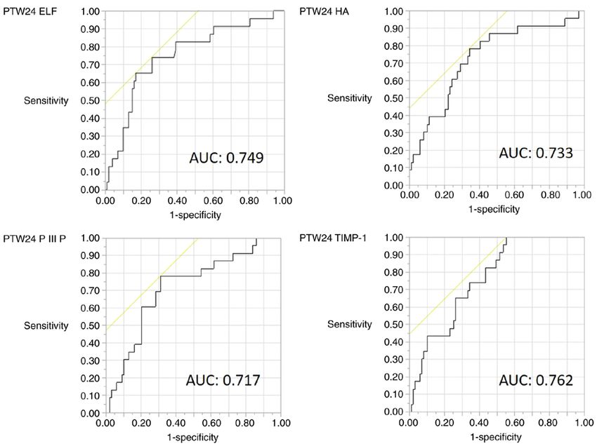
Table Ⅳ. Diagnostic accuracy of the PTW24 ELF score, PTW24 HA, PTW24 PIIINP and PTW24 TIMP‑1 for predicting
hepatocellular carcinoma development.
Diagnostic measures Cut‑off Sensitivity Specificity AUC PPV NPV P‑value
PTW24 ELF score 10.96 0.652 0.828 0.749 0.469 0.911 0.0002
PTW24 HA (ng/ml) 153.6 0.783 0.657 0.733 0.346 0.928 0.0009
PTW24 PⅢNP (ng/ml) 10.5 0.783 0.687 0.717 0.367 0.932 0.0104
PTW24 TIMP‑1 (ng/ml) 199.1 1 0.444 0.762 0.295 1 0.0013
PTW24, post‑treatment week 24; ELF, enhanced liver fibrosis; HA, hyaluronic acid; PIIINP, amino‑terminal propeptide of type‑III procollagen;
TIMP‑1, tissue inhibitor of metalloproteinase type‑1; AUC, area under the receiver operating characteristic curve; PPV, positive predictive
value; NPV, negative predictive value.
Figure 3. ROC curve of PTW24 ELF, PTW24 HA, PTW24 PIIINP and PTW24 TIMP‑1 for prediction of hepatocellular carcinoma. AUC, area under the
ROC curve; ROC, receiver operating characteristic; ELF, enhanced liver fibrosis; HA, hyaluronic acid; TIMP‑1, tissue inhibitor of metalloproteinase type‑1;
PIIINP, amino‑terminal propeptide of type‑III procollagen; PTW24, post‑treatment week 24.
association with HCC development (19). Finally, there may inflammation and fibrosis, careful follow‑up is required for
exist selection bias, as only patients with stored serum patients with a high ELF score ≥10.96 at 24 weeks after DAA
samples were selected. therapy owing to the high risk for HCC development.
In conclusion, the most useful parameter for predicting
hepatocarcinogenesis after DAA therapy for chronic hepatitis Acknowledgements
C was found to be the ELF score at 24 weeks after therapy.
In addition, the four investigated fibrosis markers decreased Not applicable.
after DAA therapy. The number of patients who achieve
SVR by DAA therapy is expected to increase in the future; Funding
therefore, the incidence of HCC development after SVR is also
expected to increase. Although DAA administration improves No funding was received.MOLECULAR AND CLINICAL ONCOLOGY 15: 215, 2021 7
Availability of data and materials 9. Yamasaki K, Tateyama M, Abiru S, Komori A, Nagaoka S,
Saeki A, Hashimoto S, Sasaki R, Bekki S, Kugiyama Y, et al:
Elevated serum levels of Wisteria floribunda agglutinin‑positive
The datasets generated and/or analyzed during the present human Mac‑2 binding protein predict the development of hepa‑
study are available from the corresponding author on reason‑ tocellular carcinoma in hepatitis C patients. Hepatology 60:
1563‑1570, 2014.
able request. 10. Vallet‑Pichard A, Mallet V, Nalpas B, Verkarre V, Nalpas A,
Dhalluin‑Venier V, Fontaine H and Pol S: FIB‑4: An inexpensive
Authors' contributions and accurate marker of fibrosis in HCV infection. Comparison
with liver biopsy and fibrotest. Hepatology 46: 32‑36, 2007.
11. Li X, Xu H and Gao P: Fibrosis index based on 4 factors (FIB‑4)
TK and TI contributed to the study concept and design; TK, predicts liver cirrhosis and hepatocellular carcinoma in chronic
TI, KA, TAH, RK, TS, SM, NO and TT contributed to data hepatitis C virus (HCV) patients. Med Sci Monit 25: 7243‑7250,
2019.
acquisition and analysis; TT revised the manuscript. TK and 12. Rosenberg WM, Voelker M, Thiel R, Becka M, Burt A,
TI have seen and can confirm the authenticity of the raw data. Schuppan D, Hubscher S, Roskams T, Pinzani M and Arthur MJ;
All the authors have read and approved the final manuscript. European Liver Fibrosis Group: Serum markers detect the pres‑
ence of liver fibrosis: A cohort study. Gastroenterology 127:
1704‑1713, 2004.
Ethics approval and consent to participate 13. Guha IN, Parkes J, Roderick P, Chattopadhyay D, Cross R,
Harris S, Kaye P, Burt AD, Ryder SD, Aithal GP, et al: Noninvasive
markers of fibrosis in nonalcoholic fatty liver disease: Validating
The present study was reviewed and approved by the Ethics the European liver fibrosis panel and exploring simple markers.
Committee of Kurume University School of Medicine (approval Hepatology 47: 455‑460, 2008.
no. 14178), Yame General Hospital (approval no. 19‑005), 14. Mayo MJ, Parkes J, Adams‑Huet B, Combes B, Mills AS, Markin RS,
Rubin R, Wheeler D, Contos M, West AB, et al: Prediction of
and Chikugo City Hospital (approval no. 2019‑09). Written clinical outcomes in primary biliary cirrhosis by serum enhanced
informed consent was obtained from all the patients enrolled liver fibrosis assay. Hepatology 48: 1549‑1557, 2008.
in the study. 15. Parkes J, Guha IN, Roderick P, Harris S, Cross R, Manos MM,
Irving W, Zaitoun A, Wheatley M, Ryder S and Rosenberg W:
Enhanced liver fibrosis (ELF) test accurately identifies liver
Patient consent for publication fibrosis in patients with chronic hepatitis C. J Viral Hepat 18:
23‑31, 2011.
16. Omran D, Yosry A, Darweesh SK, Nabeel MM, El‑Beshlawey M,
Not applicable. Saif S, Fared A, Hassany M and Zayed RA: Enhanced liver
fibrosis test using ELISA assay accurately discriminates
Competing interests advanced stage of liver fibrosis as determined by transient elas‑
tography fibroscan in treatment naïve chronic HCV patients. Clin
Exp Med 18: 45‑50, 2018.
The authors declare that they have no competing interests. 17. Loo WM, Goh GB, Wang Y, Yuan JM, Ong L, Dan YY and
Koh WP: Enhanced liver fibrosis score as a predictor of hepato‑
cellular carcinoma. Clin Chem 64: 1404‑1405, 2018.
References 18. Nagata H, Nakagawa M, Asahina Y, Sato A, Asano Y, Tsunoda T,
Miyoshi M, Kaneko S, Otani S, Kawai‑Kitahata F, et al: Effect
1. Ji F, Wei B, Yeo YH, Ogawa E, Zou B, Stave CD, Li Z, Dang S, of interferon‑based and ‑free therapy on early occurrence and
Furusyo N, Cheung RC and Nguyen MH: Systematic review with recurrence of hepatocellular carcinoma in chronic hepatitis C.
meta‑analysis: Effectiveness and tolerability of interferon‑free J Hepatol 67: 933‑939, 2017.
direct‑acting antiviral regimens for chronic hepatitis C geno‑ 19. Ioannou GN, Beste LA, Green PK, Singal AG, Tapper EB,
type 1 in routine clinical practice in Asia. Aliment Pharmacol Waljee AK, Sterling RK, Feld JJ, Kaplan DE, Taddei TH and
Ther 47: 550‑562, 2018. Berry K: Increased risk for hepatocellular carcinoma persists up to
2. Wei B, Ji F, Yeo YH, Ogawa E, Zou B, Stave CD, Dang S, Li Z, 10 years after HCV eradication in patients with baseline cirrhosis
Furusyo N, Cheung RC and Nguyen MH: Real‑world effective‑ or high FIB‑4 scores. Gastroenterology 157: 1264‑1278.e4, 2019.
ness of sofosbuvir plus ribavirin for chronic hepatitis C genotype 20. Asahina Y, Tsuchiya K, Nishimura T, Muraoka M, Suzuki Y,
2 in Asia: A systematic review and meta‑analysis. BMJ Open Tamaki N, Yasui Y, Hosokawa T, Ueda K, Nakanishi H, et al:
Gastroenterol 5: e000207, 2018. α‑fetoprotein levels after interferon therapy and risk of hepatocar‑
3. Mizokami M, Liu LJ, Fujiyama N, Littman M, Yuan J, Sekiya T, cinogenesis in chronic hepatitis C. Hepatology 58: 1253‑1262, 2013.
Hedskog C and Ng LJ: Real‑world safety and effectiveness of 21. Yasui Y, Kurosaki M, Komiyama Y, Takada H, Tamaki N,
ledipasvir/sofosbuvir for the treatment of chronic hepatitis C Watakabe K, Okada M, Wang W, Shimizu T, Kubota Y, et al:
virus genotype 1 in Japan. J Viral Hepat 28: 129‑141, 2021. Wisteria floribunda agglutinin‑positive Mac‑2 binding protein
4. Calvaruso V, Cabibbo G, Cacciola I, Petta S, Madonia S, predicts early occurrence of hepatocellular carcinoma after
Bellia A, Tinè F, Distefano M, Licata A, Giannitrapani L, et al: sustained virologic response by direct‑acting antivirals for hepa‑
Incidence of hepatocellular carcinoma in patients With titis C virus. Hepatol Res 48: 1131‑1139, 2018.
HCV‑associated cirrhosis treated with direct‑acting antiviral 22. Dvorak K, Stritesky J, Petrtyl J, Vitek L, Sroubkova R, Lenicek M,
agents. Gastroenterology 155: 411‑421.e4, 2018. Smid V, Haluzik M and Bruha R: Use of non‑invasive parameters
5. Li DK, Ren Y, Fierer DS, Rutledge S, Shaikh OS, Lo Re V III, of non‑alcoholic steatohepatitis and liver fibrosis in daily prac‑
Simon T, Abou‑Samra AB, Chung RT and Butt AA: The short‑ tice‑an exploratory case‑control study. PLoS One 9: e111551, 2014.
term incidence of hepatocellular carcinoma is not increased 23. Tanwar S, Trembling PM, Guha IN, Parkes J, Kaye P, Burt AD,
after hepatitis C treatment with direct‑acting antivirals: An Ryder SD, Aithal GP, Day CP and Rosenberg WM: Validation of
ERCHIVES study. Hepatology 67: 2244‑2253, 2018. terminal peptide of procollagen III for the detection and assess‑
6. Ioannou GN, Green PK and Berry K: HCV eradication induced ment of nonalcoholic steatohepatitis in patients with nonalcoholic
by direct‑acting antiviral agents reduces the risk of hepatocel‑ fatty liver disease. Hepatology 57: 103‑111, 2013.
lular carcinoma. J Hepatol: Sep 5, 2017 (Epub ahead of print). 24. Yilmaz Y and Eren F: Serum biomarkers of fibrosis and extracel‑
7. Kanwal F, Kramer J, Asch SM, Chayanupatkul M, Cao Y and lular matrix remodeling in patients with nonalcoholic fatty liver
El‑Serag HB: Risk of hepatocellular cancer in HCV patients disease: Association with liver histology. Eur J Gastroenterol
treated with direct‑acting antiviral agents. Gastroenterology 153: Hepatol 31: 43‑46, 2019.
996‑1005.e1, 2017. 25. McHutchison JG, Blatt LM, de Medina M, Craig JR, Conrad A,
8. Yabu K, Kiyosawa K, Mori H, Matsumoto A, Yoshizawa K, Schiff ER and Tong MJ: Measurement of serum hyaluronic acid
Tanaka E and Furuta S: Serum collagen type IV for the assess‑ in patients with chronic hepatitis C and its relationship to liver
ment of fibrosis and resistance to interferon therapy in chronic histology. Consensus interferon study group. J Gastroenterol
hepatitis C. Scand J Gastroenterol 29: 474‑479, 1994. Hepatol 15: 945‑951, 2000.8 KAWAGUCHI et al: ELF SCORE AS A PREDICTIVE MARKER FOR HCC DEVELOPMENT AFTER HCV ERADICATION
26. Leroy V, Monier F, Bottari S, Trocme C, Sturm N, Hilleret MN, 33. Shiratori Y, Imazeki F, Moriyama M, Yano M, Arakawa Y,
Morel F and Zarski JP: Circulating matrix metalloproteinases 1, Yokosuka O, Kuroki T, Nishiguchi S, Sata M, Yamada G, et al:
2, 9 and their inhibitors TIMP‑1 and TIMP‑2 as serum markers Histologic improvement of fibrosis in patients with hepatitis C
of liver fibrosis in patients with chronic hepatitis C: Comparison who have sustained response to interferon therapy. Ann Intern
with PIIINP and hyaluronic acid. Am J Gastroenterol 99: Med 132: 517‑524, 2000.
271‑279, 2004. 34. Fujita K, Kuroda N, Morishita A, Oura K, Tadokoro T, Nomura T,
27. Boeker KH, Haberkorn CI, Michels D, Flemming P, Manns MP Yoneyama H, Arai T, Himoto T, Watanabe S and Masaki T:
and Lichtinghagen R: Diagnostic potential of circulating TIMP‑1 Fibrosis staging using direct serum biomarkers is influenced by
and MMP‑2 as markers of liver fibrosis in patients with chronic hepatitis activity grading in hepatitis C virus infection. J Clin
hepatitis C. Clin Chim Acta 316: 71‑81, 2002. Med 7: 267, 2018.
28. Miyaki E, Imamura M, Hiraga N, Murakami E, Kawaoka T, 35. Hiramatsu N, Oze T and Takehara T: Suppression of hepato‑
Tsuge M, Hiramatsu A, Kawakami Y, Aikata H, Hayes CN and cellular carcinoma development in hepatitis C patients given
Chayama K: Daclatasvir and asunaprevir treatment improves interferon‑based antiviral therapy. Hepatol Res 45: 152‑161, 2015.
liver function parameters and reduces liver fibrosis markers in 36. Motoyama H, Tamori A, Kubo S, Uchida‑Kobayashi S,
chronic hepatitis C patients. Hepatol Res 46: 758‑764, 2016. Takemura S, Tanaka S, Ohfuji S, Teranishi Y, Kozuka R,
29. Ber nut h S, Yag mu r E, Schuppa n D, Sp r i n z l M F, Kawamura E, et al: Stagnation of histopathological improvement
Zimmermann A, Schad A, Kittner JM, Weyer V, Knapstein J, is a predictor of hepatocellular carcinoma development after
Schattenberg JM, et al: Early changes in dynamic biomarkers of hepatitis C virus eradication. PLoS One 13: e0194163, 2018.
liver fibrosis in hepatitis C virus‑infected patients treated with 37. Yamaguchi T, Matsuzaki K, Inokuchi R, Kawamura R,
sofosbuvir. Dig Liver Dis 48: 291‑297, 2016. Yoshida K, Murata M, Fujisawa J, Fukushima N, Sata M,
30. Yamazaki T, Joshita S, Umemura T, Usami Y, Sugiura A, Kage M, et al: Phosphorylated Smad2 and Smad3 signaling:
Fujimori N, Kimura T, Matsumoto A, Igarashi K, Ota M and Shifting between tumor suppression and fibro‑carcinogenesis in
Tanaka E: Changes in serum levels of autotaxin with direct‑acting chronic hepatitis C. Hepatol Res 43: 1327‑1342, 2013.
antiviral therapy in patients with chronic hepatitis C. PLoS
One 13: e0195632, 2018.
31. Hsu WF, Lai HC, Su WP, Lin CH, Chuang PH, Chen SH, This work is licensed under a Creative Commons
Chen HY, Wang HW, Huang GT and Peng CY: Rapid decline Attribution-NonCommercial-NoDerivatives 4.0
of noninvasive fibrosis index values in patients with hepatitis International (CC BY-NC-ND 4.0) License.
C receiving treatment with direct‑acting antiviral agents. BMC
Gastroenterol 19: 63, 2019.
32. Bachofner JA, Valli PV, Kröger A, Bergamin I, Künzler P,
Baserga A, Braun D, Seifert B, Moncsek A, Fehr J, et al:
Direct antiviral agent treatment of chronic hepatitis C results in
rapid regression of transient elastography and fibrosis markers
fibrosis‑4 score and aspartate aminotransferase‑platelet ratio
index. Liver Int 37: 369‑376, 2017.You can also read