MiR 939 3p promotes epithelial mesenchymal transition and may be used as a prognostic marker in hepatocellular carcinoma
←
→
Page content transcription
If your browser does not render page correctly, please read the page content below
ONCOLOGY LETTERS 19: 2727-2732, 2020

miR‑939‑3p promotes epithelial‑mesenchymal transition and may
  be used as a prognostic marker in hepatocellular carcinoma
                     Fei Chen1, Xiaoying Ni1, Lingxiu Chen1, Xiaoyan Wang1 and Ji Xu2‑4

     1
      Department of Digestive Center, Tiantai Branch of Zhejiang Provincial Peoples' Hospital, Taizhou, Zhejiang 317200;
               2
                 Department of Gastrointestinal and Pancreatic Surgery, Zhejiang Provincial Peoples' Hospital;
                      3
                        Key Laboratory of Gastroenterology of Zhejiang; 4School of Clinical Medicine,
                  Peoples' Hospital of Hangzhou Medical College, Hangzhou, Zhejiang 310000, P.R. China

                                      Received April 16, 2019; Accepted December 13, 2019

                                                     DOI: 10.3892/ol.2020.11361

Abstract. Hepatocellular carcinoma (HCC) is one of the most           have been performed, the carcinogenesis and progression of HCC
common types of cancer worldwide with a high morbidity and            remains unclear (4‑6). Therefore, identifying and clarifying the
mortality rate. An increasing number of studies have demon-           molecular mechanisms involved in development and progression
strated that microRNAs (miRNAs) serve an important role in            of HCC may improve prognostic outcomes.
HCC. The present study investigated the role of miR‑939‑3p in             It has been reported that microRNAs (miRNAs/miRs), which
HCC. It was demonstrated that miR‑939‑3p was upregulated in           are highly conserved, small non‑coding RNAs, 19‑25 nucleotides
HCC cell lines and HCC tissues compared with normal liver             in length and abundantly expressed in animals (7,8), may bind to
cell lines and paired normal tissues, respectively. It was also       the 3'‑untranslated region (UTR) of target genes and inhibit the
found that upregulation of miR‑939‑3p expression levels in HCC        expression of these genes through post‑transcriptional regula-
tissues was associated with a less favorable prognosis. Moreover,     tion of mRNAs (9). A number of studies have demonstrated that
the overexpression of miR‑939‑3p in LM3 cells enhanced                miRs, including miR‑21, miR‑197‑3p and miR‑497‑5p, serve an
the metastatic capacity of these cells and promoted epithe-           important role in apoptosis, cell proliferation, differentiation
lial‑mesenchymal transition (EMT). In contrast, miR‑939‑3p            and metastasis (10‑13). A previous study reported that inhibi-
inhibition decreased the invasive capacity of HCC cells and           tion of miR‑939‑3p may suppress the development of human
EMT. Potential binding target of miR‑939‑3p to estrogen               non‑small cell lung cancer (NSCLC) via the upregulation of
receptor 1 (ESR1) were predicted using TargetScan. The expres-        metalloproteinase 2 (14). However, to the best of our knowledge,
sion levels of miR‑939‑3p were negatively associated with ESR1        the function of miR‑939‑3p in HCC remains unknown.
in HCC tissues based on data from The Cancer Genome Atlas.                Estrogen receptor 1 (ESR1), a ligand‑activated transcrip-
A luciferase reporter assay was used to confirm ESR1 as a direct      tion factor, may directly bind to the transcription factor (TF)
downstream target of miR‑393‑3p. The miR‑939‑3p/ESR1 axis             complex and lead to altered functions of proteins in the cyto-
may be a potential novel target for the treatment of HCC.             plasm, for examples activation of eNOS or regulation of gene
                                                                      expression through phosphorylation (15). Numerous studies
Introduction                                                          have demonstrated that ESR1 acts as a tumor suppressor in
                                                                      various cancer types. Yang et al (16) reported that ESR1 directly
Hepatocellular carcinoma (HCC) is a type of primary liver             regulates the hypoxia‑inducible factor 1 or the pathway associ-
cancer and is one of the most common malignant types of cancer        ated with the anti‑estrogen response in breast cancer. An ESR
worldwide, with high morbidity and cancer associated mortality        α inhibitor activated the unfolded protein response, blocked
rates (1). The incidence rate of HCC in China is the highest in the   protein synthesis and induced tumor regression in HCC (17).
world due to an increased rate of hepatitis B virus infection (2).    Hishida et al (18) predicted that ESR1 is a tumor suppressor
Furthermore, the overall survival rate has remained unsatisfac-       gene in HCC by triple‑combination array analysis. Additionally,
tory for the last decade at 22‑35% (3). Although numerous studies     Tu et al (19) demonstrated that ESR1 overexpression medi-
                                                                      ated apoptosis in Hep3B cells by binding with SP1 proteins.
                                                                      However, to the best of our knowledge, the effect of ESR1 on
                                                                      the metastasis of HCC cells has not been studied. Therefore,
                                                                      the aim of the present study was to determine the potential gene
Correspondence to: Dr Ji Xu, Department of Gastrointestinal
and Pancreatic Surgery, Zhejiang Provincial Peoples' Hospital,        binding of miR‑939‑3p and the function of miR‑939‑3p in HCC.
158 Shangtang Road, Hangzhou, Zhejiang 310000, P.R. China
E‑mail: xuji120@163.com                                               Materials and methods

Key words: hepatocellular carcinoma, microRNA‑939‑3p, estrogen        Tissue samples. The present study was approved by The
receptor 1                                                            Institutional Ethics Committee of Zhejiang Provincial People's
                                                                      Hospital (Hangzhou, China). The clinical data were obtained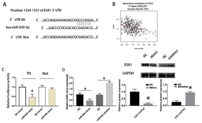
2728 CHEN et al: miR-939-3p ACTS AS A TUMOR SUPPRESSOR IN HEPATOCELLULAR CARCINOMA
from The Cancer Genome Atlas (TCGA, portal.gdc.cancer. room temperature. Signals were visualized using ECL substrate
gov/). (Pierce; Thermo Fisher Scientific, Inc.).
Cell culture. The HCC cell line (HCCLM3) was obtained from Dual‑luciferase reporter assay. PmirGLO plasmids containing
the American Type Culture Collection. Cells were cultured at the wild‑type (Wt) or mutant (Mut) 3'UTR of ESR1 were
37˚C with 5% CO2 in Minimum Essential Medium (MEM; purchased from Shanghai GenePharma Co., Ltd. PmirGLO
Thermo Fisher Scientific, Inc.) containing 10% FBS (Thermo plasmids were transfected into LM3 cells with miR‑939‑3p
Fisher Scientific, Inc.). This cell line was authenticated by mimic or inhibitor with Lipofectamine® 2000 reagent (Thermo
short tandem repeats profiling. Fisher Scientific, Inc.). Cells were cultured for 48 h prior to
measurement of luciferase intensity. At 48 h post‑transfection,
Reverse transcription‑quantitative (RT‑q)PCR. Total RNA the cells were lysed using radioimmunoprecipitation assay
was extracted using TRIzol® reagent (Invitrogen; Thermo buffer (cat. no. P0013C; Beyotime Institute of Biotechnology)
Fisher Scientific, Inc.). RNA was then reverse transcribed to according to the manufacturer's protocol. A F‑4500 fluores-
cDNA using PrimeScript™ RT Master mix (cat. no. RR036A; cence spectrophotometer (Hitachi, Ltd.) was used to measure
Takara Bio, Inc.), according to the manufacturer's protocol. the luciferase intensity according to the manufacturer's protocol
qPCR was performed using an ABI 7500 (Thermo Fisher and normalized to that of Renilla luciferase.
Scientific, Inc.). The primer sequences were as follows:
miR‑939 forward, 5'‑TGG G GAG CTGAG G CTC TG‑3' and Migration and invasion assays. Cell migration and invasion
reverse, 3'‑AGTG CAG GGTCCGAGGTAT T‑5'; U6 forward, ability was evaluated using a Transwell assay. LM3 cells (5x10⁴)
5'‑CTCG CTTCGG CAG CACA‑3' and reverse, 3'‑AACG CT were seeded in the upper chamber with FBS‑free MEM and
TCACGAATTTGCGT‑5'; and ESR1 forward, 5'‑CCGG CT the lower chamber contained MEM supplemented with 10%
CCGTAAATGCTACG‑3' and reverse, 3'‑TCCAGCAGACCC FBS. For invasion assays, membranes were coated with 50 µl
CACTTCAC‑5'. U6 was used as the internal control. growth factor‑reduced Matrigel (BD Biosciences). Cell migra-
tion and invasion were measured after incubation for 24 h.
Transfection. miR‑939‑3p mimic, miR‑939‑3p inhibitor Cells were stained with crystal violet dye solution for 5 min
and ESR1 small interfering (si) RNA were obtained from at room temperature, and the number of cells were counted in
Shanghai GenePharma Co., Ltd. Cells were seeded in 6‑well five randomly selected fields with a light inverted microscope at
plates (3x10⁵/well) and cultured for 24 h before transfection. x200 magnification. Each experiment was repeated three times.
Lipofectamine® 3000 (Invitrogen; Thermo Fisher Scientific,
Inc.) was used for transfection, according to the manufac- Bioinformation analysis. We predict the target gene of miRNA
turer's protocol. The sequences of the miRNAs were as follows: with TargetScan (version 5.0; genes.mit.edu/targetscan). The
hsa‑miR‑939 mimic sense, UGG G GAG CU GAG G CUCUG level of miR‑939‑3p in the adjacent normal tissues and HCC
GGGGUG and antisense, CCCCCAGAGCCUCAGCUCCCC tissues and the Kaplan‑Meier survival curve analysis in HCC
AUU; mimics negative control (NC) sense, UUCUCCGAACGU patients in The Cancer Genome Atlas (TCGA) were analyzed
GUCACGUTT and antisense, ACGUGACACGUUCGGAGA with miRpower (kmplot.com/analysis/) (20).
ATT; and hsa‑miR‑939 inhibitor, CACCCCCAGAGCCUCAGC
UCCCCA; and inhibitor NC, CAGUACUUUUGUGUAGUA Statistical analysis. Data are presented as the mean ± standard
CAA. The sequences of the ESR1 siRNA were as follows: sense, deviation unless otherwise shown. Statistical analysis was
GCAAGUUGAUCUUAGUUAAGU and antisense, UUAACU performed using SPSS 19.0 (IBM, Corp.). Significance between
AAGAUCAACUUGCUG; siRNA NC (cat. no. siN05815122147; groups was analyzed using an unpaired Student's t‑test. The
Guangzhou RiboBio Co., Ltd.,) was used as the siRNA negative correlation between miR‑939‑3p and ESR1 expression levels
control, but the sequence was not provided by the supplier. was examined using a Pearson's correlation coefficient. The
log rank test was used for survival analysis. P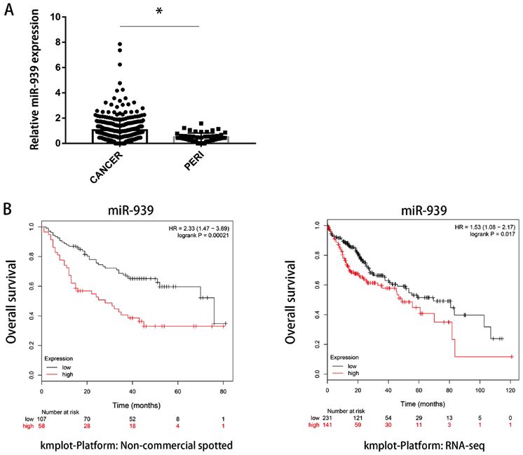
ONCOLOGY LETTERS 19: 2727-2732, 2020                                                         2729

Figure 1. miR‑939‑3p expression is upregulated in HCC tissues. (A) miR‑939‑3p expression levels were significantly higher in HCC tissues compared with
paired normal tissues. (B) Low miR‑939‑3p expression levels were associated with improved overall survival in two different datasets obtained from The
Cancer Genome Atlas. *P
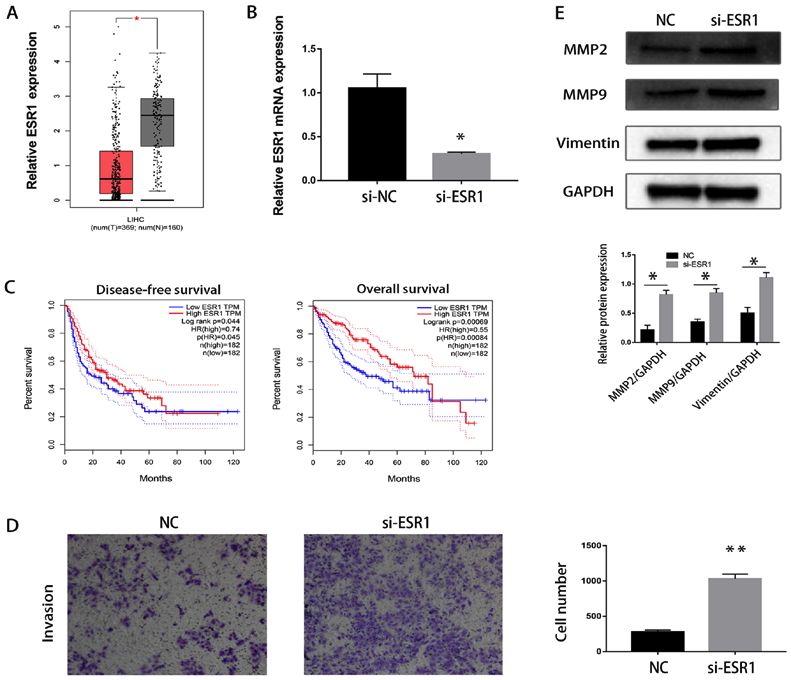
2730                    CHEN et al: miR-939-3p ACTS AS A TUMOR SUPPRESSOR IN HEPATOCELLULAR CARCINOMA

Figure 3. ESR1 is downregulated in HCC tissues. (A) ESR1 mRNA expression levels in HCC tissues and paired normal tissues. *P
ONCOLOGY LETTERS 19: 2727-2732, 2020                                                       2731

Figure 4. miR‑939‑3p directly binds to the 3'UTR of ESR1. (A) Wt and Mut sequences of the ESR1 3'UTR and the binding sequences of miR‑939‑3p.
(B) Correlation of ESR1 and miR‑939‑3p in The Cancer Genome Atlas. (C) miR‑939‑3p suppressed the luciferase activity of the Wt ESR1 3'UTR, whereas the
Mut miR‑939‑3p sequence did not in LM3 cells. *P
2732                   CHEN et al: miR-939-3p ACTS AS A TUMOR SUPPRESSOR IN HEPATOCELLULAR CARCINOMA

Authors' contributions                                                      14. Chen A, Liu S, Lu X, Wei L and Chen Y: Inhibition of microRNA939
                                                                                suppresses the development of human nonsmall cell lung cancer
                                                                                via the upregulation of tissue inhibitor of metalloproteinases 2.
JX designed the study. FC, XYN, LXC and XYW performed                           Mol Med Rep 18: 4831‑4838, 2018.
the experiments and analyzed the data. FC and XYN wrote the                 15. Bjornstrom L and Sjoberg M: Mechanisms of estrogen receptor
                                                                                signaling: Convergence of genomic and nongenomic actions on
manuscript. XYN and XYW revised the manuscript.                                 target genes. Mol Endocrinol 19: 833‑842, 2005.
                                                                            16. Yang J, AlTahan A, Jones DT, Buffa FM, Bridges E, Interiano RB,
Ethics approval and consent to participate                                      Qu C, Vogt N, Li JL, Baban D, et al: Estrogen receptor‑alpha
                                                                                directly regulates the hypoxia‑inducible factor 1 pathway associ-
                                                                                ated with antiestrogen response in breast cancer, Proc Natl Acad
The present study was approved by The Institutional Review                      Sci USA 112: 15172‑15177, 2015.
Board of the Zhejiang Provincial Peoples' Hospital (Taizhou,                17. Andruska ND, Zheng X, Yang X, Mao C, Cherian MM, Mahapatra L,
China). All patients gave written informed consent to partici-                  Helferich WG and Shapiro DJ: Estrogen receptor α inhibitor activates
                                                                                the unfolded protein response, blocks protein synthesis, and induces
pate in the study and the data were anonymized.                                 tumor regression. Proc Natl Acad Sci USA 112: 4737‑4742, 2015.
                                                                            18. Hishida M, Nomoto S, Inokawa Y, Hayashi M, Kanda M,
Patient consent for publication                                                 Okamura Y, Nishi kawa Y, Tana ka C, Kobayashi D,
                                                                                Yamada S, et al: Estrogen receptor 1 gene as a tumor suppressor
                                                                                gene in hepatocellular carcinoma detected by triple‑combination
Not applicable.                                                                 array analysis. Int J Oncol 43: 88‑94, 2013.
                                                                            19. Tu CC, Kumar VB, Day CH, Kuo WW, Yeh SP, Chen RJ, Liao CR,
                                                                                Chen HY, Tsai FJ, Wu WJ and Huang CY: Estrogen receptor alpha
Competing interests                                                             (ESR1) over‑expression mediated apoptosis in Hep3B cells by
                                                                                binding with SP1 proteins. J Mol Endocrinol 51: 203‑212, 2013.
The authors declare that they have no competing interests.                  20. Nagy Á, Lánczky A, Menyhárt O and Győrffy B: Validation of
                                                                                miRNA prognostic power in hepatocellular carcinoma using
                                                                                expression data of independent datasets. Sci Rep 8: 9227, 2018.
References                                                                  21. Wan J, Liu H, Yang L, Ma L, Liu J and Ming L: JMJD6 promotes
                                                                                hepatocellular carcinoma carcinogenesis by targeting CDK4. Int
 1. Bray F, Ferlay J, Soerjomataram I, Siegel RL, Torre LA and                  J Cancer 144: 2489‑2500, 2019.
    Jemal A: Global cancer statistics 2018: GLOBOCAN estimates              22. Hu D, Hu Y, Xu W, Yu H, Yang N, Ni S and Fu R: miR203 inhibits
    of incidence and mortality worldwide for 36 cancers in 185 coun-            the expression of collagenrelated genes and the proliferation of
    tries. CA Cancer J Clin 68: 394‑424, 2018.                                  hepatic stellate cells through a SMAD3dependent mechanism.
 2. Tan A, Yeh SH, Liu CJ, Cheung C and Chen PJ: Viral hepato-                  Mol Med Rep 16: 1248‑1254, 2017.
    carcinogenesis: From infection to cancer. Liver Int 28: 175‑188,        23. Wan X, Cheng C, Shao Q, Lin Z, Lu S and Chen Y: CD24 promotes
    2008.                                                                       HCC progression via triggering Notch‑related EMT and modula-
 3. Ren FH, Yang H, He RQ, Lu JN, Lin XG, Liang HW, Dang YW,                    tion of tumor microenvironment. Tumour Biol 37: 6073‑6084, 2016.
    Feng ZB, Chen G and Luo DZ: Analysis of microarrays of                  24. Hu Y, Yang C, Yang S, Cheng F, Rao J and Wang X: miR‑665
    miR‑34a and its identification of prospective target gene signa-            promotes hepatocellular carcinoma cell migration, invasion, and
    ture in hepatocellular carcinoma. BMC Cancer 18: 12, 2018.                  proliferation by decreasing Hippo signaling through targeting
 4. Brondfield MN, Dodge JL, Hirose R, Heimbach J, Yao FY and                   PTPRB. Cell Death Dis 9: 954, 2018.
    Mehta N: Hepatocellular carcinoma (HCC) patients listed in              25. Zhang XP, Jiang YB, Zhong CQ, Ma N, Zhang EB, Zhang F, Li JJ,
    short wait regions remain advantaged for liver transplant (LT)              Deng YZ, Wang K, Xie D and Cheng SQ: PRMT1 promoted HCC
    following 2015 HCC policy change. Liver Transpl: Dec 13, 2019               growth and metastasis in vitro and in vivo via activating the STAT3
    doi: 10.1002/lt.25701 (Epub ahead of print).                                signalling pathway. Cell Physiol Biochem 47: 1643‑1654, 2018.
 5. Bakheet AMH, Zhao C, Chen JN, Zhang JY, Huang JT, Du Y,                 26. Yu H, Shen H, Zhang Y, Zhong F, Liu Y, Qin L and Yang P:
    Gong LP, Bi YH and Shao CK: Improving pathological early                    CAV1 promotes HCC cell progression and metastasis through
    diagnosis and differential biomarker value for hepatocellular               Wnt/β‑catenin pathway. PLoS One 9: e106451, 2014.
    carcinoma via RNAscope technology. Hepatol Int: Dec 12, 2019            27. Li Y, Chen L, Chan TH, Liu M, Kong KL, Qiu JL, Li Y, Yuan YF
    doi: 10.1007/s12072‑019‑10006‑z (Epub ahead of print).                      and Guan XY: SPOCK1 is regulated by CHD1L and blocks
 6. Hu J, Wang E, Liu L, Wang Q, Xia D, Bai W, Tie J, Li X, Yuan J,             apoptosis and promotes HCC cell invasiveness and metastasis in
    Yang S, et al: Sorafenib may enhance antitumour efficacy in                 mice. Gastroenterology 144: 179‑191 e174, 2013.
    hepatocellular carcinoma patients by modulating the proportions         28. Feng L, Jing L, Han J, Wang G, Liu Y, Zhang X, Wang Y, Wang F,
    and functions of natural killer cells. Invest New Drugs: Dec 13,            Ma H and Liu Y: MicroRNA 486‑3p directly targets BIK and
    2019 doi: 10.1007/s10637‑019‑00885‑2 (Epub ahead of print).                 regulates apoptosis and invasion in colorectal cancer cells. Onco
 7. Berezikov E, Guryev V, van de Belt J, Wienholds E, Plasterk RH              Targets Ther 11: 8791‑8801, 2018.
    and Cuppen E: Phylogenetic shadowing and computational iden-            29. Liang M, Shi B, Liu J, He L, Yi G, Zhou L, Yu G and Zhou X:
    tification of human microRNA genes. Cell 120: 21‑24, 2005.                  Downregulation of miR203 induces overexpression of PIK3CA
 8. Rana TM: Illuminating the silence: Understanding the structure              and predicts poor prognosis of gastric cancer patients. Drug Des
    and function of small RNAs. Nat Rev Mol Cell Biol 8: 23‑36,                 Devel Ther 9: 3607‑3616, 2015.
    2007.                                                                   30. Wang ZY, Zhu Z, Wang HF, Qin B, Liu J, Yao XH, Li WC and
 9. Loosen SH, Schueller F, Trautwein C, Roy S and Roderburg C:                 Chen KS: Downregulation of circDYNC1H1 exhibits inhibitor
    Role of circulating microRNAs in liver diseases. World                      effect on cell proliferation and migration in hepatocellular carci-
    J Hepatol 9: 586‑594, 2017.                                                 noma through miR‑140‑5p. J Cell Physiol 234: 17775‑17785, 2019.
10. Huang X, Wang L, Liu W and Li F: MicroRNA‑497‑5p inhibits               31. Yu W, Deng W, Zhao Q, Zhuang H, Zhang C and Jian Z: miR‑501
    proliferation and invasion of non‑small cell lung cancer by regu-           acts as an independent prognostic factor that promotes the
    lating FGF2. Oncol Lett 17: 3425‑3431, 2019.                                epithelial‑mesenchymal transition through targeting JDP2 in
11. Ni JS, Zheng H, Huang ZP, Hong YG, Ou YL, Tao YP, Wang MC,                  hepatocellular carcinoma. Hum Cell 32: 343‑351, 2019.
    Wang ZG, Yang Y and Zhou WP: MicroRNA‑197‑3p acts as a prog-
    nostic marker and inhibits cell invasion in hepatocellular carcinoma.                    This work is licensed under a Creative Commons
    Oncol Lett 17: 2317‑2327, 2019.                                                          Attribution-NonCommercial-NoDerivatives 4.0
12. Tang Z, Fang Y and Du R: MicroRNA‑107 induces cell cycle                                 International (CC BY-NC-ND 4.0) License.
    arrests by directly targeting cyclin E1 in ovarian cancer. Biochem
    Biophys Res Commun 512: 331‑337, 2019.
13. Wang J, Chu Y, Xu M, Zhang X, Zhou Y and Xu M: miR‑21
    promotes cell migration and invasion of hepatocellular carci-
    noma by targeting KLF5. Oncol Lett 17: 2221‑2227, 2019.
You can also read