Original Article The correlation between exposure to BPA and the decrease of the ovarian reserve
←
→
Page content transcription
If your browser does not render page correctly, please read the page content below
Int J Clin Exp Pathol 2018;11(7):3375-3382
www.ijcep.com /ISSN:1936-2625/IJCEP0069241

Original Article
The correlation between exposure to BPA
and the decrease of the ovarian reserve
Yuming Cao1,2, Xinlan Qu1,2, Zhang Ming1,2, Yanru Yao1,2, Yuanzhen Zhang1,2
1
  Reproductive Medicine Center, 2Department of Obstetrics and Gynecology, Zhongnan Hospital of Wuhan Univer-
sity, Wuhan, Hubei Province, People’s Republic of China
Received November 16, 2017; Accepted April 24, 2018; Epub July 1, 2018; Published July 15, 2018

Abstract: Objective: This study aimed to evaluate whether exposure to bisphenol A (BPA) affects the ovarian reserve.
Methods: Follicular fluid (FF) was collected from diminished ovarian reserve (DOR) and non-DOR patients who un-
derwent in vitro fertilization or intracytoplasmic sperm injection. ELISA was used to detect the BPA and hormones
levels in 54 cases of DOR and 67 cases of non-DOR. A total of 64, five-week-old SPF C57BL/6 mice were randomly
divided into four groups, of which three were exposed to 5, 50, and 500 µg/kg/day of BPA solution, and one was ex-
posed to con oil only as the control. The weight and estrus of each mouse were recorded daily, and the E2 hormone
and anti-Müllerian hormone (AMH) in the serum were detected by ELISA. The expression levels of AMH mRNA and
protein were also detected. Results: The BPA levels in the FF of DOR patients were significantly higher than those of
non-DOR patients (234.048±81.736 ng/L vs. 193.300±67.225 ng/L, P
Effect of BPA on the ovarian reserve

thinning, amenorrhea, infertility, and other sym-    before we collected the specimens. During the
ptoms, all of which lead to the reduced fertility    entire experiment, the mice were humanely
of women. The clinical indicators of DOR include     treated. Before being sacrificed, all mice were
decreased levels of estrogen and anti-Mülleri-       anesthetized, and all operations were designed
an hormone (AMH) and a reduced number of             to minimize suffering. All experimental proce-
antral follicles (antral follicle count, AFC). The   dures were approved by the A3 Animal User,
incidence of DOR accounts for 10% of infertility     Wuhan University.
cases, and DOR typically develops into prema-
ture ovarian failure within 1-6 years. Although      Study objects and sampling
in vitro fertilization (IVF) and embryo transplan-
tation have become routine methods for add-          A total of 54 DOR patients and 67 non-DOR
ressing infertility, DOR patients generally pres-    patients who underwent IVF or intracytoplas-
ent a lower number of oocytes, a low pregnancy       mic sperm injection (ICSI) were enrolled in the
rate, a high cycle cancelation rate, a high abor-    reproductive medicine center of Zhongnan Ho-
tion rate, and increased aneuploidy risk, all of     spital of Wuhan University from November
which severely affect women’s reproductive           2015 to November 2016.
health. The etiology of DOR remains unclear to
date. DOR may be related to genetic and meta-        Incision and exclusion criteria
bolic abnormalities, autoimmune diseases, iat-
rogenic injury, infection, and environment. AMH      For the experimental group, the inclusion crite-
is a glycoprotein produced by the granulosa          ria included patients who met the DOR diagnos-
cells of preantral and small antral follicles. Its   tic criteria and provided informed consent. The
expression is flanked by two major regulatory        exclusion criteria included patients who used
steps of folliculogenesis, first appearing in the    hormone drugs within the past three months
granulosa cells of primary follicles and being       and those who have hyperprolactinemia, PCOS,
the strongest in preantral and small antral folli-   endometriosis, ovarian surgery, and thyroid or
cles. AMH can be directly used to reflect the        adrenal endocrine diseases.
state of the ovary and assess the ovarian func-
tion [11, 12]. Associated with the growth and        For the control group, the exclusion criteria in-
development of follicular fluid (FF), AMH can be     cluded patients in the experimental group who
used to accurately assess the ovarian reserve        used hormone drugs in the past three months
capacity.                                            and have hyperprolactinemia, PCOS, endome-
                                                     triosis, ovarian surgery, and thyroid or adrenal
In recent years, the incidence of DOR has been       endocrine diseases. The inclusion criterion was
increasing among the younger population. The         patients who did not receive received IVF/ICSI
influence of environmental factors on reproduc-      treatment.
tive health has been extensively studied. Stu-
dies have shown that poor environmental fac-         Collected samples and outcome index
tors can accelerate the decline of the ovarian
reserve in females. However, few studies have        Oocytes were collected by vaginal puncture
explored the relationship between BPA and            under the guidance of a transvaginal ultra-
DOR. Thus, we chose FF as an ideal biological        sound. Clear FF without blood was collected
specimen. The correlation between BPA and            using pickling glass bottles. The supernatant
DOR was investigated by detecting the concen-        was centrifuged and stored at 80°C for test-
tration of BPA and hormone levels in FF. The         ing. The serum levels of basic hormones (AMH
findings were validated on animal specimens.         and E2) were examined by the laboratory of our
We intend to provide insight into and clinical       hospital. The BPA and AMH levels in the FF
evidence for the mechanism of the occurrence         were detected by using an ELISA Kit (Wuhan
and development of DOR.                              Hualian Biological Corporation), and the opera-
Materials and methods                                tion was conducted in accordance with the
                                                     kit’s instructions.
Ethical approval
                                                     Collection of mouse samples
This study was approved by the Ethics Com-
mittee of the Zhongnan Hospital of Wuhan Un-         Five-week-old SPF C57BL/6 female mice were
iversity. All patients provided informed consent     purchased and housed at the A3 animal experi-

3376                                                    Int J Clin Exp Pathol 2018;11(7):3375-3382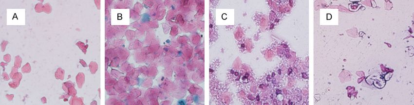
Effect of BPA on the ovarian reserve

Table 1. Primer sequence of fluorescent quantitative             was smeared onto a labeled glass slide,
PCR                                                              which was then dyed with a pap dyeing
Primer        Primer sequence                           Bp       liquid and observed under a light micro-
AMH       M   5’-AGC CAG TTT CCG CAT CTA CC-3’         244       scope.
          R   5’-GTC AGG TAG CGG TTG AAA TGG-3’
B-actin   M   5’-TGA AGG GTG GAG CCA AAA G-3’          150       Collection of serum samples and hor-
          R   5’-AGT CTT CTG GGT GGC AGT GAT-3’                  mone assays

                                                                 Blood samples were collected from the
Table 2. Analysis of hormone concentration in fol-               angular vein after anesthesia and stored
licular fluid                                                    at 4°C static. After 24 h, each blood sam-
Hormone        Non-DOR          DOR         P                    ple was centrifuged at 4°C and 3000 r/
BPA (ng/L)  193.300±67.225 234.050±81.736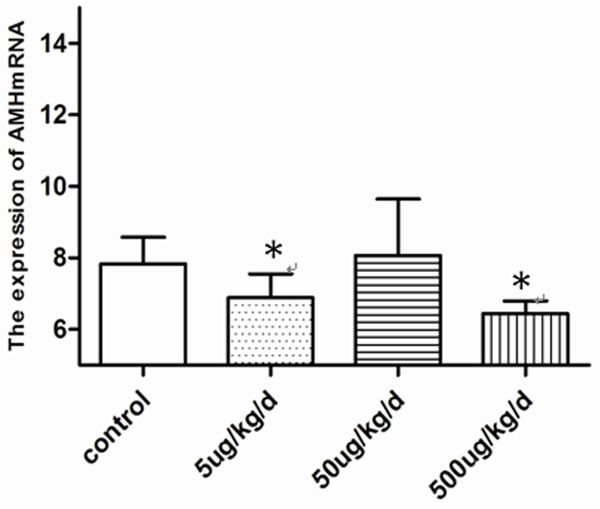
Effect of BPA on the ovarian reserve

Figure 1. Effects of chronic exposure to BPA on the estrous cycle in mice. Picture magnification 20×. A: Proestrus:
much nucleated epithelial cells, and less keratin epithelial cells; B: Estrus: keratin epithelial cells from scattered
to agglomerate, less nucleated epithelial cells; C: Metaestrus: keratin epithelial cell accumulation, many nucleated
epithelial cells and white blood cells; D: Diestrus: white blood cells and less mucus.

Table 4. Effects of BPA on estrous cycle in female rat                             Immunohistochemistry
Groups           n       Proestrus   Estrus        Metestrus      Diestrus         The paraffin blocks were cut
Control         16      0.7±0.67   2.7±0.67        2.3±0.48      4.2±1.32          into sections and mounted on
5 ug/kg/d       16      0.2±0.42* 2.1±0.57*        2.2±0.63     5.8±1.03*          slides. The sections were in-
50 ug/kg/d      16      0.6±0.42   2.6±0.32        2.1±0.32      4.3±0.63          cubated with the polyclonal
500 ug/kg/d     16      0.1±0.32* 1.4±0.84**       2.1±0.88     6.2±1.14**         AMH antibody (1:100 dilution;
*P
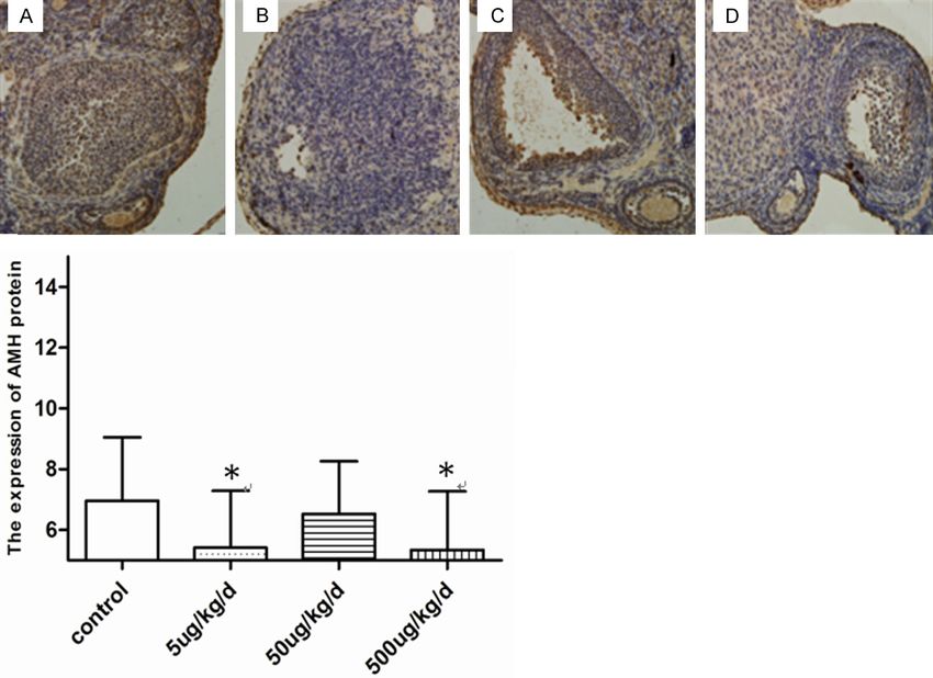
Effect of BPA on the ovarian reserve
Figure 3. Effects of Pathological changes of ovary by chronic exposure to BPA in mice. A: Control; B: 5 ug/kg/d BPA;
C: 50 ug/kg/d BPA; D: 500 ug/kg/d BPA; Picture magnification 20×.
Correlation analysis of BPA level and hormone
concentration in FF
As shown in Table 3, the BPA concentration
was negatively correlated with the AMH (r=-
0.290) and E2 concentrations (r=-0.312) in the
FF of the DOR patients.
Effect of BPA on the estrous cyclicity of the
mice
The estrus of the mice is presented in Figure 1.
Compared with the control group, the experi-
mental groups spent a longer time in diestrus
but less time in estrus and proestrus. The cy-
Figure 4. Effects of BPA on Changes in the mRNA ex- clicity of the 5 and 500 µg/kg/day of the BPA
pression of AMH, the 5 ug/kg/d and 500 ug/kg/d of exposure groups was significantly decreased
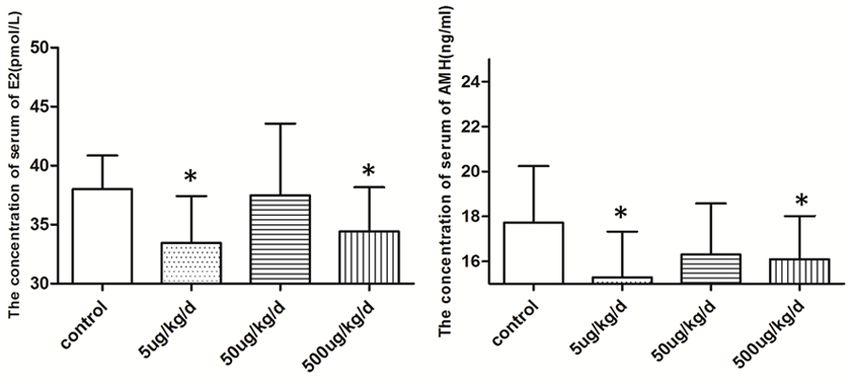
BPA expose group was decrease obviously compared (PEffect of BPA on the ovarian reserve Table 6. Effects of BPA on expression of AMH speculated that the high BPA level in the Group Num AMH mRNA P AMH protein P FF may influence the onset of DOR. The Control 16 7.84±0.74 6.96±2.09 animal experiment revealed that BPA 5 ug/kg/d 16 6.89±0.67* 0.05 the ovarian reserve when ovarian func- 500 ug/kg/d 16 6.45±0.35*
Effect of BPA on the ovarian reserve

                                                         Figure 5. The protein expression of AMH in ova-
                                                         ry exposed to BPA. A: Control; B: 5 ug/kg BPA; C:
                                                         50 ug/kg BPA; D: 500 ug/kg BPA. Picture mag-
                                                         nification 20×. The expression at the 5 ug/kg/d
                                                         and 500 ug/kg/d BPA exposure was obviously
                                                         lower than the control (*P
Effect of BPA on the ovarian reserve
same litters. The primordial follicles in the ovary [4] Calafat AM, Kuklenyik Z, Reidy JA, Caudill SP,
were depleted with the growth of the mouse. Ekong J, Needham LL. Urinary concentrations
BPA can affect the secretion hormones of gran- of bisphenol a and 4-nonylphenol in a human
ulosa cells and the meiotic process to acceler- reference population. Environ Health Perspect
2005; 113: 391-5.
ate the apoptosis of ovarian granulosa cells
[5] Zhang T, Sun HW, Kannan K. Blood and urinary
and follicular atresia. BPA may accelerate the bisphenol a concentrations in children, adults
apoptosis of granulosa cells and the atresia of and pregnant women from China: partitioning
follicles by interfering with the meiosis of germ between blood and urine and maternal and fe-
cells, which reduces the expression of AMH. tal cord blood. Environ Sci Technol 2013; 47:
With the lack of AMH, primordial follicles would 4686-94.
be increased at a faster rate, leading to the pre- [6] EPA, 1988. health assessment information on
mature maturation of the follicular pool and a bisphenol a (CASRN80-05-7). http://cfpub.
shortened reproductive lifespan. epa.gov/ncea/iris/index.cfm?fuseaction=iris.
showQuickview&substance_nmbr=0356#re-
In summary, our study determined that BPA is foral (1988).
involved in the reduction of the ovarian reserve. [7] Forte M, Mita L, Cobellis L, Merafina V, Spec-
Even sub-chronic exposure to low doses of BPA chio R, Rossi S, Mita DG, Mosca L, Castaldi
MA, De Falco M, Laforgia V, Crispi S. Triclosan
can decrease the ovarian reserve. However, the
and bisphenol a affect decidualization of hu-
effects of BPA on the number of small antral
man endometrial stromal cells. Mol Cell Endo-
follicles and the composition of follicles at all crinol 2016; 15: 74-83.
levels require further study. [8] Bruner-Tran KL, Gnecco J, Ding T, Glore DR,
Pensabene V, Osteen KG. Exposure to the envi-
Acknowledgements ronmental endocrine disruptor TCDD and hu-
man reproductive dysfunction: translating les-
This work was supported by grants from the sons from murine models. Reprod Toxicol
National Natural Science Foundation of china 2017; 68: 59-71.
(Grant No.81370707). [9] Stanley JA, Arosh JA, Burghardt RC, Banu SK. A
fetal whole ovarian culture model for the evalu-
Disclosure of conflict of interest ation of crvi-induced developmental toxicity
during germ cell nest breakdown. Toxicol Appl
None. Pharmacol 2015; 289: 58-69.
[10] Vahedi M, Saeedi A, Poorbaghi S, Sepehri-
manesh M, Fattahi M. Metabolic and endo-
Address correspondence to: Yuanzhen Zhang, Re-
crine effects of bisphenol a exposure in market
productive Medicine Center, Zhongnan Hospital of seller women with polycystic ovary syndrome.
Wuhan University, Wuhan 430071, Hubei Province, Environ Sci Pollut Res Int 2016; 23: 23546-
People’s Republic of China; Department of Obs- 23550.
tetrics and Gynecology, Zhongnan Hospital of Wu- [11] Ota T, Asahina H, Park SH, Huang Q, Minegishi
han University, Wuhan 430071, Hubei Province, T, Auersperg N, Leung PC. HOX cofactors ex-
People’s Republic of China. Tel: +86-27-67813009; pression and regulation in the human ovary.
Fax: +86-27-67813009; E-mail: zhangyuanzhen@ Reprod Biol Endocrinol 2008; 6: 49.
[12] Kristensen SG, Ebbesen P, Andersen CY. Tran-
vip.sina.com
scriptional profiling of five isolated size-
matched stages of human preantral follicles.
References Mol Cell Endocrinol 2015; 5: 189-201.
[13] Zala SM, Penn DJ. Abnormal behaviours in-
[1] Ganesan S, Keating AF. Bisphenol a-induced duced by chemical pollution: a review of the
ovotoxicity involves DNA damage induction to evidence and new challenges. Anim Behav
which the ovary mounts a protective response 2004; 68: 649-664.
indicated by increased expression of proteins [14] Wang W, Hafner KS, Flaws JA. In utero bisphe-
involved in DNA repair and xenobiotic biotrans- nol a exposure disrupts germ cell nest break-
formation. Toxicol Sci 2016; 52: 169-80. down and reduces fertility with age in the
[2] Berger A, Ziv-Gal A, Cudiamat J, Wang W, Zhou mouse. Toxicol Appl Pharmacol 2014; 276:
C, Flaws JA. The effects of in utero bisphenol a 157-164.
exposure on the ovaries in multiple genera- [15] Durlinger AL, Kramer P, Karels B, de Jong FH,
tions of mice. Reprod Toxicol 2016; 60: 39-52. Uilenbroek JT, Grootegoed JA, Themmen AP.
[3] Flint S, Markle T, Thompson S, Wallace E. Bi- control of primordial follicle recruitment by an-
sphenol a exposure, effects, and policy: a wild- ti-mullerian hormone in mouse ovary. Endocri-
life perspective. J Environ Manage 2012; 15: nology 1999; 140: 5789-5796.
19-34.
3382 Int J Clin Exp Pathol 2018;11(7):3375-3382You can also read