Clinical and Histopathological study of black and red grape seed extracts (Vitis Vinifera) effects on the Albino Mice - Open Journal Systems
←
→
Page content transcription
If your browser does not render page correctly, please read the page content below
2024 Indian Journal of Forensic Medicine & Toxicology, January-March 2021, Vol. 15, No. 1
Clinical and Histopathological study of black and red grape
seed extracts (Vitis Vinifera) effects on the Albino Mice
Snur M. A. Hassan1, Shno N. Hassan2, 3Nian N.N.Maarof
1Assist. Prof., Department of Anatomy and Pathology, College of Veterinary Medicine, Sulaimani University,
Kurdistan-Iraq, 2 Assist. Lecturer, Ministry of education, Kurdistan-Iraq, 3 Assist. Lecturer, Department of
Chemistry, College of Education, Sulaimani University, Kurdistan-Iraq.
Abstract
Grape seed extract (GSE) is a complex mixture of several compounds, mostly represented by polyphenols
and phenolic acids. The goal of the pilot study was to illustrate the safe dose of GSE in mice model and
to assess toxicity that may be initiated by the different concentrations of this plant. Forty-two mice were
divided equally into 7 groups; groups 1 attended as control, were only received water, whereas animals of
groups 2, 3, 4 were treated orally with 200, 400, and 800 mg/kg b.w. of black grape seed extract respectively,
and the remaining groups including 5, 6, and 7 were treated orally with 200, 400, and 800 mg/kg b.w. of
red grape seed extract respectively. The animals were observed daily for any sign revealing for activity
alterations and toxicity along with their body weight measurement during the experiment for 21 days with
histopathological examination. The results gained from this pilot study were recording that the 200 and 400
mg/kg b.w. doses of GSE were safe compared to the 800 mg/kg b.w.in both black and red grape seed extracts
because the higher dose led to a reduction in body weight gain and produce changes in the mentioned organs.
Keywords: Albino mice, Black grape seed, Histopathology, Red grape seed, Sharbazher village.
Introduction in industry and consist of protein, carbohydrates,
lipids, and 5-8% of polyphenols reliant on the type
Plants can produce a large number of chemical of grapes 7. Different cultivars have a different grape
compounds with significant biological effects and they seed composition 8. Additionally, revenues and seed
have been used to manufacture numerous kinds of quality affected by several environmental and biological
medicines since the creation of human being 1,2 factors, such as light, drought, high salinity, cold, metal
Grapes, Vitis vinifera berries, consider as conven ions, pollutants, xenobiotics, toxins, experimental
tional valued fruits in the world 3. The primary composition manipulations, pathogenic infection, and aging of plants
9. A multitude of flavonoids is contained in GSE 10. The
of grape pomace is skin, steam, and seeds 4. The fresh
grape chemical composition is nearby 70-80% water and most abundant of these are the proanthocyanidins, which
dissolved solids such as sugars, phenolic compounds, are oligomers of monomeric flavan-3-of units linked by
nitrogenous compounds, organic acids, minerals, aroma carbon-carbon bonds 11.
compounds, pectic substances 5. A variety of bioactive The most plentiful biologically active phytonutrients
compounds such as simple phenolics, flavonoids, among the polyphenols found in grapes are flavonoids,
anthocyanins, stilbenes, proanthocyanidins, and vitamin which are possessing cardioprotective, neuroprotective,
E are considered a distinctive mixture of phytochemicals antimicrobial, anti-aging, antioxidant, anti-inflammatory,
in grapes 6. Grape seeds consider as a waste product and anti-cancer properties 12,13.
Oral toxicity studies dealing with grape seed safety
Corresponding author: in experimental animals are few in the Iraq/Kurdistan
Snur M. A. Hassan region. Therefore, the main objective of this pilot study
E-mail address: snur.amin@univsul.edu.iq was to determine the oral toxicity of acetone-extracted
Indian Journal of Forensic Medicine & Toxicology, January-March 2021, Vol. 15, No. 1   2025

grape seed (black and red) extract at different doses in             Consequently, the mice were assigned into 7 groups
mice, through clinical observations and evaluation of           (6 mice per group) as follows: Group 1 (control): the
histopathological changes in multiple organs.                   animal of which were received normal saline without
                                                                treatment; Group 2, 3, and 4, the animal of which treated
              Materials and methods                             orally with 200, 400, and 800 mg/kg b.w. of black GSE,
    Grape seed sampling and extraction                          respectively; Group 5, 6, and 7, the animal of which
                                                                were treated orally with 200, 400, and 800 mg/kg b.w.
    The work was carried out on two different varieties         of red GSE respectively, All treatments were given as a
of grape (Vitis Vinifera) the red and black, which were         single daily dose by oral gavages every two days with
taken manually from Sulaimani (Sharbazher-Kurdistan             a third-day free treatment and this study continued for
region) in the middle of July. The grapes were isolated         about 21 days.
manually from the skin, dried in the open air away
from direct sunlight, and grounded into powder by the              Clinical observations               and     body      weight
electrical grinder. The powders were stored in dark glass       measurements
containers at -20℃ until the use.                                   The animals were examined daily during this pilot
      Each sample of (72.2 g) of red grape seed and (40.73      study (for 21 days) for signs of acute toxicity such as
g) of black grape seed powders was suspended with 202           diarrhea, curved tail, falling of hair, mortality and any
mL and 114 mL of (70%) aqueous acetone respectively             other sign indicative for activity alterations, and the
in a 500 mL Erlenmeyer flask. The mixtures were left on         body weights of mice were recorded 2 times throughout
a magnetic stirrer for 24 hours at room temperature. The        the pilot study (day 0 and at the third week).
resultant extracts were filtered on a Buchner funnel then          Tissue   sampling               and       histopathological
firstly evaporated with a rotatory evaporator to remove         examination
acetone, finally were freeze-dried for 24 hours to remove
the water 14.                                                       At the end of the experimental period, the mice were
                                                                euthanized with (Xylazine-Ketamine: 0.1 mL/10 gm of
    GC–MS analysis                                              body weight) as recommended dose intraperitoneally
    Gas Chromatography-Mass Spectrometry (GC-MS)                and cervical dislocation 16. The liver, kidney, spleen, and
analysis was used to ascertain the compounds present in         lung tissues of the sacrificed mice were excised, cleaned
the purified samples of the red and black types of grape        by normal saline, cut into the 4mm, fixed in 10% neutral
seed (Vitis Vinifera) from Sulaimania (Sharbazhear-             buffered formalin (PH 7.6) for 24 hours and undergone
Kurdistan region). Our natural plant was evaluated by           a series of histopathological processes, tissue slices of
(GCMS-QP2010 Ultra) GC systems combined with a                  4 μm thick were attained and stained using the standard
mass spectrometer 15.                                           H and E technique and envisioned by light microscope
                                                                (Leica, Germany), connected with an image analyzer
    Animals and grape seed extract treatments                   software (Am Scope, AmView, MU1000B) 17.

     Forty-two mice (Male and female Mus Muscular                                  Statistical Analysis
species, BALB/c strain) weighing 25-30 g, at 4
weeks age were purchased from the Animal House                       Statistical analysis was performed using the
at the College of Veterinary Medicine, University of            ANOVA (One-way) analyses of variance. Results were
Sulaimani (Sulaimani, Iraq/Kurdistan region), the mice          presented as a mean±standard error (Mean±SE) and P
were provided tap water and standard food ad libitum            values less than 0.05 were considered significant. All
and were permitted to acclimate for one week before the         statistical explorations were accomplished using the
start of the experiment accommodated in temperature             SPSS software version 22 (SPSS Inc., USA).
and light-controlled environment. All of the in vivo
experimentation in this pilot study was performed
                                                                                            Results
humanely according to and the ethical approval that was              Gas chromatography-MASS Spectroscopy
obtained from the Ethics Committee at the College of
Veterinary Medicine, the University of Sulaimani in                 The secondary metabolites present in red and black
number (01589).                                                 grape seeds were detected in GC-MS (Table 1and 2).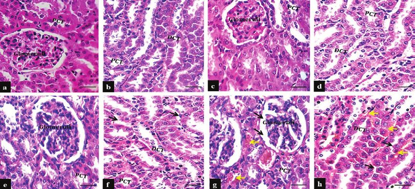
2026       Indian Journal of Forensic Medicine & Toxicology, January-March 2021, Vol. 15, No. 1

The Grape Seed (GSD) peak region percentage and peak area coverage are specified below.

                                        Table 1: G-CMS components for red grape seed

 Peak no.          RT (Min.)                Compound Name                       Peak Area         Correct area   Peak Area (%)

       1              3.439                 Heptane, 3-methyl                      4280             562353         19.620%

       2              3.808                        Octane                          9767             1039837        36.279%

       3              7.345         2-N-PROPYL-1-D1-AZIRIDINE                      5538             168753          5.888%

       4             11.304                 Tridecane, 2-methyl                    4642              87857          3.065%

       5             14.960                 Eicosane, 2-methyl-                    3572              66809          2.331%

       6             18.239               Cyclobutanone, oxime                     3308              68064          2.375%

       7             19.749                Di-isodecyl phthalate                   5223             365445         12.750%

       8             21.187               Octane, 2,4,6-trimethyl-                 4653              93120          3.249%

                                    1,2-Benzenedicarboxylic acid, bis
       9             22.193                                                        9067             212306          7.407%
                                         2-methylpropyl) ester

       10            22.554              4,4-Dimethylcyclooctene                   3692              76959          2.685%

       11            23.444              2-Acetyl-N-methylaniline                  2537              58464          2.040%

       12            23.856                      Docosane                          2972              66227          2.311%

                                       Table 2: G-CMS components for black grape seed
                                                                                                                  Peak Area
 Peak no.          RT (Min.)                Compound Name                       Peak Area         Correct area
                                                                                                                     (%)

       1              3.445                 Heptane, 3-methyl                      4267             579464          3.736%

       2              3.822                        Octane                          9823             998433          6.438%

       3              7.349         2-N-PROPYL-1-D1-AZIRIDINE                      5775             161076          1.039%

       4             11.305               Heptadecane, 2-methyl-                   5492             106189          0.685%

       5             14.961               Hexadecane, 2-methyl-                    4376              85425          0.551%

       6             25.428             Bis(2-ethylhexyl) phthalate               115510            7027490        45.312%

       7             26.876                Di-isodecyl phthalate                  140559            6550999        42.240%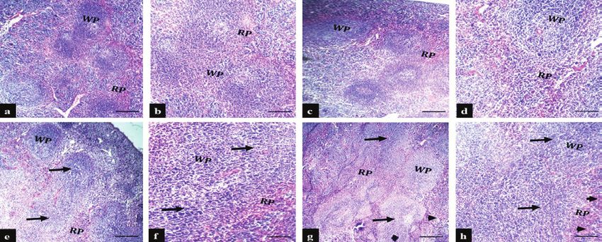
Indian Journal of Forensic Medicine & Toxicology, January-March 2021, Vol. 15, No. 1   2027

    Clinical observations                                      increase (P
2028 Indian Journal of Forensic Medicine & Toxicology, January-March 2021, Vol. 15, No. 1
Histopathological Results the kidney showed dilation of Bowman's capsule,
glomerular atrophy with the segmentation of glomerular
Regarding histopathological findings, the liver capillaries and increasing the mesangial cells, also
section of control, and 200 mg/kg b.w. treated moderate swollen of the epithelial lining of convoluted
groups in the black and red GSE exhibited normal tubules with interstitial hemorrhage (Figure- 2e-h).
histomorphological features including, central vein, Microscopical section of the spleen revealed normal
sinusoidal capillaries with normal appearance of histological appearance in control and 200mg/kg b.w. in
hepatocytes (Figure- 1a-d), in comparison to 400mg/ black and red GSE treated groups, whereas in 400mg/kg
kg b.w. treated groups in the black and red GSE, the b.w. in both types of GSE treated groups showed mild-
liver cells showed karyolitic features (Figure- 1e and f), moderate lymphocytic hyperplasia in the white pulp
while the hepatocytes in 800mg/kg b.w. treated groups region and congestion in the red pulp area if compared
in both types of GSE undergo coagulative necrosis, to the 800mg/kg b.w. in black and red GSE treated
characterized by eosinophilic cytoplasm with features of groups that showed moderate lymphocytic hyperplasia
karoryolysis and karyorrhexis of the nucleus and kupffer in white pulp region and congestion in the red pulp area
cell proliferation (Figure- 1g and h). The microscopical (Figure- 3a-h and 6a-h).The histopathological finding of
section of the kidney in the control group showed; lung parenchyma in black and red GSE treated groups
normal and intact appearance of glomeruli, proximal and revealed normal histological structures of bronchi,
distal convoluted tubules with normal renal vasculature bronchioles, alveolar ducts, alveolar sac, and alveoli
in control, and 200mg/kg b.w. in black and red GSE with normal vasculatures in control and 200mg/kg b.w.
treated groups (Figure- 2a-d). While in 400 mg/kg b.w. treated groups, but the minimum-mild proteinous fluid
in black and red GSE treated groups the epithelial lining was found in the alveolar lumen of 400 and 800mg/kg
of collecting tubules showed slightly swollen. In the b.w. black and red GSE treated groups in addition to
800mg/kg b.w. in both types of GSE treated groups, vascular congestion particularly in the 800mg/kg b.w.
black and red GSE treated groups.
Figure 1- Histomicrograph of liver sections in control, black and red GSE treated groups. a and b: The
normal liver histology in the control group, c and d: The normal histological features of liver parenchyma
in 200mg/kg b.w. treated groups, e, and f: The hepatocytes showed slightly swollen (black arrows) with
karyolysis in few hepatocytes (yellow arrows) in 400mg/kg b.w. treated groups, g and h: The hepatic cells
undergo coagulative necrosis as indicated by black arrows in 800mg/kg b.w. treated groups, (S) sinusoidal
capillaries, (H&E stain, scale bar 50 μm, scale bar 20 μm).Indian Journal of Forensic Medicine & Toxicology, January-March 2021, Vol. 15, No. 1 2029
Figure 2- Histomicrograph of kidney sections in control, black and red GSE extract treated groups. a and b: The normal
kidney histology in the control group, c and d: The normal histological structures of kidney parenchyma in 200mg/kg b.w.
treated groups, e, and f: The epithelial lining of PCT and DCT showed slightly swollen (black arrows) in 400mg/kg b.w.
treated groups, g and h: The swollen of the Bowmans’ space (blackhead arrows), segmentation of glomerular capillary tuft
with mesangial hypercellularity and glomerular atrophy, the epithelial lining of PCT and DCT showed moderately swollen
(black arrows) with interstitial hemorrhage indicated by yellow arrows in 800mg/kg b.w. treated groups, (DCT) convoluted
tubules, and (DCT) distal convoluted tubules, (H&E stain, scale bar 50 μm, scale bar 20 μm).
Figure 3- Histomicrograph of spleen parenchyma in control, black and red GSE treated groups. a and b:
The normal spleen structures in the control group, c and d: Normal microscopical features of the spleen
in 200mg/kg b.w. the treated groups, e, and f: Slightly lymphocytic hyperplasia (black arrows) in the
white pulp regions (black arrows) in 400mg/kg b.w. treated groups, g and h: Mild-moderate lymphocytic
hyperplasia (black arrows) in the white pulp regions and congestion of the red pulp sinusoids as indicated by
blackhead arrows in 800mg/kg b.w. treated groups, (WP) white pulp, and (RP) red pulp, (H&E stain, scale
bar 50 μm, scale bar 20 μm).2030 Indian Journal of Forensic Medicine & Toxicology, January-March 2021, Vol. 15, No. 1
Figure 4- Histomicrograph of the lung parenchyma of control, black and red GSE treated groups. a and
b: In the control, and 200mg/kg b.w. treated groups, the lung showed normal histological constructions of
bronchi, bronchioles, and alveoli with normal vascular appearance, c: There is slight transudate fluid in
the alveolar lumen (Inset and yellow arrows) in 400mg/kg b.w. treated groups, d: The lung parenchyma
exhibited mild accumulation of eosinophilic fluid in the alveolar lumen (Inset and yellow arrows) with
vascular congestion as indicated by black arrows in 800mg/kg b.w. treated groups, (H&E stain, scale bar 50
μm, scale bar 20 μm).
Discussion results of other studies that revealed using of the GSE in
experimental animals did not result in any disturbances
For the continuous development of phytochemicals in clinical activities or body weight loss 22,23.
under-regulated plant cell cultures, prospective
replacements are generally considered 18,19. In recent However, the significant decrease in body weight
years, plant cell biotechnology of grapes and particularly gain was found in mice of group 800mg/kg b.w. of GSE,
of grape cell suspensions have enjoyed great scientific Yamakoshi, et al, (2002), who reported a similar result
and industrial consideration 20. New research aims to in which the high dose of GSE may lead to slightly
decode the benefits of grapes as a rich source of essential significant weight loss in an animal model 24, while
phytonutrients with remarkable beneficial impacts on the observed result may disagree with the Mittal et al,
human health 21. (2003), study who documented that the high dose of GSE
did not interfere with physical activity and significant
No mortality was reported with no significant difference in body weights or other signs of clinical
changes in body weight gain or physical appearance, toxicity 25. Also, the other study on Sprague-Dawley rats
and no abnormal histopathological changes in the liver, documented no significant decrease in body weight was
kidney, spleen, and lung were observed during this pilot observed even after 90 days of oral administration of
study in that groups received the 200, and 400mg/kg GSE 26. The decrease in body weight gain may correlate
b.w. doses of both black and red GSE and the outcomes with the high antioxidant activity in induced to enhance
attained from this study are, in agreement with the the lipase effect and enhancing lipolysis 27.Indian Journal of Forensic Medicine & Toxicology, January-March 2021, Vol. 15, No. 1 2031
Additionally, it is obvious from the present study and engineering of metabolic and biosynthetic
the histopathological changes in 800mg/kg b.w. dose pathway of plant polyphenols. Current
groups of GSE represented by the mild-moderate pharmaceutical design. 2013; 19(34): 6186-206.
swollen in the hepatocytes with a moderate degree of 6. Jucá MM, Cysne Filho FMS, de Almeida JC, et al.
necrosis, segmentation of glomerular capillaries and Flavonoids: biological activities and therapeutic
moderate swollen of the epithelial lining of convoluted potential. Natural product research. 2020; 34(5):
tubules with interstitial hemorrhage in kidney organ, the 692-705.
histological changes in spleen revealed mild-moderate
7. Bijak M, Saluk J, Ponczek MB, et al., Antithrombin
lymphocytic hyperplasia in the white pulp region and
effect of polyphenol‐rich extracts from black
congestion in the red pulp area, and the lung parenchyma
chokeberry and grape seeds. Phytotherapy
showed mild accumulation of eosinophilic fluid the
Research. 2013; 27(1): 71-6.
alveolar lumen. Our findings disagree with the other
studies that mentioned no significant histopathological 8. Fernandes L, Casal S, Cruz R, et al. Seed oils of
lesions in multiple organs after administration of GSE ten traditional Portuguese grape varieties with
high dose 28,29. interesting chemical and antioxidant properties.
Food Research International. 2013; 50(1): 161-6.
Conclusion 9. Agrawal M. Air pollution: A cause of concern for
plants. Abstracts with Presidential address-2018_
Consequently, it is concluded that the results of this
Biological Sciences. 2018; 24: 71-89.
study support the health of GSE dietary components
for human use. No observed level of adverse effects 10. Amer F, Mahrose K, Basyony M. Influence of
(NOAEL) was deemed approximately in a dose of 400 grape seeds powder as a natural antioxidant on
mg/kg b. w. /day for administration in both types of growth performance, antioxidant status and carcass
GSE. While a mild-moderate changes microscopically characteristics of rabbits under hot conditionS.
was seen in the mice’s organs that treated by 800mg/kg Egyptian Journal of Rabbit Science. 2014; 24(2):
b.w. black and red GSE treated groups. 395-412.
11. Smeriglio A, Barreca D, Bellocco E, et al.
Conflict of Interest: Nil Proanthocyanidins and hydrolysable tannins:
Source of Funding: Self-funding occurrence, dietary intake and pharmacological
effects. British journal of pharmacology. 2017;
References 174(11): 1244-62.
12. Taqui SU. Fractionation of hydro-ethanolic extracts
1. Espinosa-Leal CA, Puente-Garza CA, García-
from grape pomace through membrane processing:
Lara S. In vitro plant tissue culture: means for
the effect of membrane and extracting media on
production of biological active compounds. Planta.
process performance.2014.
2018; 248(1): 1-18.
13. Georgiev V, Ananga A, Tsolova V. Recent advances
2. Cragg GM, Newman DJ. Natural products: a
and uses of grape flavonoids as nutraceuticals.
continuing source of novel drug leads. Biochimica
Nutrients. 2014; 6(1): 391-415.
et Biophysica Acta (BBA)-General Subjects. 2013;
1830(6): 3670-95. 14. Libera J, Kononiuk A, Kęska P, et al. Use of grape
seed extract as a natural antioxidant additive in dry-
3. Zhu F, Du B, Zheng L, Li J. Advance on the
cured pork neck technology. Biotechnology and
bioactivity and potential applications of dietary
Food Science. 2018; 82(2).
fibre from grape pomace. Food chemistry. 2015;
186: 207-12. 15. Azeez SH, Gaphor SM. Evaluation of antibacterial
effect against Porphyromonas gingivalis and
4. Zhu F-M, Du B, Li J. Effect of ultrafine grinding
biocompatibility of essential oil extracted from the
on physicochemical and antioxidant properties
gum of Pistacia atlantica kurdica. BioMed research
of dietary fiber from wine grape pomace. Food
international. 2019; 2019.
Science and Technology International. 2014; 20(1):
55-62. 16. Hassan SM, Remzi DO, Muhammed SF, et al.
Photo Protective Role of Wild Edible Plants on
5. Ananga A, Georgiev V, Tsolova V. Manipulation
Skin of Mice from Harmful Effects of Ultraviolet2032 Indian Journal of Forensic Medicine & Toxicology, January-March 2021, Vol. 15, No. 1
Type-B Irradiation. Current Journal of Applied iodoacetate-induced osteoarthritis. Experimental &
Science and Technology, 2016: 1-10. molecular medicine. 2011; 43(10): 561.
17. Hassan S, Hassan AH. Effect of Shogaol on the 24. Yamakoshi J, Saito M, Kataoka S, et al. Safety
Expression of Intestinal Stem Cell Markers in evaluation of proanthocyanidin-rich extract from
Experimentally Induced Colitis in BALB/c Mice. grape seeds. Food and Chemical Toxicology. 2002;
Analytical Cellular Pathology. 2019; 2019. 40(5): 599-607.
18. Altemimi A, Lakhssassi N, Baharlouei A, Watson 25. Mittal A, Elmets CA, Katiyar SK. Dietary feeding
DG, Lightfoot DA. Phytochemicals: Extraction, of proanthocyanidins from grape seeds prevents
isolation, and identification of bioactive compounds photocarcinogenesis in SKH-1 hairless mice:
from plant extracts. Plants. 2017; 6(4): 42. relationship to decreased fat and lipid peroxidation.
19. Kreis W. Exploiting plant cell culture for natural Carcinogenesis. 2003; 24(8): 1379-88.
product formation. Journal of applied botany and 26. Wren AF, Cleary M, Frantz C, et al. 90-day oral
food quality. 2019; 92: 216-25. toxicity study of a grape seed extract (IH636) in
20. Ananga A, Georgiev V, Ochieng J, et al. Production rats. Journal of agricultural and food chemistry.
of anthocyanins in grape cell cultures: a potential 2002; 50(7): 2180-92.
source of raw material for pharmaceutical, food, 27. Caimari A, Del Bas J, Crescenti A, et al. Low
and cosmetic industries. The Mediterranean doses of grape seed procyanidins reduce adiposity
Genetic Code-Grapevine and Olive: InTech. 2013. and improve the plasma lipid profile in hamsters.
21. Vislocky LM, Fernandez ML. Grapes and grape International journal of obesity 2013; 37(4): 576.
products: Their role in health. Nutrition Today. 28. Bentivegna S, Whitney K. Subchronic 3-month
2013; 48(1): 47-51. oral toxicity study of grape seed and grape skin
22. Ray S, Bagchi D, Lim PM, et al. Acute and extracts. Food and Chemical Toxicology. 2002;
long-term safety evaluation of a novel IH636 40(12): 1731-43.
grape seed proanthocyanidin extract. Research 29. El‐Ashmawy IM, Saleh A, Salama OM. Effects
communications in molecular pathology and of marjoram volatile oil and grape seed extract
pharmacology. 2001; 109(3-4): 165-97. on ethanol toxicity in male rats. Basic & clinical
23. Woo YJ, Joo YB, Jung YO, et al. Grape seed pharmacology & toxicology. 2007; 101(5): 320-7.
proanthocyanidin extract ameliorates monosodiumYou can also read