Safetyofintravitrealtriamcinoloneacetonide:an electrophysiologicandhistopathologicalstudyin rabbits
←
→
Page content transcription
If your browser does not render page correctly, please read the page content below
Safety of intravitreal triamcinolone acetonide

                                                                                                         窑Basic Research窑

Safety of intravitreal triamcinolone acetonide: an
electrophysiologic and histopathological study in
rabbits

1
 Department of Ophthalmology, Memorial Institute of                DOI:10.3980/j.issn.2222-3959.2013.06.09
Ophthalmology, 3-Al-Ahram Street, Giza,Cairo 12511, Egypt
2
 Department of Physiology, Research Institute of                   El-Shazly LH, El-Gohary AA, El-Hossary GG. Safety of intravitreal
Ophthalmology, 2-Al-Ahram Street, Giza,Cairo 12511, Egypt          triamcinolone acetonide: an electrophysiologic and histopathological
3
 Department of Pharmacology, Research Institute of                 study in rabbits.                 2013;6(6):790-795
Ophthalmology, 2-Al-Ahram Street, Giza,Cairo 12511, Egypt
Correspondence to: Laila Hassan M. El-Shazly. Memorial             INTRODUCTION
Institute of Ophthalmology, 3-Al-Ahram Street, Giza, Cairo
12511, Egypt. laihasan@gmail.com                                   T     riamcinolone         acetonide       (TA),
                                                                         glucocorticoid, is the most commonly used
                                                                                                                         a     synthetic

Received: 2013-03-20        Accepted: 2013-09-22                   glucocorticoid for intravitreal injection because its low
                                                                   intraocular solubility allows for long duration sustained
Abstract                                                           effect. Intravitreal TA (IVTA) has been used for treatment of
                                                                   intraocular diseases, such as long standing macular edema
·AIM: To evaluate the retinal safety of various doses of           due to retinal vein occlusion or branch retinal vein occlusion,
intravitreal triamcinolone acetonide (TA) in rabbits.
                                                                   diffuse diabetic macular edema, neovascular glaucoma,
· METHODS:       Thirty New Zealand albino rabbits were            ocular hypotony, chronic uveitis and age related macular
divided into five groups (six animals each). In group 1            degeneration[1-5].
(control group), each animal received a single intravitreal        Triamcinolone acetenoid suppresses inflammation               several
injection of 0.1mL phosphate buffered saline. In groups
                                                                   mechanisms. It demonstrates potent inhibitory effects on
2, 3, 4 and 5, each rabbit received a single intravitreal
                                                                   mitogen-activated protein kinase (MAPK) signaling pathways
injection of 4, 8, 16 and 32mg of TA, respectively. Each
                                                                   through the induction of MAPK-1 and this inhibits the
dose was contained in 0.1mL phosphate buffered saline.
Clinical ocular examinations were performed before the
                                                                   expression of multiple inflammatory genes [6]. In addition, TA
injection and on the 1st, 3rd, 10th and 17th post -                inhibits cyclooxygenase, interleukin-6 (IL-6) and reduces
injection    days.    A    standard     dark    adapted            vascular permeability[7]. Moreover, it increases the resorption
electroretinogram (ERG) was obtained before injection              of fluid through the retinal pigment epithelium (RPE) and
and on the 3rd, 10th and 17th post-injection days. After           down regulates the production of vascular endothelial growth
17d, animals were sacrificed and their eyes prepared for           factor (VEGF-A)[8].
pathological examination.                                          Because pevious studies suggested that the effect of
· RESULTS:     By monitoring ERG as a functional index             intravitreal triamcinolone acetonide showed a dosage
for the retina, intravitreal injection of 4mg TA showed no         dependency, many researchers attempted the use of high
significant ERG changes. At doses of 8, 16 and 32,                 doses of IVTA, aiming to reduce the frequency of required
hyper-abnormal responses in a - and b - waves of ERG               intravitreal re-injections [9,10]. Different doses of intravitreal
were detected on the 3rd post -injection day. These                TA, varying from about 4mg to 30mg, has been employed
changes gradually returned back to normal limits after             with contradictory results as regard efficacy, toxicity and
17d. Histopathological examination of the retina of all            duration of effect [10]. In rabbit's eyes, after a single intravitreal
animals showed no pathological changes.
                                                                   injection of TA, no influence on global ERG responses was
·CONCLUSION: High doses of intravitreal TA seemed to               detected [11]. On the other hand, in other studies, TA induced
have enhancing effects on the retinal function with                clear toxic effects on RPE cells, retinal M俟ller glial cells and
gradual return to normal limits with no pathological               retinal neurosensory cells [12-14]. Because IVTA is widely and
changes detected in examined eyes.                                 increasingly used in various ocular diseases, more efficient
·   KEYWORDS:         triamcinolone    acetonide;   intravitreal   and specifically targeted effects of corticosteroids are needed.
injection; electroretinogram                                       To achieve these aims, ensuring safety of the intravitreal
    790
陨灶贼 允 韵责澡贼澡葬造皂燥造熏 灾燥造援 6熏 晕燥援 6熏 Dec.18, 圆园13    www. IJO. cn
                                                                栽藻造押8629原愿圆圆源缘员苑圆   8629-82210956      耘皂葬蚤造押ijopress岳员远猿援糟燥皂
triamcinolone influence on eye tissues is essential. The            drops containing an antibiotic-corticosteroid combination
present study was undertaken to investigate the effect of           (Dexatrol, Eipico, Egypt) was applied to eyes three times
various doses of IVTA on the function (assessed by ERG)             daily for three days.
and the structure (assessed by histopathology) of the retina in     Ophthalmologic examination Ophthalmologic clinical
rabbit eyes.                                                        examinations were performed immediately before injections
MATERIALS AND METHODS                                               (baseline) and on 1st , 3rd, 10th and 17th post injection days.
Materials Thirty New Zealand albino rabbits of both sexes,          Examinations included slit lamp anterior segment
weighing between 2-2.5kg, aged between 7 to 8 months,               examination, and detailed funduscopic examination of
were used in this study. We tried to have a nearly similar age      studied eyes. A baseline standard ERG was obtained one day
in the studied groups to have same degree of retinal                before the intravitreal injection and on 3rd, 10th and 17th
maturation. Animals were used in accordance to the ARVO             postinjection days.
(Association for Research in Vision and Ophthalmology)              Electrophysiological tests Electroretinogram (ERG) was
statement for the use of animals in ophthalmic and vision           performed using the Reti-com system (Roland-Consult).
research. The experiment was approved by our institutional          After anesthesia (as described above before intravitreal
ethical committee. All through the experiment duration,             injection) rabbits were dark adapted for at least 30min after
rabbits were housed in separate cages, fed standard                 pupil dilation. The active electrode was placed near the
laboratory food and allowed free access to water in room            margin of the lower eyelid, the reference electrode was
temperature with 12h light-dark cycle in the animal house of        placed on the forehead and the earth electrode was clipped to
the Research Institute of Ophthalmology.                            the earlobe. Recording of the combined response was carried
Animals were divided into five groups randomly using                out using a mini-Ganzfeld flash stimulus with a maximum
random number generator; each comprised of six rabbits. In          intensity of 3.0cd-s/m2s with no background intensity. ERG
group 1 (control group), six rabbits were subjected to a single     signals were amplified and filtered (0.3-300Hz). Amplitude
intravitreal injection of 0.1mL phosphate buffered saline.          was measured from the baseline to the lowest point of the
Rabbits of the remaining four groups (groups 2, 3, 4 and 5)         negative peak for the a-wave and from the latter to the
were injected once with triamcinolone acetonide (Sigma-             positive peak for the b-wave. Latency was measured from the
Aldrich, Germany) by intravitreal injection at doses of 4, 8,       beginning of the stimulus to the negative peak of the a-wave
16 and 32mg respectively. Each dose was suspended in                (a latency), and to the following positive peak of the b-wave
0.1mL phosphate buffered saline. In studied animals, only           (b latency). Data were expressed as mean 依SD. Analysis of
right eyes were injected intravitreally with total of six eyes in   variance (ANOVA) with post-Hoc multiple comparisons
each group.                                                         were performed to compare responses between and within
Methods                                                             groups. value was considered significant if
Safety of intravitreal triamcinolone acetonide
Table 1 Mean values of a- and b-wave amplitudes (in microvolt) and a- and b-wave latencies (in seconds) on days 3, 10 and 17 after a
single intravitreal injection of 0.1mL phosphate buffered saline in control group 1 and injection with 4, 8, 16 and 32 mg TA/0.1mL
phosphate buffered saline in groups 2, 3, 4 and 5 respectively
Groups a-wave amplitude (μV) a-wave latency (s) b-wave amplitude (μV) b-wave latency (s)
3rd day 5.64±1.27 15.17±0.76 28.43±1.88 35.23±1.52
1 10th day 5.63±1.28 13.52±4.16 28.30±1.82 34.85±1.22
17th day 5.53±1.49 14.70±0.92 29.22±2.5 35.34±2.0
3rd day 5.54±1.14 15.13±0.74 27.38±1.74 34.92±0.96
2 10th day 5.21±0.87 15.10±0.81 28.37±1.6 35.03±1.17
17th day 5.04±0.99 15.27±1.01 29.25±1.26 35.01±0.90
1 1
3rd day 9.88±1.06 15.4±0.67 53.82±2.56 38.25±1.79
3 10th day 4.45±0.27 12.95±1.29 29.63±1.37 36.17±1.23
17th day 6.97±0.40 14.93±0.28 27.05±0.31 33.93±0.62
1 1
3rd day 9.83±0.99 15.68±1.17 54.37±2.59 38.03±1.94
1 1
4 10th day 11.0±0.21 16.02±0.35 55.03±0.28 37.73±0.30
17th day 6.87±0.51 14.63±0.42 35.30±1.04 35.82±0.79
1 1 1 1
3rd day 8.87±0.27 17.25±0.39 55.70±1.50 40.47±0.70
1 1
5 10th day 11.17±0.33 15.18±0.54 55.08±0.60 38.01±0.35
17th day 5.03±0.28 13.08±0.25 31.38±0.49 34.02±0.70
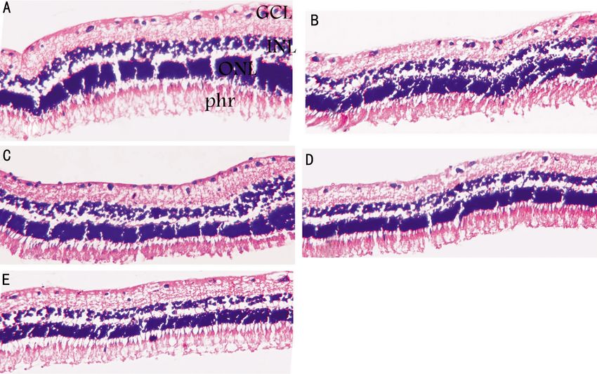
Data is expressed as mean±SD, n=6, µV: Microvolt; TA: Triamcinolone acetonide. 1significant difference (P0.05). Post-injection ERG values with no significant difference from control group 1 on
recordings are represented in Table 1 and Figure 1. Table 1 the 10th and 17th post-injection day. However,
shows the mean value of a- and b-wave amplitude and hyper-abnormal responses were still observed in group 4 and
latency in studied groups on the 3rd, 10th and 17th day after group 5 on the 10th post-injection day and a- and b-wave
injection. In control group 1, a- and b-wave amplitudes and amplitudes were significantly increased as compared to
latencies were normal on the 3rd, 10th and 17th day with no control group 1 (陨灶贼 允 韵责澡贼澡葬造皂燥造熏 灾燥造援 6熏 晕燥援 6熏 Dec.18, 圆园13 www. IJO. cn
栽藻造押8629原愿圆圆源缘员苑圆 8629-82210956 耘皂葬蚤造押ijopress岳员远猿援糟燥皂
Figure 2 Light photomicrograph of selected retinal sections A: A control eye (injected intravitreally with 0.1mL phosphate buffered
saline. It shows normal appearance of retinal layers, photoreceptors (Phr.), outer nuclear layer (ONL), ganglion cell layer (GCL); B, C, D, E:
retinas of groups 2, 3, 4 and 5 injected intravitreally with triamcinolone acetonide at doses of 4, 8, 16 and 32mg/0.1mL phosphate buffered
saline, respectively. There were no distiguishible changes between studied groups (Hematoxylin and eosin, 伊200).
significant delay in a- and b-wave latencies ( =0.002 and carboxymethylcellulose and polysorbate 80 were reported to
0.001, respectively) on the 3rd post-injection day, as produce loss of photoreceptor outer segments, RPE
compared to the control group. However, they returned to proliferation and localized vitritis [18,19]. Except for faint
normal values on the 10th and 17th post injection day with no posterior subcapsular cataract observed in one eye, such
significant difference from the control group 1. complications were not detected in the present study. It can
Figure 1 shows ERG recordings of selected eyes from groups be explained by strictly aseptic precautions during injection
1, 3, 4 and 5 (group 2 was not included as the recordings and use of preservative free TA. Also, the small sample size
were similar to the control group). and short duration of the experiment may played a role. The
Histopathological Examination Hematoxylin and eosin- low rate of complications in the present work was in
stained sections disclosed normal light microscopic agreement with McGee [20]
, who mentioned no clinical
appearance of the retina, retinal pigment epithelium and complications after single intravitreal injections of 4, 16 and
choriocapillaris in all studied animals. 25mg TA in rabbits.
Figure 2 shows photomicrographs of some retinas of ERG is a useful examination to study retinal function. The
examined groups 1, 2, 3, 4 and 5. ERG a-wave is obtained primarily from the maximal
DISCUSSION combined response and it reflects the photoreceptor function.
Because TA suspension provides longer-lasting anti- Physiologically, a-wave arises from the light evoked closure
inflammatory, antiproliferative, antiangiogenesis and of sodium channels along the outer segment plasma
antipermeability effects compared with other steroid membrane of receptor cells. The b-wave results from the
preparations, it has become more widely used in treating current flow along M俟ller cells in response to increased
wide variety of vitreoretinal diseases [15]. In the present study, extracellular potassium ion concentration. It is highly
we investigated the ERG and histopathological effect of dependent on bipolar cells within the inner nuclear layer and
increasing doses of TA after single intravitreal injection in hence on the retinal circulation[21].
albino rabbits. Many risks of intravitreal TA injection were In the present study, intravitreal TA in doses of 8, 16 and
reported which may be procedure-related (such as vitreous 32mg showed transient hyper-abnormal responses on the 3rd
hemorrhage,bacterial endophthalmitis, pseudo-endophthalmitis post-injection day with gradual return to normal responses
and retinal detachment), corticosteroid-related (such as thereafter. These hyper-abnormal responses were detected
cataract and elevated intraocular pressure) or preservative with many conditions such as albinism, atypical cone
toxicity-related of commercial preparations [16,17]. The dystrophies, optic nerve sectioning, optic neuropathies,
preservative benzyl alcohol and suspending agents sodium vascular occlusions, ischemia and uveitis [22]. Also, it was
793Safety of intravitreal triamcinolone acetonide
reported with some drugs including corticosteroids, low-dose showed that the direct toxic effect of the drug was observed
barbiturates and carbon disulphide poisoning. It was when it was injected subretinally, with no protective effect of
suggested that such responses can be due to irritation of the internal limiting membrane or vitreous It was suggested that
retina or suppression of inhibitory pathways in outer and TA in higher doses than 4mg, studies, could affect
middle retinal layers [22]. In accordance with the results of the the DNA rich mitochondria in the inner segment of
present study, Dierks [11]
, suggested that TA therapy photoreceptors and induce a non-apoptotic cell death of the
might augment rod-driven electroretinographic responses. In RPE cells and the Muller cells [27]. Thus, keeping the
addition, it was mentioned that administration of TA could preservative-free TA injection in the mid vitreous, as we did
reduce the production of VEGF-A, arachidonic acid and in this study, might add to the safety of the procedure. Some
prostaglandins allowing reactivation of fluid clearance by limitations of the present study must be considered. Firstly,
M俟ller cells. These processes could lead to decrease of TA was injected into a smaller vitreous volume, less than
M俟ller cell proteins, reduction of the osmotic swelling of 2mL in the rabbit, as opposed to 4-5mL in humans which
M俟ller cells and efflux of potassium ions which could would lead to less drug concentration if the same dose of TA
partially explain the hyper-abnormal responses encountered used in rabbits was injected in humans. Therefore,
with TA [23]. In the present investigation, ERG changes were extrapolation from animal to human studies should be done
transient and returned to normal by the 17th post-injection with caution. Additionally, histological examination was
day and this correlated well with histopathological results confined to pupil-optic nerve sections. Consequently, we
which showed no retinal toxicity manifestation. could not comment on the presence or absence of localized
Results of this study were also in agreement with results of photoreceptor damage outside these sections. Thus, further
Ruiz-Moreno [10]
, who mentioned that intravitreal TA in histopathological testing is required to evaluate retinal layers
doses of 4, 20 and 30mg did not seem to have acute toxic particularly the photoreceptor layer. Another factor was the
effects on the retinal function and structure in albino rabbits. topical antibiotic/corticosteroid drops applied drops applied
Moreover, single intravitreal injections of 4, 16 and 25mg TA post-injection could have a confounding result because
resulted in normal histological and ERG retinal findings[20]. topical eye drops may reach the retina.
McGee [20]
, observed basophilic material, which was In conclusion, based on electrophysiology and
presumed to be drug, in the vitreous with clumps adjacent to histopathology, the present study showed no evidence of
the retinal surface which was similarly detected in our study. retinal toxicity resulting from high doses of intravitreal TA in
This precipitation of triamcinolone, which is a lipophilic slow rabbits. However, further investigations are needed in animal
release large particles, could be explained by the early eyes to examine all retinal layers microscopically and in
sacrifice of animals before the crystals had cleared, as human eyes to determine the safe intravitreal TA dose.
triamcinolone was mentioned to remain in the vitreous up to REFERENCES
six months after injection [24]. It is worth to mention that 1 Ceki觭 O, Chang S, Tseng JJ, Barile GR, Del Priore LV, Weissman H,
triamcinolone crystals in direct contact with retinal cells in Del Priore LV, Schiff WM, Weiss M, Klancnik JM Jr. Intravitreal
triamcinolone treatment for macular edema associated with central retinal
cell culture have been observed to cause cell damage,
vein occlusion and hemiretinal vein occlusion. 2005;25 (7):
perhaps due to the lack of the potentially protective effect of
846-850
the ILM or vitreous[25].
2 Chen CH, Chen YH, Wu PC, Chen YJ, Lee JJ, Liu YC, Kuo HK.
In the present work we prepared TA; firstly to ensure Treatment of branch retinal vein occlusion induced macular edema in
consistent dosing and secondly to avoid the toxic effect of the treatment-naive cases with a single intravitreal triamcinolone or
solvent agent benzyl alcohol in commercially available bevacizumab injection. 2010;33(4):424-435
solutions, since several reports have recommended removal 3 Jonas JB, Akkoyun I, Kreissig I, Degenring RF. Diffuse diabetic macular
of the solvent agent [18]. It was mentioned that commercial edema treated by intravitreal triamcinolone acetonide: a comparative
triamcinolone applied to cultured primary rat retinal cells non-randomized study. 2005;89(3):321-326
induced retinal oxidative injury suggesting toxic potential [26]. 4 Jonas JB, Kreissig I, Hugger P, Sauder G, Panda-Jonas S, Degenring R.
Additionally, it was reported that cases injected with Intravitreal triamcinolone acetonide for exudative age related macular
degeneration. 2003;87(4):462-468
preservative free TA had lower rate of noninfectious
5 Gillies MC, Simpson JM, Luo W, Penfold P, Hunyor AB, Chua W,
endophthalmitis[13].
Mitchell P, Billson F. A randomized clinical trial of a single dose of
It was mentioned that commercial preservative-containing
intravitreal triamcinolone acetonide for neovascular age-related macular
TA injected intravitreal in doses of 4, 8, and 20mg induced degeneration: One-year results. 2003;121(5):667-673
prominent retinal damage manifested by damage to the 6 Clark AR. Review MAP kinase phosphatase 1: A novel mediator of
photoreceptor outer segments and RPE [12]. However, when biological effects of glucocorticoids. 2003;178(1):5-12
preservative-free TA was injected subretinally , Maia [13]
, 7 Nehm佴 A, Edelman J. Dexamethasone inhibits high glucose-, TNF-α-
disclosed disturbance in photoreceptor segments This and IL-1β-induced secretion of inflammatory and angiogenic mediators
794陨灶贼 允 韵责澡贼澡葬造皂燥造熏 灾燥造援 6熏 晕燥援 6熏 Dec.18, 圆园13 www. IJO. cn
栽藻造押8629原愿圆圆源缘员苑圆 8629-82210956 耘皂葬蚤造押ijopress岳员远猿援糟燥皂
from retinal microvascular pericytes. 2008;49 morbidity associated with intravitreal triamcinolone acetonide.
(5):2030-2038 2007;21(3):317-320
8 Zhang X, Bao S, Lai D, Rapkins RW, Gillies MC. Intravitreal 18 Kai W, Yanrong J, Xiaoxin L. Vehicle of triamcinolone acetonide is
triamcinolone acetonide inhibits breakdown of the blood retinal barrier associated with retinal toxicity and transient increase of lens density.
through differential regulation of VEGF-A and its receptors in early 2006;244(9):1152-1159
diabetic rat retinas. 2008;57(4):1026-1033 19 Morrison VL, Koh HJ, Cheng L, Bessho K, Davidson MC , Freeman
9 Bae SJ, Park SJ, Ham R, Lee TG. Dose dependent effects of intravitreal
WR. Intravitreal toxicity of the kenalog vehicle (benzyl alcohol) in rabbits.
triamcinolone acetonide on diffuse diabetic macular edema.
2006;26(3):339-344
2009;23(2):80-85
20 McGee DH, Dembinska O, Gruebbel MM. Evaluation of triamcinolone
10 Ruiz-Moreno JM, Montero JA, Bayon A, Rueda J, Vidal M. Retinal
acetonide following intravitreal injection in New Zealand white rabbits.
toxicity of intravitreal triamcinolone acetonide at high doses in the rabbit.
2005;24(6):419-425
2007;84(2):342-348
21 Rosolen SG, Kolomiets B, Varela O, Picaud S. Retinal
11 Dierks D, Lei B, Zhang K, Hainsworth DP. Electroretinographic effects
of an intravitreal injection of triamcinolone in rabbit retina. electrophysiology for toxicology studies: Applications and limits of ERG in
2005;123(11):1563-1569 animals and ex vivo recordings. 2008;60(1):17-32
12 Yu SY, Damico FM, Viola F, D'Amico DJ , Young LH. Retinal toxicity 22 Heckenlively JR, Tanji T, Logani S. Retrospective study of
of intravitreal triamcinolone acetonide: A morphological study. hyperabnormal (supranormal) electroretinographic responses in 104
2006;26(5):531-536 patients. 1994;92:218-33
13 Maia M, Farah ME, Belfort RN, Penha FM, Lima Filho AA, Aggio FB, 23 Reichenbach A, Wurm A, Pannicke T, Iandiev I, Wiedemann P,
Belfort R Jr. Effects of intravitreal triamcinolone acetonide injection with Bringmann A. Muller cells as players in retinal degeneration and edema.
and without preservative. 2007;91(9):1122-1124 2007;245(5):627-636
14 Penha FM, Rodrigues EB, Maia M, Furlani BA, Regatieri C, Melo GB, 24 Jonas JB, Hayler JK, S觟fker A, Panda-Jonas S. Intravitreal injection of
Magalh觔es O Jr, Manzano R, Farah ME. Retinal and ocular toxicity in crystalline cortisone as adjunctive treatment of proliferative diabetic
ocular application of drugs and chemicals: Part II: Retinal toxicity of
retinopathy. 2001;131(4):468-426
current and new drugs. 2010;44(4):205-224
25 Szurman P, Sierra A, Kaczmarek R, Jaissle GB, Wallenfels-Thilo B,
15 Chang YS, Wu CL, Tseng SH, Kuo PY, Tseng SY. Cytotoxicity of
Grisanti S, L俟ke M, Bartz-Schmidt KU, Spitzer MS. Different
triamcinolone acetonide on human retinal pigment epithelial cells.
biocompatibility of crystalline triamcinolone deposits on retinal cells
2007;48(6):2792-2798
and . 2007;85(1):44-53
16 Albini TA, Abd-El-Barr MM, Carvounis PE, Iyer MN, Lakhanpal RR,
Pennesi ME, Chevez-Barrios P, Wu SM, Holz ER. Long-term retinal 26 Narayanan R, Mungcal JK, Kenney MC, Seigel GM, Kuppermann BD.
toxicity of intravitreal commercially available preserved triamcinolone Toxicity of triamcinolone acetonide on retinal neurosensory and pigment
acetonide (Kenalog) in rabbit eyes. 2007;48(1): epithelial cells. 2006;47(2):722-728
390-395 27 Torriglia A, Valamanesh F, Behar-Cohen F. On the retinal toxicity of
17 Konstantopoulos A, Williams CPR, Newsom RS, Luff AJ. Ocular intraocular glucocorticoids. 2010;80(12):1878-1886
795You can also read