Lyme Carditis: A Rare Presentation of Sinus Bradycardia Without Any Conduction Defects - Cureus
←
→
Page content transcription
If your browser does not render page correctly, please read the page content below
Open Access Case
Report DOI: 10.7759/cureus.5554
Lyme Carditis: A Rare Presentation of Sinus
Bradycardia Without Any Conduction
Defects
Brittney A. Grella 1 , Mihir Patel 1 , Satish Tadepalli 2 , Christopher W. Bader 1 , Kenneth
Kronhaus 1
1. Family Medicine, Hackensack Meridian Health, Ocean Medical Center, Brick, USA 2. Internal Medicine,
Hackensack Meridian Health, Ocean Medical Center, Brick, USA
Corresponding author: Brittney A. Grella, brittney.grella@hackensackmeridian.org
Disclosures can be found in Additional Information at the end of the article
Abstract
Lyme carditis is a rare cardiac manifestation of Lyme disease that occurs when bacterial
spirochetes infect the pericardium or myocardium triggering an inflammatory response. The
most common electrocardiogram (EKG) findings in these patients include atrioventricular (AV)
conduction abnormalities (first, second, and third degree heart block).
A 56-year-old male with a history of hypothyroidism, from the Northeastern region of the
United States, presented to the emergency department with lightheadedness and chest
pain. His EKG revealed sinus bradycardia with a heart rate of 49 beats per minute, without ST
segment elevation, T wave inversions, or signs of heart block. An enzyme-linked
immunosorbent assay (ELISA) Lyme titer was elevated, and confirmatory Western blot was
positive for IgG and negative for IgM. He was treated with intravenous (IV) ceftriaxone;
however, he continued to have persistent bradycardia with his heart rate dropping to 20 to 30
beats per minute throughout the night. Additionally, he had several sinus pauses while
sleeping, with the longest lasting for 6.1 seconds. A pacemaker and an additional three-week
course of IV ceftriaxone was determined to be the best treatment for his resistant bradycardia
secondary to Lyme carditis. No symptoms were present at his one month follow-up
appointment, as an outpatient, after completing ceftriaxone therapy. The patient follows up
with cardiology regularly to have his pacemaker checked.
Here we present a unique case of Lyme carditis, without the classical findings of Lyme disease
or common EKG findings of AV conduction abnormalities. A high clinical suspicion of Lyme
carditis is required when someone from a Lyme endemic region presents with unexplained
cardiac symptoms and electrocardiogram abnormalities. This case report aims to add to the
knowledge gap between suspicion of Lyme carditis and sinus bradycardia as the only presenting
Received 02/14/2019
symptom.
Review began 04/25/2019
Review ended 08/26/2019
Published 09/02/2019
© Copyright 2019 Categories: Infectious Disease, Cardiology, Family/General Practice
Grella et al. This is an open access Keywords: lyme carditis, lyme disease, sinus bradycardia, bradycardia
article distributed under the terms of
the Creative Commons Attribution
License CC-BY 3.0., which permits Introduction
unrestricted use, distribution, and
reproduction in any medium, provided Lyme carditis is a rare cardiac manifestation of Lyme disease that can be fatal if left untreated.
the original author and source are An estimated 4%-10% of untreated Lyme disease cases progress to Lyme carditis, and in the
credited. United States 1.5%-10% of untreated Lyme disease presents as Lyme carditis. The actual
How to cite this article
Grella B A, Patel M, Tadepalli S, et al. (September 02, 2019) Lyme Carditis: A Rare Presentation of Sinus
Bradycardia Without Any Conduction Defects. Cureus 11(9): e5554. DOI 10.7759/cureus.5554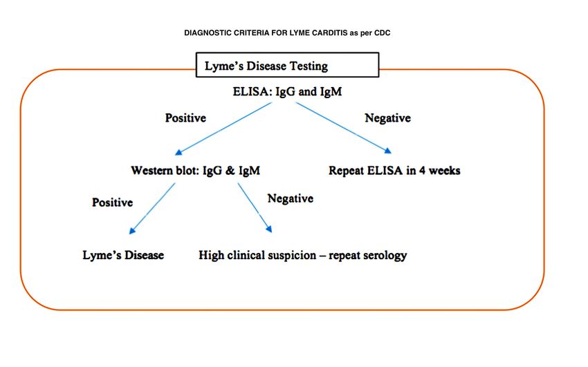
incidence of Lyme carditis is unknown; the carditis usually goes undiagnosed because heart
biopsies are rarely performed [1].
Previous studies suggest that only up to 50% of cases present with the characteristic erythema
migrans rash [2]. The postulated mechanism is that bacterial spirochetes (Borrelia burgdorferi or
Borrelia mayonii) infect the pericardium or the myocardium, triggering an inflammatory
response, which results in Lyme carditis [3]. Although only a few cases of untreated Lyme
disease develop cardiac manifestations, Lyme carditis is notorious for causing sudden cardiac
death. Therefore, a high clinical suspicion of Lyme carditis is required when someone from a
Lyme endemic region presents with unexplained cardiac symptoms and has EKG findings
suggestive of carditis.
Here we present a case of a 56-year-old male who presented with lightheadedness, chest pain,
and bradycardia.
Case Presentation
A 56-year-old male with a history of hypothyroidism, from the Northeastern region of the
United States, presented to the emergency department with lightheadedness, chest pain, and
bradycardia with a heart rate of 33 beats per minute. On the morning of admission, the patient
walked to the bathroom and began experiencing intermittent left-sided chest pain prompting
him to come to the hospital. The chest pain was rated 2 out of 10 in intensity, was localized to
the left side, was described as a heavy sensation, and was non-radiating. The patient denied
having shortness of breath, cough, nausea, vomiting, or diaphoresis. He never experienced
previous episodes of chest pain on exertion or at rest. A few weeks prior to admission, the
patient began experiencing occasional mild headaches and lightheadedness upon standing and
walking. He began taking 50 mcg of levothyroxine four months ago. He did not have a family
history of sudden cardiac death or coronary artery disease. He denied any history of smoking,
alcohol, or drug use. He denied any recent travel. A ten-point review of systems was completed
and negative except as above.
On presentation, his vitals were as follows: temperature 98.2˚F, a pulse of 33 beats per minute,
respiratory rate of 18 breaths per minute, blood pressure of 131/71 mmHg, and oxygen
saturation of 97% on room air. On exam, the patient was in no acute distress. He had no jugular
vein distention (JVD) or carotid bruits. His S1 and S2 heart sounds were normal and no
murmurs were heard on auscultation. The remainder of the physical exam was within normal
limits. His laboratory results were as follows: first troponin of 0, a second troponin (six hours
after the first troponin) of < 0.01, a third troponin (six hours after the second troponin) of <
0.01, a neutrophil percentage of 44.9 (50% -70%), lymphocyte percentage of 43.4 (25% - 43%),
monocyte percentage of 9.3 (0.0% - 0.9%), free T4 of 0.93 ng/dl (0.5 - 1.26 ng/dl ), and TSH of
1.188 IU/ml (0.3 - 4.5 IU/ml). A lipid panel showed a high-density lipoprotein (HDL) level of 34
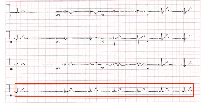
(39 - 79 mg/dl), very low-density lipoproteins (VLDL) of 29 (FIGURE 1: Patient’s electrocardiogram (EKG) revealing a 1.8
second sinus pause and sinus bradycardia without any
atrioventricular (AV) conduction defects.
His chest X-ray showed no acute pulmonary disease. A transthoracic echocardiogram was
performed, which showed a mildly reduced left ventricular systolic function with an ejection
fraction of 51%-54%. As there was a low probability of acute coronary syndrome in this patient
with negative cardiac enzymes and no suggestive EKG changes, ST-elevation myocardial
infarction and non-ST-elevation myocardial infarction were ruled out. Considering that the
patient had unexplained cardiac symptoms, with persistent bradycardia, normal thyroid
function studies, and lives in a Lyme endemic region, an ELISA Lyme titer was obtained and
found to be elevated at 2.89 (0-0.9). A confirmatory Lyme Western blot was completed, which
showed the patient was positive for IgG (positive bands included: 18, 28, 39, 41, 45, 58, 66, 93)
and negative for IgM (band 23 was positive), based on the Centers for Disease Control and
Prevention (CDC) criteria [4].
At this point, a detailed retrospective history from the patient unveiled that he remembered
having ticks crawling on him. However, he denied any noticeable insect bites, fever, rash, and
joint pains. Infectious disease was consulted, and it was presumed that the patient had Lyme
carditis and was treated with intravenous (IV) ceftriaxone for the next seven days. His heart
rate remained consistently low, dropping to 20-30 beats per minute throughout the night with
several sinus pauses while sleeping, the longest having lasted for 6.1 seconds. It was
determined that a pacemaker would be the preferred choice of treatment for his resistant
bradycardia secondary to Lyme carditis. The pacemaker was placed and the patient received an
additional three-week course of ceftriaxone therapy. The patient was discharged and asked to
follow-up with his primary care physician, cardiology, and infectious disease as an outpatient.
At his follow-up appointment, no symptoms were reported. He denied having chest pain,
lightheadedness, fever, or joint pains. His heart rate was stable at 62 beats per minute. The
patient completed the one-month course of antibiotics and is following up with cardiology
regularly to check his pacemaker.
Discussion
In 1980, Steer et al. first described Lyme carditis in a case series of 20 patients who presented
with fluctuating degrees of heart block [3]. In addition to having heart block, the patients also
2019 Grella et al. Cureus 11(9): e5554. DOI 10.7759/cureus.5554 3 of 11presented with symptoms that mimicked acute rheumatic fever. These patients were ultimately
found to have Lyme carditis, a tick-borne disease caused by infection of the heart muscle by the
spirochete bacteria Borrelia burgdorferi and Borrelia mayonii. The study showed that Lyme
carditis usually develops two to three months after an untreated tick bite [5].
According to Fish et al., Lyme carditis is commonly seen between the months of June and
December. Though 4%-10% of untreated Lyme disease progresses to Lyme carditis, an
estimated 30% have asymptomatic carditis [1].
Clinical manifestations
Patients with Lyme disease may present with the characteristic erythema migrans rash and flu-
like symptoms including fever, headache, myalgias, and arthralgias. If left untreated, cardiac
manifestations can develop within 21 days of onset of erythema migrans. Approximately 4%-
10% of people infected with Lyme disease develop symptoms from cardiac involvement. A
review of Lyme carditis cases carried out by the CDC revealed that the patients reported the
following symptoms: palpitations (69%), conduction abnormalities (19%), myocarditis (10%),
left ventricular systolic dysfunction (5%), and hospitalization (21%) [1].
The typical clinical manifestation of Lyme carditis is attributed to a self-limiting AV conduction
abnormality that presents as dizziness, syncope, and shortness of breath with or without chest
pain [6]. Atrioventricular blocks can vary from a first-degree heart block to a fatal third-degree
heart block or asystole. The spirochete can affect any cardiac muscle layer and the patient may
present with pericardial effusion, congestive heart failure, tachyarrhythmia, or QT
prolongation. Approximately 35% of patients present with bradycardia and 15% present with
tachycardia. Other common cardiac manifestations include myopericarditis, intraventricular
conduction disturbances, and bundle branch blocks. Patients who develop Lyme carditis can
present with subtle symptoms including lightheadedness, fainting, palpitations, chest pain,
and shortness of breath. On physical examination, it is common to have bradycardia, signs of
congestive heart failure with canon “a” waves in jugular venous pressure, erythema migrans,
monoarthritis, and sometimes central nervous system manifestations including
meningoencephalitis, cranial nerve palsies, etc.
Serology
Determining if a patient has Lyme disease requires serologic testing. First, an enzyme-linked
immunosorbent assay (ELISA) or enzyme-linked fluorescent immunoassay (ELFA) is performed
[7]. Presence of IgM antibodies on ELISA indicates an active infection that has been present for
up to four weeks. Presence of IgG antibodies on ELISA indicates a chronic infection that has
been present for a more extended period (four to six weeks). It is important to note that there
may be false positives, especially because ELISA can be positive in other medical conditions.
Next, Lyme disease is confirmed using a Western Blot [4]. This is a highly specific test used for
the diagnosis of Lyme disease. Although serological testing initially can give false negative
results, repeat serology must be performed within two to six weeks in a patient with a strong
suspicion of Lyme carditis [8].
According to the CDC, a positive IgM Western blot occurs when at least two of three bands are
positive (21-24, 39, and 41kDa) and a positive IgG Western blot occurs when at least five of 10
bands are positive (18, 21-24, 28, 30, 39, 41, 45, 58, 66, and 93kDa). According to German
Borreliosis Society guidelines, the presence of any one of following highly specific antibodies is
interpreted as Lyme disease: p18, p21, p22, p23, p24, p25, p39, p58 (Tables 1-2).
2019 Grella et al. Cureus 11(9): e5554. DOI 10.7759/cureus.5554 4 of 11CDC Western Blot Criteria
A positive IgM is sufficient to diagnose in
Positive 2 of the following 3 bands are present: 24 kDa (OspC) 39 kDa
early Lyme disease (< 4 weeks of onset of
IgM (BmpA) 41 kDa (Fla)
symptoms).
5 of the following 10 bands are present: 18 kDa 21 kDa (OspC) 28
Positive At any point in infection, a positive IgG is
kDa 30 kDa 39 kDa (BmpA) 41 kDa (Fla) 45 kDa 58 kDa (not
IgG diagnostic of Lyme disease.
GroEL) 66 kDa 93 kDa
TABLE 1: CDC interpretation and diagnostic criteria for Lyme disease.
CDC - Centers for Disease Control and Prevention.
Borrelia Antigen Specificity
p18 High
p21 High
p22, 23, 24, 25 High
p39 High
p41 Unspecific
p58 High
p66 Unspecific
TABLE 2: Antigen specificity in Western blot (Deutsche Borreliose-Gesellschaft e.v.,
2010)*.
* Refer [2].
However, it is crucial to keep in mind not to interpret the presence of fewer bands as a positive
serology in Lyme disease, as antibodies to several borrelial antigens are cross-reactive with
non-borrelial antigens. Therefore, having less than two IgM bands or less than four IgG bands
are not indicative of Lyme carditis by CDC criteria.
Diagnostic criteria for Lyme carditis
Below are the diagnostic criteria for Lyme carditis [9].
1. Clinical picture: New AV conduction defects or arrhythmias. Signs and symptoms of
perimyocarditis, history of tick bite and erythema chronicum migrans.
2019 Grella et al. Cureus 11(9): e5554. DOI 10.7759/cureus.5554 5 of 112. Laboratory testing: Borrelia burgdorferi antibodies in serum.
3. Specific testing: Myocardial biopsy - presence of Borrelia DNA in the polymerase chain
reaction (PCR)
Below are the diagnostic criteria for Lyme carditis as per the CDC (Figure 2).
FIGURE 2: CDC's diagnostic criteria for Lyme disease.
CDC - Centers for Disease Control and Prevention.
The suspicious index in Lyme carditis (SILC) score
In a recent article, Besant et al. proposed a new scoring system after retrospectively reviewing
84 cases of confirmed Lyme carditis (Table 3). The suspicious index in Lyme carditis (SILC)
score was developed to evaluate the possibility that a patient's high‐degree heart block can be
attributed to Lyme carditis. A SILC score of 0-1 is low, 3-6 is intermediate, and 7-12 is high
suspicion for Lyme carditis. The sensitivity of SILC risk of Lyme carditis is 93.2%, but no
specificity was reported. Our patient’s score was four (male, endemic area, presyncope), which
puts him under intermediate risk of Lyme carditis [10, 11].
2019 Grella et al. Cureus 11(9): e5554. DOI 10.7759/cureus.5554 6 of 11Patient's Characteristics Score
Age < 50 1
Male 1
Outdoor activity/endemic area 1
Constitutional symptoms (malaise, fever, arthralgias, dyspnea, pre syncope and syncope) 2
Tick bite 3
Erythema migrans 4
TABLE 3: The suspicious index in Lyme carditis scoring system as proposed by
Besant et al.*.
*Refer [11].
Histopathology
Endomyocardial biopsy is the gold standard used to diagnosis Lyme carditis [12]. A typical
biopsy specimen will show characteristic transmural inflammatory infiltrates, with a band-like
endocardial lymphocyte infiltration [9]. Occasionally the spirochete can be found within the
muscle fibers, within the myocardium, or in the inflammatory infiltrates. Polymerized chain
reaction (PCR) of the endomyocardial biopsy specimen can also establish the diagnosis of Lyme
carditis, but a negative PCR does not exclude Lyme carditis.
The possible mechanism for cardiac damage in Lyme carditis is attributed partly to the
inflammatory reaction induced by the spirochete bacteria in addition to the direct cytotoxic
effect of the spirochete itself.
The original understanding of the pathogenesis of Lyme carditis has been through rat models.
Spirochete inoculated mice show peak inflammatory changes in the first two to three weeks of
infection. After a month of inoculation, inflammatory infiltrates were found in the aortic root,
the base of the heart, the atrial and ventricular epicardium, the endocardium, the myocardium,
and in perivascular spaces especially the AV junction. Unlike neutrophil predominance as seen
in Lyme arthritis, macrophage predominance is the typical histopathological picture seen in
murine Lyme cardiac models. Immunofluorescence technique can be used to demonstrate the
presence of spirochete in the cardiac muscle [1, 13].
EKG
According to Steer et al., a 12-lead EKG and Holter monitor can show ST-segment depression,
T-wave inversions in inferolateral leads, and AV conduction abnormalities [5, 9]. However, the
most common findings are AV conduction abnormalities. First degree AV block was seen in 90%
of patients, and complete heart block was observed in 44% of patients. A progression of first-
degree AV block to complete AV block was very common when the PQ interval was > 300 msec.
Our literature search revealed that 87% of patients with Lyme carditis developed a first degree
AV block, and 53% of patients had a complete AV block or Mobitz type block.
2019 Grella et al. Cureus 11(9): e5554. DOI 10.7759/cureus.5554 7 of 11In our literature review, we found that very few cases of Lyme carditis have undergone
electrophysiological studies as a part of the workup for Lyme carditis [12]. The cases that have
undergone electrophysiological studies showed that heart block (AV block) occurred above the
bundle of His and at the same time simultaneous multiple blocks were also noted in SA node,
atrium, bundle branches, and fascicles. Furthermore, Lyme carditis patients can also present
with a sino-atrial block, temporary bundle block, paroxysmal atrial fibrillation, and prolonged
QT interval. AV block development in Lyme carditis patents is attributed to the host's immune
response to B. burgdorferi infection of the myocardial tissue.
Imaging
Echocardiography can provide valuable information on wall motion abnormalities [9]. Though
echocardiographic findings are not explicit for Lyme carditis, echocardiography is an excellent
diagnostic tool for evaluating the presence and degree of cardiac dysfunction and therefore can
provide essential information for the management of these patients. Left ventricular wall
motion dysfunction, especially abnormal ventricular wall kinetics, on echocardiogram can
indicate myocarditis.
Cardiac MRI is another diagnostic modality that can provide valuable information about
pericardial involvement [14]. For example, wall edema, which presents as increased signal
density on T1 weighted images, corresponds to the inflammatory process triggered by the
spirochete.
Treatment
A gray zone exists in regards to the treatment of Lyme disease because every patient does not
present with the characteristic rash and symptoms of Lyme disease. Consequently, previous
studies and their results cannot be generalized due to the high variability of clinical
presentation and population heterogeneity [15].
There are two separate guidelines put forth in regards to Lyme disease treatment. The
Infectious Diseases Society of America (IDSA) recommends that patients be started on a short
course of antibiotics as the persistent infection is infrequent or non-existent [16]. However, the
International Lyme and Associated Diseases Society (ILADS) recommends the Grading of
Recommendations, Assessment, Development, and Evaluation (GRADE) system. This system
emphasizes that a prolonged course of antibiotics be required, keeping in mind the high failure
rates from a short course of antibiotics and the high prevalence of disseminated disease in a
large number of cases.
Treatment of Lyme carditis is principally based on the severity of conduction disease [17].
Patients with Lyme carditis who do not have high-grade heart block are managed conservatively
with oral antibiotics. The duration of antibiotic management depends primarily on adequate
shortening of the PR interval and the normalization of AV block.
On the other hand, patients with a high-grade heart block should be hospitalized [16], closely
monitored, and treated with IV ceftriaxone 2 grams or IV penicillins for second/third-degree AV
block or prolonged for PR interval > 300 ms. As per the European Federation of Neurological
Societies (EFNS), ceftriaxone or cefotaxime for two weeks is the recommended standard of care
in acute Lyme carditis [18].
Although many Lyme carditis patients often recover well within a week of antibiotic treatment,
it is often recommended to treat acute cases of Lyme carditis with a two-week course of
ceftriaxone to eliminate the spirochete from blood and heart muscle [19]. Temporary pacing
2019 Grella et al. Cureus 11(9): e5554. DOI 10.7759/cureus.5554 8 of 11may be required in patients with an advanced degree of heart block who cannot be managed
conservatively.
The following antibiotic regimens are recommended by the CDC, ILADS, IDSA, and German
Borreliosis Society (Tables 4-5).
Lyme Carditis Treatment Regimens
Centers for Disease Control and Prevention (CDC)
Doxycycline 100 mg orally twice daily for 10-21 days or Cefuroxime Axetil 500 mg orally twice daily for 14-21 days or
Amoxicillin 500 mg orally three times daily for 14-21 days
International Lyme and Associated Diseases Society (ILADS)
Amoxicillin 1500-2000 mg orally daily in divided doses for 4-6 weeks or Cefuroxime 500 mg orally twice daily for 4-6
weeks or Doxycycline 100 mg orally twice daily for for 4-6 weeks or Azithromycin 250-500 mg orally daily for 21 days
Infectious Disease Society of America (IDSA)
Preferred
Ceftriaxone 2 grams once per day via IV for 14 days with a range of 10-28 days
Treatment
Cefotaxime 2 grams IV every 8 hours or Penicillin G 18-24 million units per day in patients with normal
Alternative
renal function divided into doses given every 4 hours or Doxycycline 200-400 mg per day in 2 divided doses
Treatments
orally for 10-28 days for patients intolerant of B-lactam antibiotics
TABLE 4: Antibiotic regimens recommended by the CDC, ILADS, and IDSA for the
treatment of Lyme carditis.
CDC - Centers for Disease Control and Prevention; ILADS - International Lyme and Associated Diseases Society; IDSA - Infectious
Disease Society of America.
2019 Grella et al. Cureus 11(9): e5554. DOI 10.7759/cureus.5554 9 of 11Antibiotics applicable in Lyme disease (Deutsche Borreliose-Gesellschaft e.v., 2010)[2]
Beta lactams: Ceftriaxone, Cefotaxime, Cefuroxime Axetil, Benzathine benzylpenicillin, Phenylmethyl Penicillin,
Amoxicillin
Tetracyclines and glycylcyclines: Doxycycline, Minocycline
Macrolides: Clarithromycin, Azithromycin
Nitromidazoles: Metronidazole
Co-drugs: Hydroxychloroquine
TABLE 5: German Borreliosis Society recommendations for the treatment of Lyme
disease.
Prognosis
There should be a high clinical suspicion of Lyme carditis in patients with unexplained
syncopal episodes, dyspnea, and chest pain, who live in a region where Lyme disease is
prevalent [19]. Patients with second- or third-degree AV block or first-degree AV block with a
prolonged PR > 300 ms should be admitted and closely monitored because these patients can
deteriorate rapidly [20]. In almost 35% of cases, patients require temporary heart pacing.
However, heart block associated with Lyme carditis resolves with antibiotics and seldom
requires a permanent pacemaker. Typically, after one to five weeks of treatment one can expect
resolution of symptoms.
Several studies funded by the National Institutes of Health (NIH) on the treatment of Lyme
disease have shown that most people recover well within a few weeks of initiation of oral
antibiotics. A few cases have reported mortality from sudden cardiac death due to lymphocytic
infiltration of the heart [5, 6]. However, if prompt admission and antibiotic treatment are
initiated, the overall prognosis of Lyme carditis is good.
Conclusions
Here we present a unique presentation of Lyme carditis, without any classical findings of Lyme
disease such as rash or any common EKG findings of heart block. This case report adds
knowledge to the gap between suspicion of Lyme carditis and sinus bradycardia as the only
presenting symptom.
Additional Information
Disclosures
Human subjects: Consent was obtained by all participants in this study. Conflicts of interest:
In compliance with the ICMJE uniform disclosure form, all authors declare the following:
Payment/services info: All authors have declared that no financial support was received from
any organization for the submitted work. Financial relationships: All authors have declared
that they have no financial relationships at present or within the previous three years with any
organizations that might have an interest in the submitted work. Other relationships: All
authors have declared that there are no other relationships or activities that could appear to
have influenced the submitted work.
2019 Grella et al. Cureus 11(9): e5554. DOI 10.7759/cureus.5554 10 of 11Acknowledgements
We would like to thank Dr. Pramil Cheriyath and Dr. Kelly Ussery-Kronhaus for their
contributions to this article.
References
1. Fish AE, Pride YB, Pinto DS: Lyme carditis. Infect Dis Clin North Am. 2008, 22:275-288.
10.1016/j.idc.2007.12.008
2. Diagnosis and treatment of Lyme borreliosis (Lyme disease). [Article in German] . Duetsche
Borreliose-Gesellschaft e.V. 2010, Accessed: February 6, 2019: http://www.borreliose-
gesellschaft.de/assets/files/Leitlinien.pdf.
3. Steere AC, Grodzicki RL, Kornblatt AN, et al.: The spirochetal etiology of Lyme disease . N Engl
J Med. 1983, 308:733-740. 10.1056/NEJM198303313081301
4. Steere AC, Batsford WP, Weinberg M, Alexander J, Berger HJ, Wolfson S, Malawista SE: Lyme
carditis: cardiac abnormalities of Lyme disease. Ann Intern Med. 1980, 93:8-16. 10.7326/0003-
4819-93-1-8
5. Torbey E, Nalluri N, Boutros K, Agarwal V, Bekheit S: Conduction riddles of Lyme carditis: a
case series. J Innov Card Rhythm Manag. 2015, 2004-2007. 10.19102/icrm.2015.060505
6. Tugwell P, Dennis DT, Weinstein A, et al.: Laboratory evaluation in the diagnosis of Lyme
disease. Ann Intern Med. 1997, 127:1109-1123. 10.7326/0003-4819-127-12-199712150-00011
7. Dressler F, Whalen JA, Reinhardt BN, Steere AC: Western blotting in the serodiagnosis of
Lyme disease. J Infect Dis. 1993, 167:392-400. 10.1093/infdis/167.2.392
8. Hinckley AF, Connally NP, Meek JI, et al.: Lyme disease testing by large commercial
laboratories in the United States. Clin Infect Dis. 2014, 59:676-681. 10.1093/cid/ciu397
9. Scheffold N, Herkommer B, Kandolf R, May AE: Lyme carditis--diagnosis, treatment and
prognosis. Dtsch Arztebl Int. 2015, 112:202-208. 10.3238/arztebl.2015.0202
10. Jiménez-Castillo RA, Carrizales-Sepúlveda EF, Vera-Pineda R, et al.: A tick beat in the
electrocardiogram: persistent third degree block as only manifestation of Lyme disease. J
Electrocardiol. 2019, 52:109-111. 10.1016/j.jelectrocard.2018.12.005
11. Besant G, Blakely C, Wan D, et al.: Suspicious index in Lyme carditis (SILC) score . Can J
Cardiol. 2018, 34:S188.
12. Reznick JW, Braunstein DB, Walsh RL, et al.: Lyme carditis. Electrophysiologic and
histopathologic study. Am J Med. 1986, 81:923-927. 10.1016/0002-9343(86)90370-0
13. Barthold SW, Hodzic E, Tunev S, Feng S: Antibody-mediated disease remission in the mouse
model of Lyme borreliosis. Infect Immun. 2006, 74:4817-4825. 10.1128/IAI.00469-06
14. Jacobs JC, Rosen JM, Szer IS: Lyme myocarditis diagnosed by gallium scan . J Pediatr. 1984,
105:950-952. 10.1016/S0022-3476(84)80086-4
15. Johnson L, Stricker RB: The Infectious Diseases Society of America Lyme guidelines: a
cautionary tale about the development of clinical practice guidelines. Philos Ethics Humanit
Med. 2010, 5:9.
16. Wormser G, Dattwyler R, Shapiro E, et.al: Erratum: The clinical assessment, treatment, and
prevention of Lyme disease, human granulocytic anaplasmosis, and babesiosis: clinical
practice guidelines by the Infectious Diseases Society of America. Clin Infect Dis. 2007,
45:941. 10.1086/521856
17. Olson LJ, Okafor EC, Clements IP: Cardiac involvement in Lyme disease: manifestations and
management. Mayo Clin Proc. 1986, 61:745-749. 10.1016/S0025-6196(12)62775-X
18. Mygland A, Ljostad U, Fingerle V, Rupprecht T, Schmutzhard E, Steiner I: EFNS guidelines on
the diagnosis and management of European Lyme neuroborreliosis. Eur J Neurol. 2010, 17:8-
16. 10.1111/j.1468-1331.2009.02862.x
19. Rosenfeld ME, Beckerman B, Ward MF, Sama A: Lyme carditis: complete AV dissociation with
episodic asystole presenting as syncope in the emergency department. J Emerg Med. 1999,
17:661-664. 10.1016/S0736-4679(99)00061-X
20. Xanthos T, Lelovas P, Kantsos H, Dontas I, Perrea D, Kouskouni E: Lyme carditis: complete
atrioventricular dissociation with need for temporary pacing. Hellenic J Cardiol. 2006, 47:313-
316.
2019 Grella et al. Cureus 11(9): e5554. DOI 10.7759/cureus.5554 11 of 11You can also read