Kawasaki Disease in the neonate: case report and literature review
←
→
Page content transcription
If your browser does not render page correctly, please read the page content below
Altammar and Lang Pediatric Rheumatology (2018) 16:43
https://doi.org/10.1186/s12969-018-0263-8

 CASE REPORT                                                                                                                                     Open Access

Kawasaki Disease in the neonate:
case report and literature review
Fajer Altammar1 and Bianca Lang2*

  Abstract
  Background: Kawasaki Disease (KD), the leading cause of acquired heart disease in children in the developed
  world, is extremely rare in neonates. We present a case of incomplete KD in a neonate and a review of the
  literature on neonatal KD.
  Case presentation: A previously healthy full term 15 day old Caucasian male with an unremarkable antenatal and
  perinatal history, presented on Day 2 of illness with fever, rash, irritability, and poor feeding. Examination revealed
  fever (39.6C), tachycardia, tachypnea, extreme irritability, and a generalized maculopapular rash, but was otherwise
  normal. His complete blood count, CRP and ESR were normal. Empiric intravenous antibiotics and acyclovir resulted
  in no improvement. On day 4, he had ongoing fever and developed recurrent apnea, required supplemental
  oxygen, and was transferred to the pediatric intensive care unit. On day 6, he developed bilateral non-purulent
  conjunctivitis, palmar erythema, bilateral non-pitting edema and erythema of his feet, and arthritis. His full septic
  work-up and viral studies were negative. On Day 7 he was treated with intravenous immunoglobulin, and over the
  next 48 h his symptoms including extremity edema resolved, he no longer required supplemental oxygen, and
  fever did not recur. On day 9 of illness he had marked thrombocytosis. Subsequently, he developed distal extremity
  desquamation. Repeated echocardiograms excluded the presence of coronary artery aneurysms (CAA).
  Conclusions: We believe this to be a rare case of incomplete KD in a neonate, in which timely IVIG administration
  led to resolution of the acute illness and may have prevented CAA. A comprehensive English-language medical
  literature review of KD presenting in the neonatal period revealed only fifteen case reports. Cases often presented
  with incomplete KD, and a number had atypical laboratory features including a normal CRP in the acute phase,
  similar to what was seen in our patient. This case and our literature review should increase awareness that KD can
  rarely occur in neonates, often presenting atypically. Recognizing KD in a neonate enables appropriate treatment
  that can result in resolution of symptoms and may decrease the risk of cardiac complications.
  Keywords: Kawasaki, Infant, Diagnosis, Newborn, Neonate

Background                                                                            conjunctivitis, a nonspecific rash, erythema and edema
Kawasaki Disease (KD), a vasculitic syndrome of early                                 of the feet and hands or periungual desquamation (sub-
childhood with a predilection for the coronary arteries,                              acute phase), and cervical lymphadenopathy (≥1.5 cm).
is now the leading cause of acquired heart disease in                                 Incomplete KD is diagnosed in patients with unex-
children in the developed world. The underlying etiology                              plained prolonged fever and less than 4 criteria, if they
is still unknown, and there is no definitive diagnostic                               have typical echocardiographic findings of KD or typical
test. The diagnosis of complete KD is based on the                                    laboratory features [1]. Prompt treatment of KD patients
presence of ≥ 5 days of fever and 4 or more of the prin-                              with IVIG has been shown to reduce the incidence of cor-
cipal clinical criteria including erythematous changes of                             onary artery aneurysms from 25% to less than 5% [1–3].
the lips and oral mucosa, bilateral non-purulent                                        The age of onset of KD is usually between 6 months
                                                                                      and 5 years of age. It is much less common under
* Correspondence: bianca.lang@iwk.nshealth.ca                                         6 months of age and rare in children under 3 months of
2
 Division of Rheumatology, Department of Pediatrics, IWK Health Centre,               age. It is especially rare in the neonatal period, with only
Dalhousie University, 5980 University Ave, Halifax, NS B3K 6R8, Canada
Full list of author information is available at the end of the article                a few neonatal cases ever reported in the literature. Even

                                        © The Author(s). 2018 Open Access This article is distributed under the terms of the Creative Commons Attribution 4.0
                                        International License (http://creativecommons.org/licenses/by/4.0/), which permits unrestricted use, distribution, and
                                        reproduction in any medium, provided you give appropriate credit to the original author(s) and the source, provide a link to
                                        the Creative Commons license, and indicate if changes were made. The Creative Commons Public Domain Dedication waiver
                                        (http://creativecommons.org/publicdomain/zero/1.0/) applies to the data made available in this article, unless otherwise stated.
Altammar and Lang Pediatric Rheumatology (2018) 16:43 Page 2 of 6
in Japan, where KD is much more prevalent than else-
where in the world, very few neonates with KD have
been identified. Data on 105,755 KD patients from the
first 12 Japanese nationwide surveys of KD (1970–1992),
identified 1768 KD cases (1.67%) 90 days old or younger.
Of these, only six cases were of age 30 days old or youn-
ger, with the youngest being 26 days old [4]. In a review
of the Japanese nationwide surveys of KD from 2001 to
2012, 23 neonates were identified, representing 0.02% of
KD patients overall [5]. We present a case of suspected
incomplete KD in a neonate and a review of the litera-
ture on neonatal KD with the aim to increase awareness
that KD can occur in the first month of life and may
present with atypical features.
Case presentation
A previously healthy full term 15 day old Caucasian
male with an unremarkable antenatal, perinatal, and
family history presented on Day 2 of illness with a 24 h
history of poor feeding, irritability, fever, and rash.
Examination revealed fever (39.6 C), tachycardia (HR
180–210), tachypnea (RR 68), extreme irritability, acro-
cyanosis, and a generalized maculopapular rash, but
otherwise was normal. Admission blood work revealed a
normal complete blood count, slightly elevated serum
transaminase levels, mild hypoalbuminemia (30 g/L),
and a normal c-reactive protein (CRP) of 2.6 mg/L. Em-
piric treatment with intravenous ampicillin, cefotaxime,
and acyclovir was given for presumed neonatal sepsis
and possible encephalitis. On day 4 of illness, in addition
to ongoing fever, he developed recurrent apnea and re-
quired supplemental oxygen and transfer to the tertiary
care hospital pediatric intensive care unit. His blood
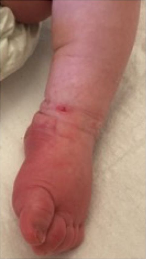
Fig. 1 Photograph demonstrating extremity changes including
work showed mild neutropenia, elevation of serum
marked non-pitting edema and erythema of foot
transaminase levels (alanine transaminase 86 U/L, aspar-
tate transaminase 220 U/L), a normal ESR (1 mm/hr)
and hypoalbuminemia. An infectious etiology was con- he no longer required supplemental oxygen, and fever
sidered unlikely given negative bacterial cultures from did not recur. His CRP remained normal throughout his
blood, cerebral spinal fluid, and urine, and negative viral illness. Laboratory values improved and his platelet
studies for herpes simplex virus 1 and 2, Respiratory count progressively increased from 246,000 on Day 7 to
Syncytial Virus, Influenza A and B, cytomegalovirus, Ep- 852,000 on Day 13 of illness. In the subacute phase, he
stein Barr virus, rubella, adenovirus, and rotavirus. The was treated with low dose aspirin. He had persistent
urine and blood cultures were collected before the start thrombocytosis followed by typical extremity desquam-
of antibiotics. His chest radiograph was normal. On day ation. Echocardiograms on weeks 1, 2, and 6, and at
6 of illness, he developed bilateral non-purulent con- 12 months were normal with no carditis or coronary ar-
junctivitis, palmar erythema, striking bilateral tery abnormalities.
non-pitting edema and erythema of his feet, and ery-
thema and swelling of several proximal interphalangeal Discussion and conclusions
joints (Fig. 1). He continued to require supplemental We report a rare case of incomplete KD in a neonate,
oxygen for suspected pneumonitis. On Day 7 of illness, where timely IVIG administration resulted in clinical
after 5 days of fever and meeting 3 out of 5 criteria for improvement and may have prevented cardiac complica-
KD, with no other obvious diagnosis, he was given IVIG tions. Our patient had fever documented for 5 days and
(2 g/kg) for suspected incomplete KD. Over the next 3 additional principal clinical features including rash,
48 h, his symptoms including extremity edema resolved, bilateral non-purulent conjunctivitis, and very typicalAltammar and Lang Pediatric Rheumatology (2018) 16:43 Page 3 of 6
and pronounced non-pitting edema and erythema of his relevant publications were also reviewed to identify fur-
feet, as well as palmar erythema, and desquamation of ther cases that were not included in the databases. The
the feet in the subacute phase. Other clinical features in- neonatal period was defined as patients from birth to
cluded extreme irritability, arthritis, apnea, and respira- 28 days of age.
tory distress. Because of the rarity of KD in the neonatal Our literature search revealed only a few reported
period, a bacterial or viral etiology was suspected on ad- cases of suspected neonatal KD worldwide (15 cases)
mission, and the diagnosis of KD was not initially con- (Table 1), confirming the rarity of this disorder, and con-
sidered. However, after he had failed to improve with sistent with the very few cases identified in Japan
antibiotic and antiviral therapy, there was dramatic im- through nationwide surveys [4–15].
provement of clinical features following IVIG. Although The infrequent occurrence of neonatal KD is consist-
our patient did not have echocardiographic abnormal- ent with the current theory of an infectious trigger for
ities to confirm a diagnosis of KD, he did have 3 typical KD and the protection that most very young infants
laboratory findings including elevated ALT and AST have through passive immunity transferred from their
levels, hypoalbuminemia, and marked thrombocytosis mother. The demographic and clinical features of the
starting on day 9 of illness. Our literature review encom- babies with suspected KD identified in our literature
passed a thorough search of the electronic databases of review are presented in Table 1. In all the presented
PubMed and Embase for KD presenting in the neonatal cases including ours, most common infectious etiologies
period. The key terms used in the search included new- were excluded and all patients were empirically treated
borns, neonates, week-old, weeks old baby, babies, and for sepsis. Four patients had complete KD, all with docu-
Kawasaki or mucocutaneous lymph node syndrome. mented CAA. Of the 12 patients with fewer than 4 of 5
Only English articles were reviewed. The references of KD criteria, 6 infants, including our case, had fever with
Table 1 Literature Review: Summary of the Case Reports of Neonatal Kawasaki Disease (0–28 days of age)
Pt Ref Age at Sex Fever Rash Oral Extremity Red Cervical CAA Other CRP IVIG res- CA outcome
onsetb duration changes changesd eyes adenitis c
(high/ ponse (last F/U)
(days) (days) WNL)
1 4 20 F 10 + + + + – + DIC high + WNL (8mos)
2 6 10 M 5 + + + + – + Apnea, seizures WNL + CAA (2.5 yrs)
3 7 8 F 9 + + + + – + MR/AR high + WNL (6 wks)
4 8 16 F 13 + + + + – + MR/TR high + N/A
5 9 20 F N/A + + + – – + MR/AR high + CAA (8 wks)
6 10 8 M >9 + – + – – + Myocardial high + CAA (9 wks)
ischemiaa
7 5 22 F 4 + – + – – – – high + WNL (3 mos)
8 11 18 M 6 + + + – – – Pneumonitis WNL + WNL (6 wks)
9 11 16 M 7 + + + – – – Cough, high + WNL (11 yrs)
diarrhea
10 12 21 F 4 + + + – – – Abdominal WNL + WNL (6 wks)
pain vomiting
11 12 14 F 3 + + + – – – Hyperemia WNL + WNL (6 mos)
12 12 16 M 4 + + + – – – Hyperemia WNL + WNL
13 13 5 M 1 – – – – – + CHF, MR/AR high + WNL(12mos)
14 14 1 M 0 – – – – – + DIC, pericardial N/A N/A WNL(12mos)
effusion
15 15 1 F 0 – – – – – + MI, CA N/A N/A death (day1)
vasculitis
16 PR 15 M 5 + – + + – – Apnea WNL + WNL(12mos)
pneumonitis
CAA coronary artery abnormality, CRP C-reactive protein, IVIG intravenous immunoglobulin, DIC disseminated intravascular coagulation, CHF congestive heart
failure, AR aortic regurgitation, TR tricuspid regurgitation, MI myocardial infarction, PR present report, N/A not available, MR mitral regurgitation, WNL within
normal limits
a
Patient 6 underwent successful coronary thrombolysis
b
All patients were full term with the exception of patient #14 who was 35.5 weeks
c
Patients 2, 4, 5, 6, and 15 had CA aneurysms; patients 1, 2, 3, 13 and 14 had CA dilatations
d
Patients 1, 2, 3, 4, 5, 8, 9, 10, 11, 12 and 16 had distal edema; patients 1, 2, 3, 5, 6, 7, 8, 11 and 16 had desquamationAltammar and Lang Pediatric Rheumatology (2018) 16:43 Page 4 of 6
2 or 3 criteria, and either CAA, or typical desquamation to the lower likelihood of young infants meeting KD
or thrombocytosis in the sub-acute phase, as well as criteria [19, 20]. Our case report and literature review
resolution of symptoms following IVIG therapy. Three also demonstrate that a significant number of neonates
patients presented with 3–4 days of fever and 3 criteria, with KD display unusual laboratory manifestations, in-
including striking extremity edema and erythema, and cluding a normal CRP in the acute phase in 6/14 (43%),
had rapid resolution of all symptoms with IVIG treat- and thrombocytopenia in the acute phase of the illness in
ment at 3–4 days, despite not meeting KD criteria [12]. 4/12 (27%). In our case, the presence of a normal CRP on
Three patients with suspected neonatal KD presented days 2 and 4 of his illness led to the early assumption that
within the first 1–5 days of life with CAA suggestive of the diagnosis of KD was unlikely. Elevated inflammatory
KD without prolonged fever or other principal criteria. markers are reported in the vast majority of older infants
While it is not possible to confirm their diagnosis, they with KD, and presence of a normal CRP generally suggests
may represent the spectrum of neonatal KD. Our case an alternative diagnosis [1, 22].
and literature review illustrate that KD in neonates com- Several studies have suggested that infants under the
monly presents with incomplete features (75%). Table 2 age of 6 months not only present more commonly with
summarizes the frequency of the clinical and laboratory incomplete KD, but are also at higher risk for coronary
manifestations of KD in our neonatal cases. None of the artery abnormalities and death [16, 20, 23–25]. Accord-
cases reviewed met the criteria for cervical lymphaden- ing to Hangai et al., from nationwide Japanese surveys,
opathy, and few developed conjunctivitis. Fever, rash, the risk of coronary abnormalities was 17% among the
and extremity changes were reported in most infants, 23 neonates diagnosed with Kawasaki disease [5].
oral changes in close to two thirds, and desquamation Among the neonatal cases we reviewed, the frequency of
during the subacute phase was also common. Our find- cardiac complications was even higher (56%). This may
ings are consistent with the high rate of incomplete KD in part be due to the fact that the diagnosis of KD in
reported in neonates identified in the Japanese nation- three cases was made based on the presence of typical
wide surveys between 2001 and 2012, where only 8 of 23 cardiac abnormalities alone. Krapf et al. described a neo-
infants had complete KD [5]. Our findings are also con- nate who presented with fatal myocardial infarction
sistent with the high frequency of incomplete KD re- without any clinical features of KD, who at autopsy, was
ported in infants less than 6 months of age who are found to have an acute/subacute necrotizing coronary
beyond the neonatal period [16–20]. Moreover, the clin- vasculitis typical of KD [15]. In a more recent case
ical manifestations in young infants can be short-lived report by Parashar et al., bilateral coronary artery dilata-
and may not all manifest at any given time. Patients with tions were detected on the first day of life in the absence
incomplete KD who are less than 6 months of age, espe- of other KD features [14]. Bolz et al. described a baby
cially those lacking eye or oral mucosal changes appear who presented during his first week of life in congestive
to be particularly at risk for delays in diagnosis [21]. The heart failure who was found to have dilatation of his left
absence of cervical lymphadenopathy in patients re- coronary arteries without other KD features [13]. In the
ported with neonatal KD supports the conclusions of latter two cases, the coronary dilatation resolved; in the
Lee and Manlhiot that this criterion is not a sensitive in- second case this occurred after IVIG therapy. As KD is a
dicator of KD in infants under 6 months of age, and sug- clinical diagnosis, it is not possible to confirm the diag-
gests that this may be an important factor contributing nosis of KD in these three infants. However, they did not
have another known cause of CA dilatation, such as
asphyxia, sepsis, congenital cardiac anomaly, other rec-
Table 2 Frequency of key clinical and laboratory features of 16
cases of NKD
ognized systemic vasculitis or systemic juvenile arthritis,
and they did not have prolonged fever, which itself may
Clinical and Laboratory Features Frequency (%)
result in CA dilatation, although this is usually transient,
Fever (any duration) 14/16 88
with Z scores less than 2.5 [1]. The fatal neonatal case
Duration of fever ≥ 5 daysa 8/15 53 reported by Krapf et al. is strikingly similar to cases of
Rash 13/16 81 infantile polyarteritis nodosa (IPN), a uniformly fatal
Extremity changes 13/16 81 vasculitis of infancy with a predilection for CAs. A
Oral changes 10/16 63 review of 20 cases of IPN from North America published
Conjunctivitis 5/16 31
four years before Dr. Kawasaki reported the criteria for
KD in Japan described the clinical features seen in many,
Cervical lymphadenopathy 0/16 0
but not all, infants with IPN, including fever, rash, con-
Cardiac complications 9/16 56 junctivitis, abnormal urinary sediment, and cardiomegaly
Normal CRPb 6/14 43 [26]. A decade after the description of KD, Landing and
a
Duration of fever not reported in 1 case. b
CRP not reported in 2 cases Larson reviewed the clinical and pathologic features ofAltammar and Lang Pediatric Rheumatology (2018) 16:43 Page 5 of 6
20 patients with IPN and compared these with two fatal infants may be the most important intervention for these
cases of KD, and concluded that the two conditions were patients.
clinically and pathologically indistinguishable [27]. Fol- In conclusion, this case, in addition to the cases pre-
lowing the worldwide recognition of KD, reports of IPN sented in our review, illustrate the importance of consid-
essentially disappeared [28]. The neonate reported by ering the diagnosis of KD in the first month of life, as
Krapf et al. had CA pathology typical of IPN and KD, appropriate treatment can result in resolution of symp-
supporting the concept that IPN is the severe end of the toms and a decreased risk of cardiac complications. It is
spectrum of incomplete KD. The absence of fever was important to recognize that neonatal KD, while rare,
recognized years ago in some infants with IPN, and has often poses a diagnostic challenge. Incomplete KD is
recently been reported in three patients suspected to common, and unusual features such as a normal CRP in
have “afebrile KD” [29, 30]. Two of these patients from the acute phase can be seen in a significant number of
Japan had bilateral conjunctivitis, erythema at their BCG cases. It is essential to maintain a high index of suspi-
sites, and CA Z scores ≥ 8.0, while one 3-month infant cion for such a diagnosis in febrile neonates who fail to
from North America had no clinical features of KD respond to antibiotics, while also recognizing the limita-
other than CAA (Z score ≥ 6.0). These cases, as well as tions of applying the AHA guidelines for diagnosis in
the NKD cases we present, challenge us to keep a high this age group.
index of suspicion for KD, especially in young infants, as
Abbreviations
the criteria Dr. Kawasaki developed were not designed ALT: Alanine transaminase; AST: Aspartate transaminase; CAA: Coronary
to identify infants at risk of CAA, but rather, to make a Artery Aneurysms; CRP: C-reactive protein; ESR: Erythrocyte sedimentation
diagnosis of what he believed at the time was a benign, rate; IPN: Infantile polyarteritis nodosa; IVIG: Intravenous immunoglobulin;
KD: Kawasaki Disease; NKD: Neonatal Kawasaki Disease
self-limited febrile illness.
To help clinicians recognize incomplete cases of KD Acknowledgements
and reduce the delay in diagnosis that predisposes to Joseph Saunders provided technical assistance and reviewed the final
written manuscript prior to submission.
CAA, particularly in high-risk infants under the age of
6 months, the American Heart Association (AHA) 2017 Authors’ contributions
guidelines provide recommendations for the evaluation FA and BL made substantial contributions to conception of the report, chart
review, literature review, and manuscript drafting and revisions. Both authors
of suspected incomplete KD [1]. According to the AHA read and approved the final manuscript. FA and BL agree to be accountable
diagnostic algorithm, in children with 5 or more days of for all aspects of the case report.
fever and 2 or 3 compatible clinical criteria, or in infants
Ethics approval and consent to participate
with fever for 7 or more days without another explan- The local research ethics board at the IWK Health Centre was consulted prior
ation, the presence of an elevated CRP or ESR warrants to undertaking this case report. Formal ethics approval for the project was
further laboratory and/or echocardiographic testing for waived; however, as is standard at the IWK Health Centre, written informed
consent was obtained from the parent of the infant presented in the case
KD [1]. In the subgroup of patients whose CRP is less report.
than 3.0 mg/dl and/or ESR < 40 mm/hour, serial clinical
and laboratory re-evaluation is warranted only if fever Consent for publication
As per the IWK Health Centre research ethics board recommendation,
persists, and an echocardiogram is recommended if typ- written “consent to be included in a case report” was obtained from the
ical peeling develops. While this approach may success- parent of the infant reported. The consent form states that information
fully identify older infants with KD, the AHA diagnostic about the child’s illness will be published and available to other people,
including health care professionals and the general public.
algorithm may result in a delay in diagnosis and treat-
ment of neonates with KD, as our literature review and Competing interests
case report illustrate that incomplete features are com- The authors declare that they have no competing interests.
mon and a normal CRP may occur in the acute illness.
Ideally, a diagnostic test for KD will be found, otherwise Publisher’s Note
Springer Nature remains neutral with regard to jurisdictional claims in
more inclusive diagnostic criteria may be needed for this published maps and institutional affiliations.
age group so that the diagnosis of KD is not delayed.
Pannaraj et al. surveyed general pediatricians and Author details
1
Department of Pediatrics, IWK Health Centre, Dalhousie University, 5980
pediatric infectious disease physicians in San Diego University Ave, Halifax, NS B3K 6R8, Canada. 2Division of Rheumatology,
using a questionnaire to understand physician practices Department of Pediatrics, IWK Health Centre, Dalhousie University, 5980
in diagnosing Kawasaki disease and showed that 71 of University Ave, Halifax, NS B3K 6R8, Canada.
124 (57.3%) general pediatricians and 86 of 324 (26.5%) Received: 24 March 2018 Accepted: 20 June 2018
pediatric infectious disease specialists did not consider
the diagnosis of KD in children younger than 6 months
References
of age in their differential diagnosis [31]. Increasing 1. McCrindle BW, Rowley AH, Newburger JW, Burns JC, Bolger AF, Gewitz M,
awareness of KD and its unusual presentation in young American Heart Association Rheumatic Fever, Endocarditis, and KawasakiAltammar and Lang Pediatric Rheumatology (2018) 16:43 Page 6 of 6
Disease Committee of the Council on Cardiovascular Disease in the Young; Comparison of 20 patients from North America with patients from Hawaii
Council on Cardiovascular and Stroke Nursing; Council on Cardiovascular and Japan. Pediatrics. 1977;59(5):651–62.
Surgery and Anesthesia; and Council on Epidemiology and Prevention, et al. 28. Burns JC. History of the worldwide emergence of Kawasaki disease. Int J
Diagnosis, treatment, and long-term Management of Kawasaki Disease: a Rheum Dis. 2018;21:13–5.
scientific statement for health professionals from the American Heart 29. Yoshino A, Tanaka R, Takano T, Oishi T. Afebrile Kawasaki disease with
Association. Circulation. 2017;135(17):e927–99. coronary artery dilatation. Pediatr Int. 2017;59:375–7.
2. Furusho K, Nakano H, Shinomiya K. High-dose intravenous gammaglobulin 30. Pinches H, Dobbins K, Cantrell S, May J, Lopreiato J. Asymptomatic Kawasaki
for Kawasaki disease. Lancet. 1984;2:1055–7. disease in a 3-month-old infant. Pediatrics. 2016:e20153936. https://doi.org/
3. Newburger JW, Takahashi M, Burns JC. Treatment of Kawasaki syndrome 10.1542/peds.2015-3936.
with intravenous gamma globulin. N Engl J Med. 1986;315:341–7. 31. Pannaraj PS, Turner CL, Bastian JF, Burns JC. Failure to diagnose Kawasaki
4. Tsuchida S, Yamanaka T, Tsuchida R, Nakamura Y, Yashiro M, Yanagawa H. disease at the extremes of the pediatric age range. Pediatr Infect Dis J.
Epidemiology of infant Kawasaki disease with a report of the youngest 2004;23(8):789–91.
neonatal case ever reported in Japan. Acta Paediatr. 1996;85(8):995–7.
5. Hangai M, Kubota Y, Kagawa J, Yashiro M, Uehara R, Nakamura Y, et al.
Neonatal Kawasaki disease: case report and data from nationwide survey in
Japan. Eur J Pediatr. 2014;173(11):1533–6.
6. Stanley TV, Grimwood K. Classical Kawasaki disease in a neonate. Arch Dis
Child Fetal Neonatal Ed. 2002; https://doi.org/10.1136/fn.86.2.F135.
7. Nakagawa N, Yoshida M, Narahara K, Kunitomi T. Kawasaki disease in an 8-
day-old neonate. Pediatr Cardiol. 2009;30(4):527–9.
8. Thapa R, Pramanik S, Dhar S, Kundu R. Neonatal Kawasaki disease with
multiple coronary aneurysms and thrombocytopenia. Pediatr Dermatol.
2007;24(6):662–3.
9. Bhatt M, Anil SR, Sivakumar K, Kumar K. Neonatal Kawasaki disease. Indian J
Pediatr. 2004;71(4):353–4.
10. Karia VR, Hescock GC, Gedalia A, Ross-Ascuitto N. Successful emergent
coronary thrombolysis in a neonate with Kawasaki's disease. Pediatr Cardiol.
2010;31(8):1239–42.
11. Mitchell S, Francis J, Burgner D. Kawasaki disease in the neonatal period. J
Pediatr Infect Dis. 2011;6(4):265–7.
12. Nasir A, Al Tatari H, Hamdan MA. Very high serum ferritin levels in three
newborns with Kawasaki-like illness. Paediatr Child Health. 2012;17(4):201–4.
13. Bolz D, Arbenz U, Fanconi S, Bauersfeld U. Myocarditis and coronary
dilatation in the 1st week of life: neonatal incomplete Kawasaki disease? Eur
J Pediatr. 1998;157(7):589–91.
14. Parashar R, Lysecki PJ, Mondal T. Diffuse coronary artery dilatation in a
neonate: a case report. J Neonatal Perinatal Med. 2013;6(3):263–6.
15. Krapf R, Zimmerman A, Stocker F. Lethal vasculitic of coronary arteries in a
neonate and two infants: possible neonatal variant of the MLNS/IPN
complex? Helv Paediatr Acta. 1981;36:589–98.
16. Burns AC, Wiggins JW, Toews WH, Newburger JW, Leung DYM, Wilson H, et
al. Clinical spectrum of Kawasaki disease in infants younger than 6 months
of age. J Pediatr. 1986;109:759–63.
17. Rosenfeld EA, Corydon KE, Shulman ST. Kawasaki disease in infants less than
one year of age. J Pediatr. 1995;126:524–9.
18. Joffe A, Kabani A, Jadavji T. Atypical and complicated Kawasaki disease in
infants – do we need criteria? West J Med. 1995;162:322–7.
19. Lee KY, Hong JH, Han JW, Lee JS, Lee JS, Burgner D. Features of Kawasaki
disease at the extremes of age. J Paediatr Child Heath. 2006;42(7–8):423–7.
20. Manlhiot C, Yeung R, Clarizi N, Chaha N, McCrindle B. Kawasaki disease at
the extremes of the age spectrum. Pediatrics. 2009;124(3):E410–5.
21. Minich LL, Sleeper LA, Atz AM, McCrindle BW, Lu M, Colan SD, et al.
Pediatric heart network investigators. Delayed diagnosis of Kawasaki
disease: what are the risk factors? Pediatrics. 2007; https://doi.org/10.
1542/peds.2007-0815.
22. Koyanagi H, Yanagawa H, Nakamura Y, Yashiro M. Serum C-reactive protein
levels in patients with Kawasaki disease: from the results of nation-wide
surveys of Kawasaki disease in Japan. Acta Paediatr. 1997;86:613–7.
23. Chang FY, Hwang B, Chen SJ. Characteristics of Kawasaki disease in infants
younger than six months of age. Pediatr Infect Dis J. 2006;25(3):241–4.
24. Singh S, Agarwal S, Bhattad S, Gupta A, Suri D, Rawat A, et al. Kawasaki
disease in infants below 6 months: a clinical conundrum? Int J Rheum Dis.
2016;19(9):924–8.
25. Salgado AP, Ashouri N, Berry EK, Sun X, Jain S, Burns JC, et al. High risk of
coronary artery aneurysms in infants younger than 6 months of age with
Kawasaki disease. J Pediatr. 2017; https://doi.org/10.1016/j.jpeds.2017.03.025.
26. Roberts FB, Fetterman GH. Polyarteritis Nodosa in infancy. J Pediatr. 1963;
63(4):519–29.
27. Landing BH, Larson EJ. Are infantile Periarteritis Nodosa with coronary artery
involvement and fatal Mucocutaneous lymph node syndrome the same?You can also read