Case Report of a pathologically confirmed vascular parkinsonism with early cognitive impairment and Behavioral disturbance
←
→
Page content transcription
If your browser does not render page correctly, please read the page content below

Zhang et al. BMC Neurology       (2021) 21:15
https://doi.org/10.1186/s12883-020-02038-y

 CASE REPORT                                                                                                                                          Open Access

Case Report of a pathologically confirmed
vascular parkinsonism with early cognitive
impairment and Behavioral disturbance
Shouzi Zhang1*, Yuanyuan Wang2, Lixin Liu1, Li Zhang1, Li Ma1, Haiyan Wu1, Xuelin He1, Mingwei Zhu2,
Luning Wang2 and Fan Mei3

  Abstract
  Background: Vascular Parkinsonism(VaP) is defined as parkinsonism resulting from cerebral vascular
  disease(CVD), with presence of variable motor and non-motor signs that are corroborated by clinical,
  anatomic or imaging findings of cerebrovascular disease. Overlapping syndromes with mixed pathologies
  make VaP difficult to distinguish from primary neurodegenerative parkinsonism.To understand the clinical and
  pathological features of VaP,we report a case of autopsy confirmed vascular Parkinsonism that was clinical
  misdiagnosed as idiopathic Parkinson’s disease.Clinical features include early mixed symptoms of dementia,
  behavioral disturbance and parkinsonism that were similar to Dementia with lewy Body(DLB) and Parkinson
  disease Dementia(PDD).
  Case presentation: A 84-year-old man presented progressive parkinsonism with prominent postural instability,
  gait impairment, pseudobulbar, early cognitive impairment, irritability, hallucination, urinary symptoms and
  poor responsiveness to dopaminergic drugs. He was clinically diagnosed as Parkinson disease(PD). In the post-
  mortem study, we examined Aβ and phospho-tau as pathological biomarker for Alzheimer’s disease(AD), α-
  synucleing in medulla, pons and midbrain for PD and DLB. Hematoxylin and eosin staining in cerebral cortex,
  cerebellum and brainstem examines vascular pathological changes and microvascular lesion.Neither Lewy
  bodies in the substantia nigra ,locus ceruleus and cerebrumnor accumulation of Aβ, neurofibrillary tangles
  were noted. Instead, there were many cerebral infarctions and widespread arteriosclerosis in the brain. The
  final brain autopsy supported a diagnosis of VaP not PD.
  Conclusions: This case of pathologically confirmed VaP misdiagnosed as idiopathic PD suggested that we
  must be vigilant about the possibility of VaP for patients with parkinsonisms, cognitive impairments, early
  behavioral and psychological symptoms,imaging performances of cerebral small vessel disease and other
  vascular damages.
  Keywords: Vascular parkinsonism(VaP), Parkinson's disease(PD), Cerebral small vessel disease (CSVD), Vascular
  dementia(VaD)

* Correspondence: lanczsz@126.com
1
 Department of Psychiatry, Beijing Geriatric Hospital, 100095 Beijing, P.R.
China
Full list of author information is available at the end of the article

                                          © The Author(s). 2021 Open Access This article is licensed under a Creative Commons Attribution 4.0 International License,
                                          which permits use, sharing, adaptation, distribution and reproduction in any medium or format, as long as you give
                                          appropriate credit to the original author(s) and the source, provide a link to the Creative Commons licence, and indicate if
                                          changes were made. The images or other third party material in this article are included in the article's Creative Commons
                                          licence, unless indicated otherwise in a credit line to the material. If material is not included in the article's Creative Commons
                                          licence and your intended use is not permitted by statutory regulation or exceeds the permitted use, you will need to obtain
                                          permission directly from the copyright holder. To view a copy of this licence, visit http://creativecommons.org/licenses/by/4.0/.
                                          The Creative Commons Public Domain Dedication waiver (http://creativecommons.org/publicdomain/zero/1.0/) applies to the
                                          data made available in this article, unless otherwise stated in a credit line to the data.
Zhang et al. BMC Neurology   (2021) 21:15                                                                                        Page 2 of 6

Background                                                      and bilateral palmomental reflex and Babinski’s sign
Parkinsonism secondary to cerebrovascular diseases and          were not recognized.No other abnormal neurologic signs
systemic disease can mimic neurodegenerative disorders          were evident.
[1]. Parkinsonism of vascular origin defined as Vascular          He had a history of hypertension and hyperlipidemia
parkinsonism (VaP) accounted for 4.4–12% of all cases           for 20 years and coronary atherosclerotic heart disease
of parkinsonism.There are no specific diagnostic criteria       for 14 years. There was no history of autoimmune dis-
of VaP so far, nor accurate incidence and prevalence            ease, such as thromboembolic vasculitis.. He had 18 years
rates [2]. Pathological evidence of a vascular disease in       of education and no family history of dementia or other
the absence of typical PD lesions (e.g. Lewy bodies) is         neurologic diseases.
the gold standard.VaP is differentiated from degenerative         Apolipoprotein E (APOE) genotype was ε3/ε4. Levels
parkinsonism by neuropathological and clinicoanatomic           for folic acid, vitamin B12 and thyroxine function were
studies.It can be inferred presumably by vascular cause         normal. Evaluation of neuropsychological rating scales
with multiple strokes or vascular event history. An aut-        performed in 2004 and 2007 are reported in Table 1.
opsy for VaP would demonstrate vascular pathology de-             3T brain Magnetic Resonance Imaging (MRI, 2007)
rived from ischemic or hemorrhagic strokes involving            showed severe atrophy in the bilateral temporal and
the Substantia nigra and/or nigrostriatal pathway [3].          frontal lobes, mild atrophy in parietal lobes, as well as la-
However, a clear history of acute neurological deficits or      cunar infarction in bilateral radiation coronal, basal gan-
obvious radiological evidence of previous strokes may           glia, left thalamus and brainstem. Mild white matter
not be present, while microvascular pathology can be            hyperintensities (WMHs) can be found on both sides
still identified on autopsy.VaP may be confused with            lateral ventricleas.(Fig. 1).
other diseases such as Dementia with Lewy Body(DLB)               After hospitalization,levodopa/benserazide,Memantine
and Parkinson’s disease in clinics. To recognize the clin-      Hydrochloride and rehabilitation treatments were imple-
ial and pathological features of VaP, We report the clin-       mented for the patient. He suffered several cerebral vas-
ical and pathological features of VaP in an elderly             cular diseases on the following years. He developed
patient with progressive extrapyramidal symptoms and            progressive dysphagia requiring nasogastric tube place-
cognitive impairment, who was initially misdiagnosed            ment. On March 3, 2018, the patient suddenly developed
and treated as idiopathic Parkinson’s disease.                  coma with left limb movement deprived and unequal
                                                                pupils. A cerebral hemorrhage was suspected and he
Case presentation                                               died 12 hours later.
A 68 year old man started complaining of stiffness of his
lower limbs, nightmares, and recurrent falls from bed           Neuropathological examination
during his sleep since 2002. He was taken to an out-            Postmortem examinations were performed within 24
patient department of neurology on the following year.          hours. The brain weighted 1426 g, and brain tissue was
Brain MRI showed intracranial multiple lacunar infarc-          fixed in 10% formalin for two weeks. 8-µm thick series
tion, leukoencephalopathy, old cerebral hemorrhage in           sections were mounted, deparaffinized, dehydrated, and
medulla oblongata pons and slightly cerebral atrophy. In        stained. Pathologic examination was performed on 13
consideration of the combination of limbs stiffness and         sections using routine hematoxylin & eosin(HE) (Fig. 2),
possible rapid-eye-movement(REM) behavioural disor-
ders such as nightmares and falls from bed, Parkinson’s         Table 1 Neuropsychological tests of the patient in 2004 and
disease (PD) was diagnosed and levodopa/benserazide             2007
was prescribed with slight improvement of rigidity.             Cognitive and motor              Test                  Score          Score
   In 2004, he developed memory loss, irritability, right       Function                                               (2004)         (2007)
hand tremor, discontinuous urinary and fecal incontin-          Dementia                         MMSE                  22             9
ence. From then on, he took donepezil hydrochloride                                              MoCA                                 4
(10 mg per day) and sedative drugs (Benzodiazepines).                                            CDR                                  3
Possible visual hallucination, illusion, yelling, progressive
                                                                Language                         BNT                                  5
amnesia and gait instability were seen in 2005. In 2006,
he developed a significant decline in daily living capabil-     Parkinsonism estimation          UPDRS                                69
ity and became wheelchair bound because of frequent             BPSD                             NPI                                  32
falls. In May 2007 the patient was admitted to the Psy-         Activities of dailyliving        Barthel Index         85             20
chological department of Beijing Geriatric Hospital.            MMSE Mini-Mental State Examination; MoCA Montreal cognitive assesment;
Neurological examination showed rigidity and weakness           CDR Clinical dementia rating scale; BNT the 30-item Chinese version of the
                                                                Boston Naming Test; UPDRS Unified Parkinson disease rating scale; BPSD
of the four limbs, mild aphasia, subtle hand tremor.The         Behavioral and psychological symptoms of dementia; NPI
deep tendon reflex and muscle tonus were abnormal,              neuropsychological inventory.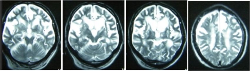
Zhang et al. BMC Neurology        (2021) 21:15                                                                                            Page 3 of 6

 Fig. 1 Patient’s brain MRI imagesSevere atrophy in the bilateral temporal and frontal lobes, mild atrophy in parietal lobes, as well as lacunar
 infarction in bilateral radiation coronal, basal ganglia, left thalamus and brainstem. Mild white matter hyperintensities (WMHs) can be found on
 both sides lateral ventricle

Luxol fast blue (LFB), modified Gallyas-Braak (GB) silver                    in the lumen of the right internal carotid artery and bilateral
staining, and immunohistochemical (IHC) staining for                         depigmentation in the substantia nigra (Fig. 3c).
Aβ (mouse monoclonal antibody; diluted 1:100; Dako,
Glostrup,Denmark), phosphorylated tau (mouse mono-
clonal clone AT8; diluted 1:500; Thermo Fisher, Roc-                         Neuropathological microscopic findings
ford,IL,USA), α-synuclein (rabbit polyclonal antibody;                       Volume shrinkage of neurons were observed in cere-
diluted 1:1000; Sigma-Aldrich, St.Louis, MO,USA;), TAR                       brum, brainstem and cerebellum due to brain edema.
DNA binding Protein-43 (TDP-43) (rat monoclonal anti-                        Marked Purkinje’s cell loss were found in the cerebel-
body; pSer409/410; diluted 1:500;Merck Millipore,Te-                         lem. We have not found Lewy bodies in the substantia
mecula,CA, Germany), and P62 (mouse monoclonal                               nigra, locus ceruleus and cerebrum by HE staining
clone ;diluted 1:1000; Abnova, Taipei China), according                      (Fig. 2b). AT8 and P62 immunoreactive neurofibrillary
to the National Institute on Aging-Alzheimer’s Associ-                       tangles and threads were seen in the hippocampus,
ation (NIA-AA) guidelines for AD neuropathologic                             which were also revealed by Gallyas-Braak staining
changes (ADNC) [4].                                                          (Fig. 2d). No accumulation of Aβ, α-synuclein or TDP-
                                                                             43 were noted in the brain (Fig. 2c). The Alzheimer’s
Neuropathological macroscopic finding                                        disease neuropathologic change was A0, B1, C0 (Braak
Severe whole brain edema was observed. There were many                       stage I) according to diagnostic criteria made by the Na-
cerebral infarctions in the middle cerebral artery. The locus                tional Institute for Aging-Alzheimer Association [3].
coeruleus was relatively preserved. There was mild midbrain,                 There were no astrocytic plaques or tufted astrocytes in
pons, basal ganglia, thalamus, hemi-ovary center (Figs. 3a                   the brain or brainstem(data not shown).Based on above
and b and 2a). Widespread arteriosclerosis was seen in large,                fingdings Vascular parkinsonism(VaP) pathologic diag-
middle and small cerebral vessels, and thrombosis was found                  nosis was made.

 Fig. 2 Gross findings: a Many micro infarctions in internal capsule, puteman, caudate nucleus and thalamus of both sides. b A micro infarction in
 the midbrain at the level of superior colliculus. c Mild depigmentation in the substantia nigra
Zhang et al. BMC Neurology (2021) 21:15 Page 4 of 6 Fig. 3 Microscopic findings. a Perivascular tissue necrosis with several hemosiderin cells. b Slightly depigmentation and no Lewy bodies in the substantia nigra. c No Lewy bodies or Lewy threads in the substantia nigra. d Neurofibrillary tangles and thresds in the CA1 segment of Hippocampus. HE stain (a, b), α-synuclein immunostain(c),AT immunostain (d). Scale bars:100 μm (a, b, c, d) Discussion and conclusions diagnosis of Vascular parkinsonism(VaP) was made and Different movement disorders have been reported after Parkinson’s disease or DLB were excluded. AD neuropath- cerebrovascular events, such as parkinsonism, tremor, ologic change should be ranked based on three Parame- dystonia, myoclonus, asterixis, stereotypies, akathisia, ters: Aβ plaque score, Braak NFT stage,and neuritic tics, choreaand freezing of gait.The frequency of cerebra- plaque score to obtain an ‘‘ABC score’’. The ABC scores vascular abnormal movements has been estimated to are transformed into one of four levels of AD neuropatho- vary from 1–4% of all strokes [5]. Vascular Parkinsonism logic change: Not, Low, Intermediate or High. In this pa- (VaP) is very commonand has been found to be present tient, mild neurofibrillary tangles(NFL) and threads were in about 3–5% of post-mortem studies of patients with seen in the hippocampus (Braak stage I), and no accumu- parkinsonism [6].The frequency of post-stroke move- lation of A β and neuritic plaques. The Alzheimer’s dis- ment disorders is likely to be underestimated. A pro- ease neuropathologic change was A0, B1, C0, and not fits spective cohort study reported the incident of VaP over the diagnostic standard of AD[4, 10]. a mean period of 5.2 years in 15 patients (3%) out of 503 An academic working group recommends definition of patients with cerebral small vessel disease who had no vascular parkinsonism into three subtype by acute or sub- parkinsonism at baseline [7], indicating that pure forms acute post-stroke VaP subtype, insidious onset VaPand of VaP are relatively rare. A previous study reported 28 mixed or overlapping syndromes of idiopathic Parkinson’s pathologically confirmed VaP cases of whom only six disease or other neurodegenerative parkinsonisms and co- were diagnosed as VaP during lifetime and the remain- morbid cerebral vescular disease (CVD) [11]. The acute or der as either PD or atypical parkinsonism [8]. Thus, VaP subacute post-stroke VaP subtype generally responds to is often misdiagnosed as in the case that we reported in dopaminergic drugs and is typically asymmetric,presenting this manuscript. acute or subacute onset of parkinsonism. Insidious onset This patient have showed rapid progressed parkinson- VaP subtype is more frequent, presenting progressive par- isms and cognitive impairment, as well as early behavioral kinsonism with prominent postural instability, gait impair- and psychological symptoms. In the neuropathological ment,corticospinal, pseudobulbar, cerebellar, cognitive examination, many cerebral infarctions can be seen in and urinary symptoms and tends to be poor responsive to various areas of the brain, especially in the basal ganglia. dopaminergic drugs.It would manisfested as a higher-level There were no Lewy bodies, α-synuclein, accumulation of gait disorder frequently in the clinical spectrum [12]. Aβ and proliferation of glial cells. The mild depigmen- Mixed or overlapping syndromes of idiopathic Parkinson’s tation in the substantia nigra can be interpreted as a result disease and comorbid CVD is diagnosed based on the mo- of aging, being comparable with the findings of aged- lecular imaging biomarkers such as dopamine transporter matched subjects with no symptoms of PD [9].Thus,A imaging in clinical practice.
Zhang et al. BMC Neurology   (2021) 21:15                                                                                       Page 5 of 6

  In the insidious onset VaP subtype, the cognitive im-      striatal presynaptic dopamine transporter (DAT) shows
pairment is very common because of an overlap between        a significant decrease in tracer uptake in PD, while VaP
Vascular Dementia(VaD) and VaP. Both VaP and VaD             was generally normal. SPECT combined with MRI can
have similar frequent occurrence of brain atrophy, white     effectively differentiate Parkinson’s disease from
matter hyperintensities and lacunes, and represent dif-      VaP.123I-metaiodobenzylguanidine(MIBG)         can    be
ferent aspects of subcortical vascular encephalopathy.       appliedto differentiate VaP from PD, which indicative of
The pathological feature of VaP is brain damage caused       function of cardiac sympathetic nerve. Total MIBG up-
by vascular factors. The manifestations were ischemia        take was decreased in patients with PD and DLB, but
with main lesions in subcortical white matter, basal gan-    VaP showed normal or mild decrease [22].
glia, thalamus and midbrain, but hemorrhage was rare
                                                             Abbreviations
[13].The pathological changes of brain tissue in VaP
                                                             VaP: Vascular Parkinsonism; DLB: Dementia with lewy Body; CVD: Cerebral
were mainly lacune and white matter lesions with severe      vascular disease; CSVD: Cerebral small vessel disease; PD: Parkinson’s disease;
oligodendrocyte loss.                                        WMHs: white matter hyperintensities; PSP: Progressive Supranuclear Paralysis;
                                                             MSA: Multiple System Atrophy; CBD: Corticobasal Degeneration;
  Cerebral small vessel disease (CSVD) can be asymp-
                                                             DAT: Dopamine transporter
tomatic or manifest as lower body parkinsonism.It refers
to a series of clinical, imaging and pathological syn-       Acknowledgements
dromes caused by affected intracerebral arterioles, capil-   We are very grateful to all the participants of the study. This work was
                                                             supported by the department of nuclear medicine in Chinese PLA General
laries and venules [12]. Clinical manifestations include     Hospital and we deeply appreciate all the works done by Dr Mingwei Zhu.
lacunar infarction, cerebral hemorrhage, subcortical
white matter lesions,cerebral microhemorrhage and            Authors’ contributions
microinfarction [14]. Age, hypertension and hyperhomo-       SZZ and LNW designed the study strategy. MWZ performed the brain
                                                             autopsy and made pathologic diagnosis. LXL, ZL, ML,HYW and XH performed
cysteinemia are recognized risk factors for cerebrovascu-    the assessment of the data collection. SZZ,YYW and FM wrote the
lar disease in many studies. In the early, middle and late   manuscript. All authors reviewed the manuscript. The author(s) read and
stage of cerebral small vessel disease, cognitive dysfunc-   approved the final manuscript.

tion and emotional changes can be observed to different
                                                             Funding
extent. Depression, gait instability, dysphagia, bladder     This work was supported by the Military Special Health Care Project (Grant
sphincter dysfunction and decline in daily living ability    No. 15BJZ38).The funds was used to purchase immunohistochemical Kits and
                                                             laboratory supplies in the process of pathological examination.
are very common [12]. Multiple studies have investi-
gated the relationship of cerebral small vessel disease      Availability of data and materials
with dementia, particularly microbleeds and WHM are          Data sharing is not applicable to this article as no datasets were generated
frequently seen in those with cognitive impairment [15,      or analysed during the current study.
16]. The association of MRI-based infarcts with an in-
                                                             Ethics approval and consent to participate
creased risk of dementia was reported in several             Not applicable.
population-based studies[17, 18].Autopsy has confirmed
that this patient had CSVD, coincident with insidious        Consent for publication
                                                             Written informed consent for publication was obtained from the patient’s
type of VaP. It is speculated that insidious type of VaP
                                                             family members.
may occur due to disruption in the extra-nigrostriatal
white or gray matter vascular lesions and connectivity       Competing interests
associated with parkinsonism, evena distinct focal           The authors declare that they have no competing interests.
nigrostriatal deficit does not occur[19].                    Author details
  Parkinsonism can also be present in patient with and       1
                                                              Department of Psychiatry, Beijing Geriatric Hospital, 100095 Beijing, P.R.
PD, DLB, Progressive Supranuclear Paralysis(PSP), Mul-       China. 2Chinese People’s Liberation Army General Hospital, Beijing 100853,
                                                             P.R. China. 3Institute of Systems Biomedicine, Peking University Health
tiple System Atrophy(MSA) or Corticobasal Degenera-          Science Center, Beijing 100191, P.R. China.
tion(CBD) that involve α-synucleinopathy,tau or other
proteinopathies[20]. The co-occurrence of clinically         Received: 26 August 2020 Accepted: 28 December 2020
manifest primary neurodegenerative parkinsonism with
vascular disease is common due to mixed pathologies          References
[21]. Similar mixed coincidence of vascular disease and      1. Mehanna R. Joseph Jankovic. Movement disorders in cerebrovascular
neurodegenerative parkinsonisms can occur in PSP,                disease. Lancet Neurol. 2013;12:597–608.
                                                             2. Thanvi B, Lo N, Robinson T. Vascular parkinsonism—an important cause of
MSA or CBD [11].Molecular imaging techniques such as             parkinsonism in older people. Age Ageing. 2005;34:114–19.
dopaminge transporter imaging is helpful to identify         3. Joaquin A, Vizcarra AE, Lang, Kapil D, Sethi, et al. Vascular Parkinsonnism:
more ‘pure’ sub-types of VaP, as well as indicative              deconstructing a syndrome. Mov Disord 2015;30(7):886–94.
                                                             4. Bradley T, Hyman CH, Phelps,Thomas G. Beach,et al.National Institute on
ofmixed or overlapping syndromes of neurodegenerative            Aging–Alzheimer’s Association guidelines for the neuropathologic
parkinsonism and comorbid CVD. SPECT imaging of                  assessment of Alzheimer’s disease.Alzheimers Dement. 2012; 8(1): 1–13.
Zhang et al. BMC Neurology (2021) 21:15 Page 6 of 6
5. Thomas J, Montine,Creighton H,et al. National Institute on Aging–
Alzheimer’s Association guidelines for the neuropathologic assessment of
Alzheimer’s disease:a practical approach. Acta Neuropathol 2012;123:1–11.
6. Handley A, Medcalf P, Hellier K, Dutta D. Movement disorders after stroke.
Age Ageing 2009;38:260–66.
7. Jellinger KA. Prevalence of cerebrovascular lesions in Parkinson’s disease. A
postmortem study.Acta Neuropathol. 2003;105:415–9.
8. Van der Holst HM, van Uden IW, Tuladhar AM, et al.Cerebral small vessel
disease and incident parkinsonism:The RUN DMC study. Neurology.2015;85:
1569–77.
9. Glass PG, Lees AJ, Bacellar A, Zijlmans J, Katzenschlager R, Silveira-Moriyama
L. The clinical features of pathologically confirmed vascular parkinsonism. J
Neurol Neurosurg Psychiatry. 2012;83:1027–9.
10. Yue Xing,Abdul Sapuan,Rob A Dineen,et al.Life span pigmentation changes
of the subtantia nigra detected by neuromelanin-sensitive MRI.Mov Disord.
2018;33(11):1792-99.
11. Ivan Rektor,Nicolaas I, Bohnen,et al. An Updated Diagnostic Approach to
Subtype Definition of Vascular Parkinsonism Recommendations from an
expert working group. Parkinsonism Relat Disord. 2018 April; 49: 9–16.
12. Korczyn AD. Vascular parkinsonism–characteristics, pathogenesis and
treatment. Nat Rev Neurol. 2015;11:319–26.
13. Foltyniet T, Barker R, Brayne C. Vascular parkinsonism:a review of the precision
and frequency of the diagnosis. Neuroepidemiology. 2002;21(1):1–7.
14. Wardlaw JM, Smith C, Dichgans M. Mechanisms of sporadic cerebral small
vessel disease: insights from neuroimaging. Lancet Neurol. 2013;12:483–97.
15. Ding J, Sigurosson S, Eiriksdottir JPV, Meirelles G, Kjartansson O. O, et al.
Space and location of cerebral microbleeds,cognitive decline, and dementia
in the community. Neurology. 2017;88:2089–97.
16. Romero JR, Beiser A, Himali JJ, Shoamanesh A, DeCarli C, Seshadri S.
Cerebral microbleeds and risk of incident dementia: the Framingham Heart
Study. Neurobiol Aging. 2017;54:94–9.
17. Sigurdsson S, Aspelund T, Kjartansson O, Gudmundsson. EF,Jonsdottir
MK Eiriksdottir G, et al. Incidence of brain infarcts,cognitive change, and risk of
dementia in the general population:The AGES-Reykjavik Study (Age Gene/
Environment SusceptibilityReykjavik. Study) Stroke. 2017;48:2353–60.
18. Kaffashian S, Soumare A, Zhu YC, Mazoyer B, Debette S. Tzourio C.Long-
term clinical impact of vascular brain lesions on magnetic resonance
imaging in older adults in the population. Stroke. 2016;47:2865–9.
19. Vizcarra JA, Lang AE, Sethi KD. Espay AJ.VascularParkinsonism:deconstructing
a syndrome.Mov Disord. 2015; 30:886–94.
20. Ghebremedhin E, Rosenberger A, Rub. U,et al.Inverse relationship
between cerebrovascular lesions and severity. of lewy body pathology in
patients with lewy body diseases. J Neuropathol Exp Neurol. 2010;69:442–8.
21. Bohnen NI, Muller ML, Zarzhevsky N, et al. Leucoaraiosis, nigrostriatal
denervation. and motor symptoms in Parkinson’s disease. Brain. 2011;134:
2358–65.
22. Nacarro-Otano J, Gaig C, Muxi A, et al. 123I-MIBG cardiac uptake,smell
identification and 123I-FP-CIT SPECT in the differential diagnosis between
vascualar parkinsonism and Parkinson’s disease [J]. Parkinsonism Relat
Disord. 2014;20(2):192–7.
Publisher’s Note
Springer Nature remains neutral with regard to jurisdictional claims in
published maps and institutional affiliations.You can also read