SPLEnic salvage and complications after splenic artery EmbolizatioN for blunt abdomINal trauma: the SPLEEN-IN study
←
→
Page content transcription
If your browser does not render page correctly, please read the page content below
Clements et al. CVIR Endovascular     (2020) 3:92
https://doi.org/10.1186/s42155-020-00185-4
                                                                                                                                  CVIR Endovascular

 ORIGINAL ARTICLE                                                                                                                                  Open Access

SPLEnic salvage and complications after
splenic artery EmbolizatioN for blunt
abdomINal trauma: the SPLEEN-IN study
Warren Clements1,2,3* , Tim Joseph1, Jim Koukounaras1,2 , Gerard S. Goh1,2,3 , Heather K. Moriarty1,2 ,
Joseph Mathew3,4 and Tuan D. Phan1

  Abstract
  Background: As an adjunct to non-operative management, splenic artery embolization (SAE) has been increasingly
  utilized throughout the world and is now the standard of care for hemodynamically stable patients. This study
  aimed to retrospectively assess the rate of splenic salvage and complications after SAE for blunt trauma at a level 1
  trauma center using the 2018 update to the AAST criteria, and further sub-stratify the role of angiography in AAST
  grade III injuries with significant hemoperitoneum.
  All patients between 1 January 2009 and 1 January 2019 who underwent blunt trauma and proceeded to
  embolization were included. Data was collected concerning initial injury grade, location of embolization, type of
  embolic material used, complications, and need for subsequent splenectomy. Technical success was defined as
  successful angiographic occlusion of the target artery at the conclusion of embolization. Clinical success was
  defined as splenic salvage at discharge. Vascular lesions were characterized including those with active bleeding,
  pseudoaneurysm, and arterio-venous fistula.
  Results: Two hundred thirty-two patients were included in the study. Treatments were performed at a median of 0
  days (range 0–28 days) and the median AAST grade was IV (range III-V). Technical success was achieved in all
  patients. There were 13 complications (5.6%) consisting of re-bleed (9, 3.9%), infarction (3, 1.3%), and access site
  haematoma (1, 0.43%). Clinical success was achieved in 97% of patients with 7 patients requiring splenectomy after
  SAE (3.0%) at a median time of 4 days (range 0–17 days). Angiography in patients with grade III injuries identified 18
  occult vascular injuries not identified at initial CT (p < 0.0001).
  Conclusions: The SPLEEN-IN study shows that treatment of intermediate-high grade blunt force traumatic splenic
  injuries using SAE resulted in a low rate of complication and splenic salvage in 97% of patients, providing a safe
  and effective treatment in stable patients. In addition, angiography of grade III injuries identified occult vascular
  lesions and may warrant treatment of select patients in this cohort.
  Level of evidence: Level 3.
  Keywords: Trauma, Spleen, SAE, Hemorrhage, Embolization

* Correspondence: W.clements@alfred.org.au; w.clements@alfred.org.au
1
 Department of Radiology, Alfred Health, 55 Commercial Road, Melbourne,
VIC 3004, Australia
2
 Department of Surgery, Monash University, Melbourne, Australia
Full list of author information is available at the end of the article

                                      © The Author(s). 2020 Open Access This article is licensed under a Creative Commons Attribution 4.0 International License,
                                      which permits use, sharing, adaptation, distribution and reproduction in any medium or format, as long as you give
                                      appropriate credit to the original author(s) and the source, provide a link to the Creative Commons licence, and indicate if
                                      changes were made. The images or other third party material in this article are included in the article's Creative Commons
                                      licence, unless indicated otherwise in a credit line to the material. If material is not included in the article's Creative Commons
                                      licence and your intended use is not permitted by statutory regulation or exceeds the permitted use, you will need to obtain
                                      permission directly from the copyright holder. To view a copy of this licence, visit http://creativecommons.org/licenses/by/4.0/.
Clements et al. CVIR Endovascular (2020) 3:92 Page 2 of 9
Introduction intermediate-grade injuries (AAST III) where there is
Splenic artery embolization (SAE) has been increasingly significant hemoperitoneum as defined by the presence
utilized throughout the world (Roy et al. 2018). Splenic of blood in three or more abdominal quadrants on CT,
salvage for hemodynamically stable trauma patients is although we acknowledge that this is an area of
now standard of care (Patil et al. 2020). Splenic controversy.
embolization is also relatively unique, in that it can offer This study aimed to retrospectively assess the rate of
both immediate hemodynamic control as well as preserv- splenic salvage after blunt trauma using our protocol. In
ing splenic function (Lukies et al. 2020; Aiolfi et al. 2017). addition, we aimed to sub-stratify the angiographic find-
Modern trauma management requires a multi- ings in patients with AAST III injuries to identify
disciplinary approach. Trauma physicians, surgeons, and whether our protocol can clarify the role of treatment in
interventional radiologists need to consider treatment in this controversial cohort of trauma patients.
the context of the clinical scenario and intended goal.
The first concept to consider is the spleen with vascular Material and methods
injury, including active bleeding or the presence of 1 or Ethics
more pseudoaneurysms. In this circumstance, treatment Approval was provided by The Alfred Hospital Human
is almost always indicated and this must be planned in Research and Ethics Committee, number 361/19. For
addition to considering the overall severity of the injury this retrospective analysis, individual patient consent was
(Quencer and Smith 2019). In focal vascular injuries, not required.
splenic embolization is often performed as distal to the
hilum as possible to treat the vascular lesion but reduce
Patient identification
the overall risk of infarction, however the choice of em-
The study covered a 10-year period from 1 January 2009
bolic location in this group is controversial particularly if
to 1 January 2019. Patients were identified through the
there are multiple vascular lesions (Quencer and Smith
Radiology Information System (RIS). Information includ-
2019). A second scenario is high-grade splenic injuries
ing demographics, treatment, and complications were
without major vascular injury. Patients are often
obtained from a combination of RIS, Picture and Com-
hemodynamically stable and treatment is targeted at re-
munications Archive (PACS), and the Electronic Medical
ducing the overall rate of delayed re-bleed (Rong et al.
Record (EMR). Splenic injuries were graded on CT by 2
2017). In these circumstances, embolization proximal to
radiologists and any discrepancies were mediated by an
the hilum is usually performed. In some cases where
independent interventional radiologist before being
there is a vascular lesion and a high-grade injury, a tan-
included.
dem embolization (both proximal and distal) may be
employed.
The severity of injury may be graded according to a Inclusion criteria and endpoints
variety of different injury classification systems. Arguably All patients over the age of 16 who underwent SAE after
the most widely used is the American Association for blunt trauma were included. Patients were excluded if
the Surgery of Trauma (AAST) which first released a the injury was penetrating, if an angiogram was per-
grading system in 1994 (Moore et al. 1989). A 2018 formed but no embolization was employed, or if SAE
manuscript which included authors from the patient as- was performed for non-traumatic reasons. Complica-
sessment committee of the AAST, put forward a revised tions were defined according to the CIRSE classification
grading system whereby it was suggested that the pres- system (Filippiados et al. 2017). Potential complications
ence of vascular injury be incorporated into the original included abscess, infarction/post-embolization syn-
AAST system to more accurately reflect the need for drome, access site complication, access vessel dissection,
NOM adjuncts such as SAE in these patients (Kozar and splenectomy.
et al. 2018).
One of the biggest problems facing practitioners is the Data collection
heterogeneity in the endovascular treatment offered to Data collected included age, gender, injury severity score
these patients. Who to treat, when to treat, which em- (ISS), time from injury to embolization, AAST trauma
bolic to use, and where to employ the chosen embolic grade (2018 classification), vascular injury at CT, vascu-
harbors considerable debate and controversy, particu- lar injury at angiogram, location of embolization, type of
larly for AAST grade III injuries. embolic material used, complication, time for complica-
At The Alfred Hospital, a level 1 trauma center in tion to occur, need for splenectomy after SAE, and time
Melbourne, Australia, we consider angiography for pa- for splenectomy to occur after SAE. Vascular injury was
tients with high grade injury (AAST IV and V). In defined as the presence of active bleed, pseudoaneurysm,
addition, we consider angiography for patients with or arteriovenous fistula. Data was collected during theClements et al. CVIR Endovascular (2020) 3:92 Page 3 of 9
index admission and up to 30 days after the traumatic Results
event. Patient demographics
During the defined study time, 232 patients met inclu-
sion criteria for the study. The mean age was 40 years
Embolization definition
and 80.1% were male. Treatments were performed at a
For the purposes of this manuscript, “proximal
median time of 0 days (day of injury) and the median
embolization” was defined as large vessel occlusion prox-
AAST injury grade was IV. On initial trauma CT, 59% of
imal to the splenic hilum but distal to the dorsal pancre-
patients had a vascular injury while at angiography 79%
atic artery (Fig. 1a and b). “Distal embolization” was
showed a vascular injury (p = 0.001). Table 1 summa-
considered embolization of selected splenic artery
rizes patient and treatment demographics.
branches distal to the hilum (Fig. 2a and b). “Tandem
Only 9 embolizations were performed in 2009 while
embolization” refers to a procedure where distal
33 were performed in 2018 with the increasing trend be-
embolization was performed for focal vascular lesion
tween these years shown in Fig. 3.
followed by concurrent proximal embolization.
Success and complications
Outcome definition Technical success was achieved in 100% of patients.
Technical success was defined as angiographic occlusion There were 13 complications in total (5.6%) including 1
of the target artery at the conclusion of the treatment. CIRSE grade 1, 3 CIRSE grade 2, 3 CIRSE grade 3, and 6
Clinical success was defined as splenic salvage after SAE. CIRSE grade 4. There were 7 patients (3.0%) who under-
went splenectomy after embolization meaning the
splenic salvage rate was 97%. Table 2 outlines the indi-
Embolization technique
vidual demographics of patients who either experienced
All procedures were performed by one of 8 fellowship-
a complication and/or underwent subsequent splenec-
trained interventional radiologists, or an advanced
tomy. Compared with the overall cohort, there was no
trainee under supervision. According to operator prefer-
significant difference in the group who experienced a
ence, embolization is performed using pushable coils
complication in terms of ISS (p = 0.52), age (p = 0.41),
(0.018“ or 0.035” Cook Nester or Cook Tornado, Cook
gender (p = 0.27), or vascular injury at CT (p = 0.29).
Medical, Bloomington, USA), amplatzer vascular plug
(St Jude Medical, Plymouth, USA), microvascular plug
Choice of embolic
(Medtronic, Dublin, Ireland), EOS (Artventive, San Mar-
The majority of procedures (84.9%) employed pushable
cos, USA), or gelatin sponge.
fibered coils as shown in Table 3. A vascular plug was
used in 6%. Cases using combination embolic (5.6%), de-
Statistical analysis tachable coils (2.2%), and gelatin sponge (1.3%) were less
Data was de-identified and analyzed using Microsoft frequent. All combination embolic cases consisted of
Excel (Microsoft, USA) with the Real Statistics Resource pushable coils and gelatin sponge. The overall rate of
Pack software (Release 6.8) (Zaiontz 2020). Data was complications was not significantly different between the
summarized using mean and standard deviation, median groups (p = 0.079).
and range, or frequency and percentage as appropriate
to the type of data. Using the Mann-Whitney U test, a Location of embolization
two-sided p-value less than 0.05 was chosen to indicate Embolizations were proximal to the hilum in 176 pa-
statistical significance. tients (75.9%) with a complication rate of 6.8% in this
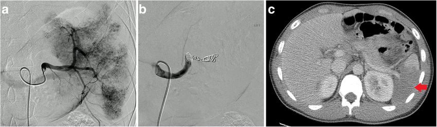
Fig. 1 Catheter angiography from the splenic artery showing high grade parenchymal injury a successfully treated with proximal embolization b
and patient with infarct after proximal embolization (arrow) cClements et al. CVIR Endovascular (2020) 3:92 Page 4 of 9
Fig. 2 Selective catheter angiography from an upper pole splenic artery showing focal parenchymal injury a, successfully treated with distal
embolization b
cohort, and 7 patients (4.0%) required subsequent splen- for other groups, p = 0.027. There was a higher rate of vas-
ectomy. There were no complications in the 35 patients cular injuries in the grade V patient group (p < 0.0001). No
(15.1%) who underwent distal embolization. Tandem significant difference was seen between the groups compar-
embolization was used in 21 patients (9.0%) and only 1 ing ISS (p = 0.21), proportion of proximal embolizations
(0.43%) experienced a complication but did not require (p = 0.55), time to embolization (p = 0.81), complication
splenectomy. No significant difference in the rate of rate (p = 0.085), or splenectomy rate (p = 0.11).
complications (p = 0.69) or splenectomy (p = 0.32) was
identified based on the embolization location. AAST grade III
Of the 41 patients who were graded as AAST III based on
AAST grading their initial CT but proceeded to angiography/SAE, 18 of
Using the updated 2018 AAST grading system, Table 4 these patients showed evidence of a vascular injury at angi-
outlines the number of treatments and patient demograph- ography compared to 0 who showed evidence of vascular
ics based on each injury grade. There were 41 patients with injury at CT (p < 0.0001). There were no complications and
AAST grade III injury, 109 patients with grade IV injury, no patients who required splenectomy in this cohort.
and 82 patients with grade V injury. Patients with grade IV
injuries had a significantly lower mean age than for the Discussion
other grades, p = 0.01. There was a higher proportion of The number of splenic embolizations has been steadily
males in the group of patients with grade III injuries than increasing in frequency at our institution, and similarly
reflected in data from other institutions including from
Table 1 Patient and procedure demographics different countries (Roy et al. 2018). There are many fac-
Number of embolizations 232 tors that may contribute to this. From the perspective of
Age (mean, SD) 40 (18.7) our network, our hospital is the largest Level 1 center in
Male gender (number, percentage) 185 (80.1%) the Victorian State Trauma system which has prepro-
grammed responses built in to patient referral and treat-
Time to embolization after injury in days (median, range) 0 (0–28)
ment processes and is supported by efficient ground and
AASTa injury grade (median, range) 4 (3–5)
air ambulance including a 24-h helipad. This has de-
Evidence of vascular injury at CT (number, percentage) 137 (59.0%) creased mortality and also time to definitive hemorrhage
Evidence of vascular injury at embolization (number, 184 (79.3%) control (Cameron et al. 2008). On arrival to the trauma
percentage) center, a multidisciplinary trauma team approach to
Proximal embolization (number, percentage) 176 (75.9%) damage control resuscitation has decreased the time to
Use of pushable coils (number, percentage) 197 (84.9%) stabilization, access to CT, and definitive hemorrhage
Injury severity score (median, range) 22 (4–66) control. This may allow for endovascular rather than
Complications (number, percentage) 13 (5.6%)
emergency operative intervention (Matsumoto et al.
2015). In addition, a dedicated interventional radiology
Time to complication in days (median, range) 2.1 (0–7)
on-call roster allows for rapid activation of services.
Splenectomy after embolization (number, percentage) 7 (3.0%) These factors support a median time to treatment of 0
Time to splenectomy (median, range) 4 (0–17) days. The increasing trend also reflects the increasing
a
AAST American Association for the Surgery of Trauma abundance of supportive data towards the role of SAE inClements et al. CVIR Endovascular (2020) 3:92 Page 5 of 9 Fig. 3 Ten-year trend of increasing number of SAE procedures at our institution for blunt abdominal trauma supporting NOM in blunt trauma including recent sys- splenectomy after embolization, in the opinion of the au- tematic reviews (Moore et al. 1989; Schnüriger et al. thors it does not reflect a complication of the 2011). embolization itself. The demographic in this study with a mean age of 40 With the update to the AAST classification in 2018 to and predominantly males, is in keeping with the ex- incorporate splenic artery vascular injury, 82% are now pected demographic of road traffic trauma (Ferrah et al. considered high-grade (AAST IV or V). This compares 2019). The median injury severity score of 22 also re- to 126 high-grade injuries (54%) if our patients were flects the complexity of high-energy multi-system graded per the previous 1994 AAST classification. The trauma at our institution (Ferrah et al. 2019). The demo- grade V cohort showed a significantly higher rate of vas- graphics in both the overall cohort and for those who cular injury than for the lower grade groups which is ex- experienced a complication were similarly matched. pected given the severity of injury to meet the grade V The overall rate of complications in this study was low criteria. The rate of splenectomy in the grade V cohort at 5.6% and occurred at a median time of 2.1 days after of 8.5% is also low and comparable to literature includ- the treatment. In addition, only 3% of patients proceeded ing the systematic review of Rong et al. which included to splenectomy at a median time of 4 days which is low, 10 studies and 876 patients (Moore et al. 1989). It is en- and in keeping with the rate shown in prior literature couraging to see that of all patients with grade IV (Moore et al. 1989; Schnüriger et al. 2011; Davies and splenic injury or lower, only 2 of 150 patients (1.3%) re- Wells 2019; Hughes et al. 2017). Of the 7 patients re- quired splenectomy after SAE. quiring splenectomy after embolization, 1 patient was a Our practice to strongly consider angiography in pa- 30-year-old male who presented after a motor vehicle tients who have AAST grade III injury and three or accident with multiple injuries (ISS 38) including grade more quadrant hemoperitoneum (41 patients, 17.6%) is V splenic injury, hollow viscus injury, and pelvic bleed- acknowledged to be a controversial decision. However, ing on CT scan. The patient was emergently transferred as shown in this study there was a significant increase in to the angiography suite before the operating theatre, the number of vascular injuries identified at angiography where pelvic embolization was performed, and a decision compared to those identified at CT in patients who were was made between the interventional radiologist and deemed to have AAST III injury based on their trauma trauma surgeon to embolize the spleen proximally to fa- CT scan. It is for this reason that considering angiog- cilitate safe transfer to the operating suite where splen- raphy in the grade III cohort is likely to identify patients ectomy was performed as a pre-planned procedure. who are at increased risk of re-bleed. The absolute risk While this has been included in the analysis as a however is difficult to quantify without a prospective
Table 2 Patients who experienced complications and/or underwent splenectomy after SAE
                                                                                                                                                                                                            Clements et al. CVIR Endovascular

Patient Age Gender ISS AAST injury Vascular lesion Vascular lesion Location of Type of embolic                                 CIRSE complication Complication            Splenectomy Days to splenectomy
number                 grade       on CT?          on angiogram? embolization used                                             status (Filippiados type                   required    after trauma
                                                                                                                               et al. 2017)
1         17     Male      16   4              No                 Yes                Proximal         Pushable coils           2                      Infarction          No          N/A
2         22     Female    33   4              Yes                Yes                Proximal         Plug (AVP IV)            2                      Infarction          No          N/A
                                                                                                                                                                                                               (2020) 3:92

3         48     Male      17   4              Yes                No                 Proximal         Pushable coils           3                      Re-bleed            No          N/A
                                                                                                                                   a                       a
4         30     Male      38   5              Yes                Yes                Proximal         Plug (AVP IV)            N/A                    N/A                 Yes         0
5         55     Male      29   5              Yes                Yes                Proximal         Plug (AVP IV)            3                      Re-bleed            No          N/A
6         54     Female    41   5              Yes                Yes                Tandem           Pushable coils           3                      Re-bleed            No          N/A
                                                                                                      and gelfoam
7         29     Male      21   5              Yes                Yes                Proximal         Pushable coils           4                      Re-bleed            Yes         0
8         89     Male      10   4              Yes                Yes                Proximal         Pushable coils           1                      Groin               No          N/A
                                                                                                                                                      hematoma
9         48     Female    34   5              No                 Yes                Proximal         Pushable coils           4                      Re-bleed            Yes         4
10        31     Male      34   5              No                 Yes                Proximal         Pushable coils           4                      Re-bleed            Yes         5
11        37     Female    57   4              No                 Yes                Proximal         Plug (Eos)               4                      Re-bleed            Yes         10
12        78     Male      20   4              No                 No                 Proximal         Pushable coils           4                      Re-bleed            Yes         17
13        53     Male      20   5              Yes                Yes                Proximal         Pushable coils           4                      Re-bleed            Yes         3
14        38     Female    16   5              Yes                Yes                Proximal         Pushable coils           2                      Infarction          No          N/A
ISS injury severity score
AAST American Association for the Surgery of Trauma
a
 patient 4 underwent planned splenectomy after pelvic embolization and the spleen was embolized pre-operatively. This was not considered a complication of embolization
                                                                                                                                                                                                                Page 6 of 9
Clements et al. CVIR Endovascular (2020) 3:92 Page 7 of 9
Table 3 Use of different embolic agents, and association with complication and need for splenectomy
Type of embolic Number Percentage Complications Splenectomy
Pushable coils 197 84.9 9* 5*
Detachable coils 5 2.2 0* 0*
Vascular plug 14 6.0 4* 2*
Gelatin sponge 3 1.3 0* 0*
Combination of embolic 13 5.6 1* 0*
*p > 0.05
**p < 0.05
and randomized design, which may not be feasible. The embolization even though a range of catheters and em-
effect of these results is to further sub stratify this pa- bolic materials are always in stock.
tient group beyond the AAST classification where it is It is encouraging to see that there is no significant dif-
possible that occult vascular injuries are the reason for ference in the rate of complication or splenectomy based
those who do subsequently re-bleed with a grade III in- on the choice of embolic material or the embolic loca-
jury. In addition, it can be argued that the low rate of tion in this study. The analysis of Rong et al. suggested a
complications in the AAST III cohort of 0% and preser- higher rate of complications with the use of gelatin
vation of splenic function (Lukies et al. 2020; Schimmer sponge and this has also been shown in other studies
et al. 2016) further warrants such consideration for (Moore et al. 1989; Abada and Golzarian 2007). How-
treatment. The presence of vascular injury in the grade ever, as the number of cases using gelatin sponge in our
III cohort from this study also supports the recent study was extremely low, it is not possible to correlate
changes to AAST to acknowledge the importance of vas- with their findings reliably. While it is unusual to see
cular injury in their classification system. In addition, zero complications in the distal embolization cohort, the
with a median ISS of 22 and as high as 59 in the grade small number of patients in this cohort (35, 15.1%)
III cohort, a diminished risk of re-bleed after which was heavily biased towards proximal embolization
embolization is often appreciated in complex multi- may account for this.
trauma patients where the cause of abnormal or chan- The authors acknowledge a number of limitations with
ging physiology can be hard to pinpoint. this study. This is a retrospective audit and as such lacks
In the whole cohort, the choice of proximal embolic patient-level clinical data in which the trauma setting it
location (75.9%) and pushable fibred coils (84.9%) re- is arguable most vital. As such, we have used AAST and
flects a decision at our institution to provide a cost- ISS to risk-stratify patients. As a single-center study, our
effective solution for our patients in an institution that is practice reflects the internal structure of our trauma and
government-funded (Yip et al. n.d.). In many cases, interventional radiology teams and is also impacted by
proximal embolization also offers a rapid option to pro- the nearby position of emergency/trauma and imaging
vide treatment even with significant anatomic tortuosity, services. This may not necessarily reflect how other hos-
anatomic variation, vasospasm, and/or underlying vascu- pitals are structurally designed. We also acknowledge
lar disease which may inhibit or prolong attempts at dis- that AAST grade was determined at initial CT and while
tal embolization. Such choices are at the discretion of treatment was performed based on a combination of CT
the treating interventional radiologist at the time of and angiographic findings, the grade was not changed
Table 4 Comparison of AAST subgroup demographics (2018 revision) and outcomes
hba Age (mean, SD) ISS Male gender Vascular injury Number of Proximal Time to Complications Splenectomy
(median, (number, at CT (number, embolizations embolization embolization (number, (number,
range) percentage) percentage) (number, (median, range) percentage) percentage)
percentage)
Grade III 44 (19.4)* 22 (9–59)* 38 (93%)** N/Aa 41 33 (87%)* 0 (0–18)* 0 (0%)* 0 (0%)*
Grade IV 36 (17.0)** 22 (4–57)* 80 (73%)* 48 (44%)* 109 85 (78%)* 0 (0–21)* 6 (5.5%)* 2 (1.8%)*
Grade V 43 (19.7)* 22 (5–66)* 67 (82%)* 74 (90%)** 82 58 (71%)* 0 (0–28)* 7 (8.5%)* 5 (8.5%)*
ISS injury severity score
AAST American Association for the Surgery of Trauma
*p > 0.05
**p < 0.05
a
vascular injury is not possible with grade III classificationClements et al. CVIR Endovascular (2020) 3:92 Page 8 of 9
after angiography for the purposes of this analysis. We Ethics approval and consent to participate
acknowledge the heterogeneity in the way embolizations Approval was provided by The Alfred Hospital Human Research and Ethics
Committee, number 361/19.
were performed in our cohort and that there has been
grouping of individual patients regardless of the fine de- Consent for publication
tail of splenic parenchymal injury variation. The choice Ethics approval for this retrospective study included a waiver of individual
patient consent.
of embolic and choice of embolic location is also heavily
skewed to proximal embolization with pushable coils, Competing interests
leaving smaller numbers for the other groups. This will The authors declare that they have no conflict of interest.
reduce the statistical reliability of analysis. In addition,
Author details
all embolics are different even for pushable coils and the 1
Department of Radiology, Alfred Health, 55 Commercial Road, Melbourne,
effectiveness between 0.018″, 0.035″, and different types VIC 3004, Australia. 2Department of Surgery, Monash University, Melbourne,
or lengths of coils in each size was not measured. Never- Australia. 3National Trauma Research Institute, Central Clinical School,
Monash University, Melbourne, Australia. 4Department of Trauma, Alfred
theless, our results indicate that proximal splenic Health, Melbourne, Victoria, Australia.
embolization is a robust strategy that can be successful
despite technical differences and this is supported in Received: 21 October 2020 Accepted: 29 November 2020
previous systematic reviews which also suffer from simi-
lar heterogeneity in data (Moore et al. 1989; Schnüriger References
et al. 2011). Abada HT, Golzarian J (2007) Gelatine sponge particles: handling characteristics
In conclusion, the SPLEEN-IN study shows that treat- for endovascular use. Tech Vasc Interv Radiol 10(04):257–260
Aiolfi A, Inaba K, Strumwarrer A et al (2017) Splenic artery embolization versus
ment of intermediate-high grade blunt force traumatic splenectomy. Analysis for early in-hospital infectious complications and
splenic injuries using SAE results in a low overall rate of outcomes. J Trauma Acute Care Surg 83(3):356–360. https://doi.org/10.1097/
complication and splenic salvage in 97% of patients, pro- TA.0000000000001550
Cameron PA, Gabbe BJ, Cooper DJ et al (2008) A statewide system of trauma
viding a safe and effective adjunct to NOM in these pa- care in Victoria: effect on patient survival. Med J Aust 189(10):546–550.
tients. Splenic salvage after SAE increases to 98.7% of https://doi.org/10.5694/j.1326-5377.2008.tb02176.x
patients with a grade IV injury or lower. These results Davies J, Wells D (2019) Splenic artery embolisation in trauma: a five-year single-
Centre experience at a UK major trauma Centre. Trauma. 21(4):280–287.
can be achieved with lower cost pushable coils and prox- https://doi.org/10.1177/1460408618781412
imal splenic embolization in most patients. Ferrah N, Cameron P, Gabbe B et al (2019) Trends in the nature and
The SPLEEN-IN study also shows that embolization of Management of Serious Abdominal Trauma. World J Surg 43:1216–1225.
https://doi.org/10.1007/s00268-018-04899-4
grade III injuries in certain patients is safe, and that Filippiados DK, Binkert C, Pellerin O et al (2017) Cirse quality assurance document
proceeding to angiography in this cohort can identify and standards for classification of complications: the Cirse classification
vascular injuries initially occult on CT. The results system. Cardiovasc Intervent Radiol 40:1141–1146. https://doi.org/10.1007/
s00270-017-1703-4
support the recent changes to the AAST classification Hughes J, Scrimshire A, Steinberg L et al (2017) Interventional radiology service
where the importance of splenic vascular injury is provision and practice for the management of traumatic splenic injury across
now acknowledged. the regional trauma networks of England. Injury. 48(5):1031–1034. https://doi.
org/10.1016/j.injury.2017.02.031
Kozar RA, Crandall M, Shanmuganathan K et al (2018 Dec) Organ injury scaling
Abbreviations
2018 update: spleen, liver, and kidney. J Trauma Acute Care Surg 85(6):1119–
NOM: Non-operative management; SAE: Splenic artery embolization;
1122. https://doi.org/10.1097/TA.0000000000002058
CT: Computed tomography; AAST: American Association for the Surgery of
Lukies MW, Kavnoudias H, Zia A, Lee R, Bosco JJ, Joseph T, Clements W (2020)
Trauma; ISS: Injury severity score; RIS: Radiology information system;
Long term immune function following splenic artery embolisation for blunt
PACS: Picture and communication archive; EMR: Electronic medical record
abdominal trauma. Cardiovasc Intervent Radiol. https://doi.org/10.1007/
s00270-020-02627-x
Acknowledgements
Matsumoto J, Lohman BD, Morimoto K et al (2015) Damage control
The authors acknowledge the assistance of Dr. Shelley Chapman and Dr.
interventional radiology (DCIR) in prompt and rapid endovascular strategies
Katherine Martin as well as the National Trauma Research Institute.
in trauma occasions (PRESTO): A new paradigm. Diagn Interv Imaging 96(7–
8):687–691. https://doi.org/10.1016/j.diii.2015.06.001
Informed consent Moore EE, Shackford SR, Pachter HL et al (1989) Organ injury scaling: spleen, liver,
Ethics approval for this retrospective study included a waiver of individual and kidney. J Trauma 29(12):1664–1666
patient consent. Patil MS, Goodin SZ, Findeiss LK (2020) Update: splenic artery embolization in
blunt abdominal trauma. Semin Interv Radiol 37(01):097–102. https://doi.org/
Authors’ contributions 10.1055/s-0039-3401845
All authors contributed to patient management, data gathering, and construction Quencer KB, Smith TA (2019) Review of proximal splenic artery embolization in
of the manuscript. The author(s) read and approved the final manuscript. blunt abdominal trauma. CVIR Endovasc 2:11. https://doi.org/10.1186/s42155-
019-0055-3
Funding Rong J-J, Liu D, Liang M et al (2017) The impacts of different embolization
This study was not supported by any funding. techniques on splenic artery embolization for blunt splenic injury: a
systematic review and meta-analysis. Military Med Res 4:17. https://doi.org/
Availability of data and materials 10.1186/s40779-017-0125-6
The datasets generated and/or analyzed during the current study are not Roy P, Mukherjee R, Parik M (2018) Splenic trauma in the twenty-first century:
publicly available as this was not a part of the IRB approval process, please changing trends in management. Ann R Coll Surg Engl 100(8):650–656.
contact the corresponding author for additional information. https://doi.org/10.1308/rcsann.2018.0139Clements et al. CVIR Endovascular (2020) 3:92 Page 9 of 9
Schimmer JAG, van der Steeg AF, Zuidema WP (2016) Splenic function after
angioembolization for splenic trauma in children and adults: a systematic
review. Injury 47(03):525–530
Schnüriger B, Inaba K, Konstantinidis A, Lustenberger T, Chan LS, Demetriades D
(2011) Outcomes of proximal versus distal splenic artery embolization after
trauma: a systematic review and meta-analysis. J Trauma 70(01):252–260
Yip H, Skelley A, Morphett L, Mathew J, Clements W The cost to perform splenic
artery embolisation following blunt trauma: analysis from a level 1 Australian
trauma Centre. Injury. https://doi.org/10.1016/j.injury.2020.09.039
Zaiontz C. (2020) Real Statistics Using Excel. Available form URL www.real-
statistics.com. Accessed 15 Jan 2020
Publisher’s Note
Springer Nature remains neutral with regard to jurisdictional claims in
published maps and institutional affiliations.You can also read


