SARS COV2 INFECTION: FUNCTIONAL AND MORPHOLOGICAL CARDIOPULMONARY CHANGES IN ELITE HANDBALL PLAYERS - NATURE
←
→
Page content transcription
If your browser does not render page correctly, please read the page content below
www.nature.com/scientificreports
OPEN SARS‑CoV2 infection:
functional and morphological
cardiopulmonary changes in elite
handball players
S. Fikenzer 1,4*, A. Kogel1,4, C. Pietsch2, D. Lavall1, St. Stöbe1, U. Rudolph1, U. Laufs1,
P. Hepp3,4 & A. Hagendorff1,4
There is increasing evidence of cardiac involvement post-SARS-CoV-2 infections in symptomatic as
well as in oligo- and asymptomatic athletes. This study aimed to characterize the possible early effects
of SARS-CoV-2 infections on myocardial morphology and cardiopulmonary function in athletes. Eight
male elite handball players (27 ± 3.5 y) with past SARS-CoV-2 infection were compared with four
uninfected teammates (22 ± 2.6 y). Infected athletes were examined 19 ± 7 days after the first positive
PCR test. Echocardiographic assessment of the global longitudinal strain under resting conditions was
not significantly changed (− 17.7% vs. − 18.1%). However, magnetic resonance imaging showed minor
signs of acute inflammation/oedema in all infected athletes (T2-mapping: + 4.1 ms, p = 0.034) without
reaching the Lake-Louis criteria. Spiroergometric analysis showed a significant reduction in VO2max
(− 292 ml/min, − 7.0%), oxygen pulse (− 2.4 ml/beat, − 10.4%), and respiratory minute volume (VE)
(− 18.9 l/min, − 13.8%) in athletes with a history of SARS-CoV2 infection (p < 0.05, respectively). The
parameters were unchanged in the uninfected teammates. SARS-CoV2 infection caused impairment
of cardiopulmonary performance during physical effort in elite athletes. It seems reasonable to screen
athletes after SARS-CoV2 infection with spiroergometry to identify performance limitations and to
guide the return to competition.
The global coronavirus disease 2019 (COVID-19) pandemic caused by severe acute respiratory syndrome coro-
navirus type 2 (SARS-CoV-2) affected all age groups, including young and physically active individuals. There is
growing evidence in the literature of cardiac involvement in symptomatic as well as in asymptomatic individuals
after SARS-CoV-2 i nfections1. Elite athletes may be at an increased risk of sudden cardiac death due to myocar-
ditis during SARS-CoV-2 infections because of their overall higher physical activity2,3. Therefore, outbreaks in
professional teams represent a potential risk for all athletes involved. To this date, a validated screening strategy
for myocardial effects of SARS-CoV-2 in elite athletes is missing. Specifically, the usefulness of spiroergometry
for the evaluation post COVID-19 has not been a ssessed4.
A recent MRI study on 26 athletes with and without symptoms reported 4 cases with signs of myocarditis.
Eight athletes showed late enhancement in MRI without an elevation in T2 w eighting5. The functional signifi-
cance of these imaging findings is still unknown. Athletes with persistent symptoms for weeks and months after
SARS-CoV-2 infection are reported in the literature6. Additionally, a cohort study of 1597 athletes of the Big
Ten American football conference identified 37 athletes (2.3%) with signs of clinical or subclinical m yocarditis4.
Cardiac MRI is very sensitive, but not readily available for screening of athletes all over the world. Therefore, the
need for additional data on screening strategies is still high.
Data on the impact of SARS-CoV-2 infection on cardiopulmonary performance are sparse. This information
is important for a safe return to sport and the athletes’ long-term health7–9. The study aims to characterize the
possible cardiac involvement of a SARS-CoV-2 infection in athletes. For this purpose, we evaluated parameters
of spiroergometry, as well as echocardiography and magnetic resonance imaging (MRI). To our knowledge, this
1
Klinik und Poliklinik für Kardiologie, Universitätsklinikum Leipzig, Liebigstr. 20, 04103 Leipzig, Germany. 2Institut
für Medizinische Mikrobiologie und Virologie ‑ Fachbereich Virologie, Universitätsklinikum Leipzig, Johannisallee
30, 04103 Leipzig, Germany. 3Klinik und Poliklinik für Orthopädie, Unfallchirurgie und Plastische Chirurgie,
Universitätsklinikum, Leipzig, Germany. 4These authors contributed equally: Sven Fikenzer, A. Kogel, P. Hepp and
A. Hagendorff. *email: sven.fikenzer@medizin.uni-leipzig.de
Scientific Reports | (2021) 11:17798 | https://doi.org/10.1038/s41598-021-97120-x 1
Vol.:(0123456789)
www.nature.com/scientificreports/
Mean (SD) Mean (SD)
Clinical parameter COVID-19 Non-COVID-19 p
N 8 4
Age,y 27 (3.5) 22 (2.6)
Weight, kg 96.7 (5.4) 96.0 (8.0) ns
Height, cm 190.8 (5.4) 193.5 (10.4) ns
Time from the positive COVID-19 test result to clinical tests, d 19 (7)
Clinical symptoms Yes No
No symptoms, No 1
Headache, No 5
Rhinitis, No 4
No sense of taste and/or smell, No 3
Chest pain, No 2
Breathing problems, No 2
Sore throat, cough, sniffles, No 1
Listlessness, No 1
Aching limbs, No 1
ECG-abnormalities rest, No 0 0
ECG-abnormalities exercise, No 1 0
Echocardiographic measurements
Global longitudinal strain (GLS), % − 18.1 (1.8) − 17.4 (1.7) ns
Pericardial effusion, No 1 0
Cardiac MRI measurements (left ventricular)
Ejection fraction, % 58.3 (4.7) 58.3 (4.1) ns
End-diastolic volume index, ml/m2 60.9 (4.8) 61.1 (6.0) ns
End-systolic volume index, ml/m2 25.6 (4.1) 25.5 (3.0) ns
Stroke volume index, ml/m2 35.4 (3.2) 35.6 (4.6) ns
Native T1 value, ms (reference: 1250 ms) 1205 (55) 1215 (24) ns
Base 1202 (53) 1216 (41) ns
Middle 1203 (61) 1205 (27) ns
Apex 1210 (54 1222 (11) ns
Number of segments above reference 2.6 (3.5) 2.5 (3.7) ns
Native T2 value, ms (reference: 45 ms) 49.1 (4.4) 48.7 (0.8) ns
Base 47.8 (4.0) 48.2 (2.8) ns
Middle 48.5 (3.5) 49.4 (1.1) ns
Apex 51.2 (4.4) 48.7 (2.0) ns
Number of segments above reference 13.6 (3.5) 13.3 (4.0) ns
Late gadolinium enhancement present, No 2 0
Table 1. Clinical information, cardiac magnetic resonance imaging, and quantitative echocardiographic data
from all athletes included in this study. ECG Electrocardiogram, ns Not significant, No Number.
is the first study to combine functional and image morphological data in the screening of a homogeneous group
of athletes after SARS-CoV-2 infection.
In addition, we obtained information on the practical aspects of spiroergometry in individuals post SARS-
CoV-2 infection and examined the potential risk for clinical personnel by performing RT-PCR analysis of swab
samples taken from the spirometry masks to assess a possible residual excretion of virus particles.
Methods
Subjects. Eight male elite handball players from the first division in Germany with SARS-CoV-2 infection
and four noninfected teammates presenting for the yearly pre-season medical check-up were enrolled at the car-
diological outpatient clinic and were retrospectively analyzed. The infected athletes were all part of one cluster
outbreak. The characteristics of the participants are shown in Table 1. In our study group, seven athletes had a
mild illness according to international g uidelines10, one remained asymptomatic. The study was conducted in
accordance with the Declaration of Helsinki on Ethical Principles and was approved by the Ethical Committee of
the Medical Faculty, University of Leipzig (reference number 036/21-ek). Informed consent was obtained from
all the participating athletes. Each subject was examined during the summer 2020 (t0) preparation phase and
either after SARS-CoV-2 infection in winter 2020 or during routine control in winter 2020 (t1).
Scientific Reports | (2021) 11:17798 | https://doi.org/10.1038/s41598-021-97120-x 2
Vol:.(1234567890)www.nature.com/scientificreports/
Figure 1. Clinical procedure after SARS-CoV-2 infection in elite athletes. In athletes who did not reach the
criteria for proceeding to the next step, the diagnostic procedure was repeated after 2 weeks.
Study design. Athletes had to be tested negative for SARS-CoV-2 by specific real-time RT-PCR in naso-
pharyngeal swabs two times in 48 h before the examinations. Before the first test, all athletes were under quar-
antine for 14 days and had to be free of symptoms. The uninfected teammates did not participate in structured
team training for at least two weeks, because due to high numbers of SARS-CoV-2 infections in the general
population team training was disbanded. In the COVID-19 group, medical history and symptoms were taken
using a questionnaire. Subjects received a physical examination and vital parameters, body measurements, and
a resting electrocardiogram (ECG). Each patient was examined via MRI and transthoracic echocardiography.
When no signs of myocarditis were found based on fulfilling Lake-Louis criteria, incremental cardiopulmonary
exertion tests (CPET) were performed. Respiratory function was quantified using spirometry (Fig. 1).
Echocardiographic examination. TTE was performed as described earlier11 using a Vivid E9 or E95
ultrasound system with an M5-S or 6-VT phased array probe (GE Healthcare Vingmed Ultrasound AS, Horten,
Norway). Echocardiographic analyses were performed with the EchoPac software (Version 204, GE Healthcare
Vingmed Ultrasound AS, https://www.gehealthcare.com/products/ultrasound/vivid/echopac, Horten, Norway)
using the quantitative analysis software package. Global (GLS) longitudinal deformation was assessed by speckle
tracking in all standardized apical views. The endocardial contour was manually adjusted, whereas only seg-
ments with accurate tracking by careful visual evaluation were accepted to exclude imaging artifacts. Tracking
areas were adjusted to enable full myocardial tracking, excluding epicardial as well as valvular or atrial structures.
Magnetic resonance imaging (MRI). A comprehensive cardiac magnetic resonance imaging (CMR)
examination was performed, including steady-state free precession cine sequences, T1 and T2 mapping, T2STIR
(T2 weighted imaging with short tau inversion recovery), early and late gadolinium enhancement (LGE) using
a T1 weighted fast gradient-echo sequence with adjusted inversion recovery pre-pulse as well as phase-sensitive
Scientific Reports | (2021) 11:17798 | https://doi.org/10.1038/s41598-021-97120-x 3
Vol.:(0123456789)www.nature.com/scientificreports/
inversion recovery sequence, on a 3.0-T scanner (Achieva, Philips Medical System, Best, The Netherlands) with
a 32-channel phased-array surface coil with dS anterior and posterior coil. A standardized protocol was used
as described earlier12. Cine images were acquired covering the entire left ventricle in contiguous short axis and
long axis using a steady-state free precession sequence (slice thickness 8 mm, no interslice gap). Quantitative
analyses were performed using Philips IntelliSpacePortal software(Version 11.1, Philips, https://www.philips.de/
healthcare/product/HC881103/intellispace-portal11 Amsterdam, The Netherlands).
Incremental cardiopulmonary exertion tests (CPET). CPET was performed on a semi-recumbent
ergometer (GE eBike, GE Healthcare GmbH, Solingen, Germany) at a constant speed of 60–70 revolutions per
minute (rpm). The test began at a workload of 50 W with an increase of 50 W every 3 min until volitional exhaus-
tion occurred. Each subject continued for an additional 5-min recovery period at a workload of 25 W. In the
CPET, spirometry data were collected using a digital spirometer (Vyntus™ CPX, Vyaire Germany, Hoechberg,
Germany). Maximum oxygen consumption (VO2max), minute ventilation (VE), and HR (GE-Cardiosoft, GE
Healthcare GmbH, Solingen, Germany) were monitored continuously at rest, during IET, and during recovery.
After IET the masks were sampled by swiping the inside surface with swabs to evaluate possible residual infec-
tious risk.
Detection and characterisation of SARS‑CoV‑2 viral genomes. Viral RNA in oropharyngeal and
mask swabs was assessed by real-time RT-PCR (Alinity m SARS-CoV-2 assay, Abbott Molecular, Des Plaines, IL,
USA) following the manufacturer’s instruction. In samples with sufficient viral RNA (Ct value ≥ 30) viral whole-
genome sequences were obtained using the EasySeq RC-PCR SARS CoV-2 kit (NimaGen B.V., Nijmegen, The
Netherlands) and a NextSeq sequencing system (Illumina, San Diego, CA, USA). Viral sequences were aligned
and evaluated using Geneious Prime software (Version 2019.2. Biomatters, https://www.geneious.com/ Auck-
land, New Zealand). Viral lineages were assessed by Pangolin COVID-19 Lineage A ssigner13.
Statistical analysis. All values are expressed as means and standard deviations unless otherwise stated, and
the significance level was defined as p < 0.05. Data were analyzed using Microsoft Office Excel® 2010 for Win-
dows (Microsoft Corporation, Redmond, Washington, USA) and GraphPad Prism 9 (Version 9.1.1, GraphPad
Software Inc., https://www.graphpad.com/scientific-software/prism/ California, USA). For distribution analysis,
the D’Agostino–Pearson normality test was used. For a normal distribution, comparisons were made using a
paired t-test. Otherwise, the Wilcoxon Sign Test was performed. MRI data was tested against reference value
using a one-sample t-test. Correlation analysis using Pearson’s r test was performed using J ASP14.
Results
At time of the cluster outbreak SARS-CoV-2 infection was confirmed by virus specific RT-PCR in all eight
athlethes. Sufficient viral RNA for whole genome sequencing was available in oropharyngeal swabs of five of
them. Sequencing analysis showed high nucelic acid identity (> 99.9%) and revealed a lineage B.1.177.86 virus
in all sequenced samples.
At time of the pre-season medical check-up presence of SARS-CoV-2 RNA was neither shown in the respira-
tory swabs nor in the mask swabs.
Imaging. Echocardiography revealed no significant change in global longitudinal strain.
Cardiac resonance imaging showed native T2 values above the reference in the athletes with a history of
SARS-COV-2 infection and the noninfected teammates.
Late gadolinium enhancement was present in two SARS-COV-2 positive athletes and none of the non-
COVID-19 group (Table 1). No athlete reached all Lake-Louis criteria for the diagnosis of acute myocarditis.
One athlete exhibited a pericardial effusion in echocardiography and MRI, which declined in the subsequent
examinations. This one was the only athlete with native T1 values above the reference and the highest T1- and
T2-values.
Spirometry. Table 2 summarizes the spirometric data. SARS-CoV-2 infection had no impact on pulmonary
parameters under resting conditions. Interestingly, the non-COVID group showed a significant decrease in peak
expiratory flow (PEF) and a significant increase in maximal expiratory flow at 25% of the vital capacity (MEF25).
Cardiopulmonary exercise test (CPET). Results of CPET are shown in Table 2 and Figs. 2 and 3. SARS-
CoV-2 infection did not affect cardiac or pulmonary parameters under resting conditions compared to the
preparation phase in summer 2020 (t0). Athletes achieved maximal exertion (RER 1.1 vs. 1.1) in the preparation
phase as well as after SARS-CoV-2 infection, while the maximum load was reduced significantly by 16 W (−
5.3%, p = 0.006). Maximal heart rate was seven beats/min (+ 3.7%, p = 0.038) higher after SARS-CoV-2 infection
than pretest. The oxygen pulse was significantly reduced by 2.4 ml/beat (− 10.4%, p = 0.015). Respiratory oxygen
uptake significantly decreased during the period by − 292 ml/min (− 7.0%, p = 0.03). These changes were not
present in the non-COVID group. Pulmonary parameters showed only a significant reduction in respiratory
minute volume by 18.9 l/min (− 13.8%, p = 0.013). One athlete showed a T-inversion under stress which declined
completely in following examinations.
Notable, the athlete who had pericardial effusion and high T1- and T2- values in magnetic resonance imaging
also had the most significant reduction in critical parameters of CPET (Fig. 2).
Scientific Reports | (2021) 11:17798 | https://doi.org/10.1038/s41598-021-97120-x 4
Vol:.(1234567890)www.nature.com/scientificreports/
COVID-19 t0 COVID-19 t1 p value Non-COVID t0 Non-COVID t1 p value
Spirometry Mean (SD) Mean (SD) Mean (SD) Mean (SD) ns
FVC, l 6.5 (0.7) 6.5 (0.6) ns 5.6 (2.5) 5.7 (2.6) ns
FEV1, l/s 5.1 (0.5) 5.1 (0.5) ns 4.1 (1.8) 4.3 (1.9) ns
PEF, l/s 11.5 (1.5) 10.7 (0.8) ns 9.3 (3.9) 8.6 (3.9) 0.036
MEF 25, l/s 2.2 (1.0) 2.1 (0.7) ns 1.8 (0.5) 2.0 (0.7) 0.013
CPET
Rest
HR, 1/min 70 (8.2) 78 (10.2) ns 76 (4.7) 79 (6.3) ns
VO2, ml/min 428 (76) 451 (76) ns 491 (37) 511 (81) ns
VE, l/min 13 (1.5) 14 (2.1) ns 14 (1.4) 15 (2.1) ns
VTex, l 0.7 (0.2) 0.8 (0.2) ns 0.7 (0.0) 0.8 (0.1) ns
O2-pulse, ml/HR 6.1 (0.7) 5.8 (0.7) ns 6.4 (0.3) 6.6 (1.2) ns
RER 0.8 (0.1) 0.9 (0.1) 0.044 0.8 (0.1) 0.8 (0.1) ns
Maximum
Pmax, Watt 309 (33) 293 (37) 0.006 286 (24) 285 (23) ns
HRmax, 1/min 183 (14) 190 (15) 0.038 187 (3.3) 183 (10) ns
VO2max, ml/min 4082 (520) 3790 (513) 0.030 4222 (427) 3911 (46) ns
VEmax, l/min 134 (15) 115 (17) 0.013 144 (44) 140 (22) ns
VTex, l 2.9 (0.4) 2.8 (0.4) ns 2.8 (0.2) 2.5 (0.2) ns
O2-pulse, ml/HR 22.3 (2.6) 19.9 (1.7) 0.015 21.6 (2.2) 21.4 (1.1) ns
RER 1.1 (0.1) 1.1 (0.1) ns 1.1 (0.1) 1.1 (0.1) ns
Table 2. Results of spirometry and incremental cardiopulmonary exercise test (CPET). FVC Forced vital
capacity, FEV1 Forced expiratory volume in the first second, PEF Peak expiratory flow, MEF Maximal
expiratory flow at 25% of the vital capacity, HR Heart rate, VO2 Respiratory oxygen uptake, VE Respiratory
minute volume, VTex Tidal volume, RER Respiratory exchange ratio, Pmax Maximal load, ns Not significant,
t0 Routine CPET during preparation phase in summer 2020, t1 post-SARS-CoV-2 infection (COVID-19) in
winter 2020 or during other cardiac outpatient diagnostics in winter 2020 (non-COVID).
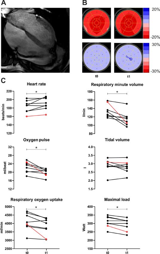
The effect of SARS-CoV-2 infection on cardiopulmonary parameters under moderate intensity (100–200 W)
is shown in Fig. 3. The moderate-intensity corresponded to 52% and 53% of maximum respiratory oxygen
uptake for COVID-19 and non-COVID groups. At this intensity level, significant changes in heart rate (+ 7.9%,
p = 0.028) and O2-pulse (− 8.2%, p = 0.0008) were only observed in athletes with a history of COVID-19. No other
parameters were changed significantly. The non-COVID group showed no significant changes during this period.
Correlation analysis. Overall reduction in maximal load was correlated with maximum respiratory minute
volume (R = 0.914, p < 0.001), maximum respiratory oxygen uptake (R = 0.838, p < 0.001), and the number of
single cardiac segments above reference value in T1-mapping (R = 0.669, p = 0.017). This number also correlated
to the decrease in maximum respiratory oxygen uptake (R = 0.711, p = 0.010) and respiratory minute volume
(R = 0.701, p = 0.01). Notably, the COVID-19 group’s increase in global longitudinal strain strongly correlates to
the rise in maximal heart rate (R = 0.778, p = 0.023) but not to other functional changes. Neither increased values
in T2-mapping nor the number of single cardiac segments above reference value in T2-mapping correlated with
functional cardiopulmonary parameters.
RT‑PCR swab of CPET‑masks. All masks were tested negative for SARS-CoV-2 RNA after CPET using
RT-PCR standard diagnostic procedure.
Discussion
The CPET showed a significant reduction of cardiopulmonary performance in elite athletes after SARS-CoV-2
infection. The affected athletes had a reduced maximal load, maximal oxygen uptake, a higher heart rate at
comparable exertion, and a significantly reduced O2 pulse. Additionally, the respiratory minute volume as a
parameter of pulmonary function was considerably reduced. The cardiac changes in heart rate and oxygen pulse
were present at moderate intensities, while the pulmonary effects became evident at higher intensity levels.
In contrast to the functional data, the morphological data only showed minor signs of inflammation that
did not reach the Lake-Louis criteria for the diagnosis of acute myocarditis. However, changes in the global
longitudinal strain strongly correlated to increasing maximal heart rate. In cardiac magnetic resonance imaging,
the number of segments with increased T1-values correlated to maximum respiratory oxygen uptake decrease.
In literature, isolated higher T1-values in athletes are also described as a possible effect of a history of intensive
training15. Notably, the athlete with pericardial effusion and highest T1- and T2-values also had the most signifi-
cant change in crucial spiroergometric parameters. His global longitudinal strain was not changed significantly,
but it must be noted that heart rate was different between both assessments (t0: 56/min vs. t1: 40/min). GLS and
Scientific Reports | (2021) 11:17798 | https://doi.org/10.1038/s41598-021-97120-x 5
Vol.:(0123456789)www.nature.com/scientificreports/
Figure 2. The athlete with the highest T1 values and pericardial effusion (red line) exhibited the greatest
reduction in functional cardiopulmonary parameters. t0: Routine CPET during preparation phase in summer
2020 t1: post-SARS-CoV-2 infection (COVID-19) in winter 2020 of athletes with a history of SARS-CoV-2
infection. (A) cardiac resonance imaging of athlete with pericardial effusion. (B) longitudinal strain (red) and
post systolic shortening index (blue) at t0 and t1. (C) key spiroergometric parameters at maximal load. The
athlete with the highest T1-mapping values in magnetic resonance imaging and with pericardial effusion during
the infection is highlighted in red. *: p < 0.05.
heart rate have been shown to be negatively c orrelated16, so that the unchanged GLS in combination with reduced
heart rate could potentially represent an impairment of left ventricular deformation. It can be assumed that
conventional echocardiographic parameters of left ventricular function such as left ventricular ejection fraction
and GLS are not suitable to detect minor cardiac involvement due to myocarditis17. Alterations of circumferential
strain and rotational LV deformation might be more sensitive to detect minor functional alterations in infectious
diseases due to distinct morphological alterations detected by cardiac M RI18,19.
Sequencing showed identical virus strains in all analysed respiratory swabs demonstrating a cluster infec-
tion. This finding is consistent with the results of contact tracing. The observed individual differences in clinical
outcomes are therefore not explained by specific viral lineages or a specific virus strain. At the time the B1.177
variant was responsible for around 10 percent of all cases in Germany20.
Risks for clinical personnel during CPET are not well evaluated. During our study, SARS-CoV-2 RNA was
not shown in mask swab samples. This finding suggests that athletes who repeatedly tested negative for SARS-
CoV-2 RNA in nasopharyngeal swabs do not pose an infectious risk for the examining staff.
The effect of infectious and especially viral disease on cardiopulmonary performance is not well characterized
outside of manifest m yocarditis21,22. While guidance for athletes with manifest myocarditis is available23–25, the
Scientific Reports | (2021) 11:17798 | https://doi.org/10.1038/s41598-021-97120-x 6
Vol:.(1234567890)www.nature.com/scientificreports/
Figure 3. Mean values of cardiac and pulmonary parameters under moderate intensity in a CPET (100–200
Watt) in athletes with or without a history of SARS-CoV-2 infection. t0: Routine CPET during preparation
phase in summer 2020 t1: post-SARS-CoV-2 infection (COVID-19) in winter 2020 or during other cardiac
outpatient diagnostics in winter 2020 (non-COVID). The athlete with the highest T1-mapping values in
magnetic resonance imaging and with pericardial effusion during the infection is highlighted in red. *: p < 0.05;
***: p < 0.001.
effect of SARS-CoV-2 infection on athletes missing this definition can still be relevant for safety while return-
ing to competition. Measuring individual performance restrictions and specific areas to target or take care of in
training is a valuable tool to avoid overload and regain optimal performance.
One could think that the fact that after diagnosis of SARS-CoV-2 infection the athletes did not participate
in an exercise of any kind and were instructed to keep their heart rate at low levels could have led to a general
change of cardiopulmonary parameters in C PET26. But contrary to this, it has been shown that after training
pauses with a duration of around two to three weeks, like in our COVID-19 group, the athletes are generally
expected to have a maintained cardiopulmonary performance with specific a daptations27–30. Additionally, the
non-COVID control group did not participate in structured team training and did not show any signs of the
effects seen in the COVID-19 group.
Scientific Reports | (2021) 11:17798 | https://doi.org/10.1038/s41598-021-97120-x 7
Vol.:(0123456789)www.nature.com/scientificreports/
A possible explanation for the observed changes could be the prevalence of thrombosis and pulmonary micro-
embolisms in COVID-1931–33, potentially contributing to reduced oxygen uptake, lower O2-pulse, and higher
heart rate. We did not see any clinical indications of thrombosis or pulmonary embolisms. However, this chain
of events cannot be discarded with current evidence, and further evaluation by prospective studies is necessary.
The early changes under moderate intensities of cardiac parameters while pulmonary changes did only occur
at maximal load could indicate a more pronounced functional effect of SARS-CoV-2 infection on the heart.
On the other hand, it is known that the cardiac reserve may generally be smaller than the pulmonary reserve
in elite a thletes34. This higher pulmonary reserve could conceal pulmonary restrictions at moderate intensities.
The findings of this study identify spiroergometry as a valuable addition to imaging studies post COVID4.
We screened athletes without myocarditis in the cardiac MRI using incremental cardiopulmonary exertion
tests. Spiroergometry identified relevant functional impairment in nearly all athletes of our homogenous group
of elite handball players. This, for the first time, shows the need for additional diagnostic measures to ensure a
safe and optimal return to training.
Limitations of the study include the low number of evaluated athletes and the retrospective non-blinded
analysis. However, the study group is highly homogeneous in terms of type of training and training status,
patient characteristics and time of infection and representative of elite athletes. The functional and imaging
measurements are highly standardized, and the parameters are well established from regular examinations in
athletes at our institution.
Conclusion
SARS-CoV2 infection caused functional impairment of cardiopulmonary performance in elite athletes. It seems
reasonable and safe to screen athletes after SARS-CoV2 infection with spiroergometry to detect performance
limitations. The knowledge of individual limitations and specific areas of higher or lower restriction can help to
tailor training to individual needs for optimal effects in regaining performance.
Received: 6 May 2021; Accepted: 12 August 2021
References
1. Chen, C., Zhou, Y. & Wang, D. W. SARS-CoV-2: A potential novel etiology of fulminant myocarditis. Herz 45, 230–232 (2020).
2. Corrado, D. & Zorzi, A. Sudden death in athletes. Int. J. Cardiol. 237, 67–70 (2017).
3. Peterson, D. F. et al. Aetiology and incidence of sudden cardiac arrest and death in young competitive athletes in the USA: A 4-year
prospective study. Br. J. Sports Med. https://doi.org/10.1136/bjsports-2020-102666 (2020).
4. Daniels, C. J. et al. Prevalence of clinical and subclinical myocarditis in competitive athletes with recent SARS-CoV-2 infection:
Results from the big ten COVID-19 cardiac registry. JAMA Cardiol. https://doi.org/10.1001/jamacardio.2021.2065 (2021).
5. Rajpal, S. et al. Cardiovascular magnetic resonance findings in competitive athletes recovering from COVID-19 infection. JAMA
Cardiol. 6, 116–118 (2021).
6. Wilson, M. G. et al. Cardiorespiratory considerations for return-to-play in elite athletes after COVID-19 infection: A practical
guide for sport and exercise medicine physicians. Br. J. Sports Med. 54, 1157–1161 (2020).
7. Budts, W. et al. Recommendations for participation in competitive sport in adolescent and adult athletes with Congenital Heart
Disease (CHD): Position statement of the Sports Cardiology & Exercise Section of the European Association of Preventive Car-
diology (EAPC), the European Society of Cardiology (ESC) Working Group on Adult Congenital Heart Disease and the Sports
Cardiology, Physical Activity and Prevention Working Group of the Association for European Paediatric and Congenital Cardiology
(AEPC). Eur. Heart J. 41, 4191–4199 (2020).
8. Tahir, F., Bin Arif, T., Ahmed, J., Malik, F. & Khalid, M. Cardiac manifestations of coronavirus disease 2019 (COVID-19): A com-
prehensive review. Cureus 12, e8021 (2020).
9. Verwoert, G. C. et al. Return to sports after COVID-19: A position paper from the Dutch Sports Cardiology Section of the Neth-
erlands Society of Cardiology. Neth. Heart J. Mon. J. Neth. Soc. Cardiol. Neth. Heart Found. 28, 391–395 (2020).
10. COVID-19 Treatment Guidelines. Clinical Spectrum | COVID-19 Treatment Guidelines. https://w ww.c ovid1 9trea tment guide lines.
nih.gov/overview/clinical-spectrum/ (2021).
11. Stöbe, S. et al. Echocardiographic characteristics of patients with SARS-CoV-2 infection. Clin. Res. Cardiol. Off. J. Ger. Cardiac
Soc. 109, 1549–1566 (2020).
12. Lenk, K. et al. Angiography-based quantitative coronary contrast-flow ratio measurements correlate with myocardial ischemia
assessed by stress MRI. Int. J. Cardiovasc. Imaging 36, 1407–1416 (2020).
13. COG-UK. Available at https://pangolin.cog-uk.io/ (2021).
14. JASP Team. JASP (Version 0.14.1)[Computer software] (2020), https://jasp-stats.org/
15. Görmeli, C. A. et al. Assessment of myocardial changes in athletes with native T1 mapping and cardiac functional evaluation using
3 T MRI. Int. J. Cardiovasc. Imaging 32, 975–981 (2016).
16. Peverill, R. E., Cheng, K., Cameron, J., Donelan, L. & Mottram, P. M. Relationships of global longitudinal strain with s′, long-axis
systolic excursion, left ventricular length and heart rate. PLoS ONE 15, e0235791 (2020).
17. Stöbe, S., Hagendorff, A., Gutberlet, M. & Tayal, B. Myocardial work: A modern tool to detect possible compensation mechanism
of deformation in acute myocarditis with preserved left ventricular function. J. Cardiovasc. Echogr. 30, 206–210 (2020).
18. Döbel, T. et al. Possible new options and benefits to detect myocarditis, right ventricular remodeling and coronary anomalies by
echocardiography in systematic preparticipation screening of athletes. Int. J. Cardiovasc. Imaging 36, 1855–1885 (2020).
19. Hofrichter, P. et al. Analysis of left ventricular rotational deformation by 2D speckle tracking echocardiography: A feasibility study
in athletes. Int. J. Cardiovasc. Imaging https://doi.org/10.1007/s10554-021-02213-3 (2021).
20. outbreak.info. outbreak.info. https://outbreak.info/situation-reports?pango=B.1.177&selected=DEU&loc=USA&loc=USA_US-
CA&loc=DEU (2021).
21. Müller, I. et al. Effect of schistosomiasis and soil-transmitted helminth infections on physical fitness of school children in Côte
d’Ivoire. PLoS Negl. Trop. Dis. 5, e1239 (2011).
22. Friman, G. & Wesslén, L. Infections and exercise in high-performance athletes. Immunol. Cell Biol. 78, 510–522 (2000).
23. McKinney, J. et al. COVID-19-myocarditis and return to play: Reflections and recommendations from a Canadian Working Group.
Can. J. Cardiol. https://doi.org/10.1016/j.cjca.2020.11.007 (2020).
24. Compagnucci, P. et al. Myocardial inflammation, sports practice, and sudden cardiac death: 2021 Update. Medicina (Kaunas) 57,
277 (2021).
Scientific Reports | (2021) 11:17798 | https://doi.org/10.1038/s41598-021-97120-x 8
Vol:.(1234567890)www.nature.com/scientificreports/
25. Phelan, D. et al. Screening of potential cardiac involvement in competitive athletes recovering from COVID-19: An expert con-
sensus statement. JACC Cardiovasc. Imaging 13, 2635–2652 (2020).
26. Fikenzer, S. et al. Impact of COVID-19 lockdown on endurance capacity of elite handball players. J. Sports Med. Phys. Fitness
https://doi.org/10.23736/S0022-4707.20.11501-9 (2020).
27. Colliander, E. B. & Tesch, P. A. Effects of detraining following short term resistance training on eccentric and concentric muscle
strength. Acta Physiol. Scand. 144, 23–29 (1992).
28. Girardi, M., Casolo, A., Nuccio, S., Gattoni, C. & Capelli, C. Detraining effects prevention: A new rising challenge for athletes.
Front. Physiol. 11, 588784 (2020).
29. Pereira, L. A. et al. The impact of detraining on cardiac autonomic function and specific endurance and muscle power performances
of high-level endurance runners. J. Sports Med. Phys. Fitness 56, 1583–1591 (2016).
30. Schlumberger, A. & Schmidtbleicher, D. Zeitlich Verzögerte Effekte Beim Krafttraining. Leistungssport 28(3), 33–38 (1998).
31. Ackermann, M. et al. Pulmonary vascular endothelialitis, thrombosis, and angiogenesis in Covid-19. N. Engl. J. Med. 383, 120–128
(2020).
32. Idilman, I. S. et al. Lung and kidney perfusion deficits diagnosed by dual-energy computed tomography in patients with COVID-
19-related systemic microangiopathy. Eur. Radiol. 31, 1090–1099 (2021).
33. Manolis, A. S., Manolis, T. A., Manolis, A. A., Papatheou, D. & Melita, H. COVID-19 infection: viral macro- and micro-vascular
coagulopathy and thromboembolism/prophylactic and therapeutic management. J. Cardiovasc. Pharmacol. Ther. 26, 12–24 (2021).
34. Weder, M. M. & Truwit, J. D. Pulmonary disorders in athletes. Clin. Sports Med. 30, 525–536 (2011).
Author contributions
S.F., A.H., P.H. and U.L. were involved in planning the work. S.F. and A.K. performed and spirometry and CPET,
S.T.S. and A.H. performed echocardiographic and magnetic resonance imaging. C.P. performed virological
diagnostics. S.F. and A.K. processed the experimental data, performed the analysis, drafted the manuscript, and
designed the figures in consultation with U.L. D.L. and U.R. aided in interpreting the results and worked on the
manuscript. A.H., P.H. and U.L. supervised the work. All authors discussed the results and commented on the
manuscript.
Funding
Open Access funding enabled and organized by Projekt DEAL. We acknowledge support from Leipzig University
for Open Access Publishing.
Competing interests
The authors declare no competing interests.
Additional information
Correspondence and requests for materials should be addressed to S.F.
Reprints and permissions information is available at www.nature.com/reprints.
Publisher’s note Springer Nature remains neutral with regard to jurisdictional claims in published maps and
institutional affiliations.
Open Access This article is licensed under a Creative Commons Attribution 4.0 International
License, which permits use, sharing, adaptation, distribution and reproduction in any medium or
format, as long as you give appropriate credit to the original author(s) and the source, provide a link to the
Creative Commons licence, and indicate if changes were made. The images or other third party material in this
article are included in the article’s Creative Commons licence, unless indicated otherwise in a credit line to the
material. If material is not included in the article’s Creative Commons licence and your intended use is not
permitted by statutory regulation or exceeds the permitted use, you will need to obtain permission directly from
the copyright holder. To view a copy of this licence, visit http://creativecommons.org/licenses/by/4.0/.
© The Author(s) 2021
Scientific Reports | (2021) 11:17798 | https://doi.org/10.1038/s41598-021-97120-x 9
Vol.:(0123456789)You can also read