CHARACTERIZATION OF AN ASYMPTOMATIC COHORT OF SEVERE ACUTE RESPIRATORY SYNDROME CORONAVIRUS 2 (SARS- COV-2) INFECTED INDIVIDUALS OUTSIDE OF WUHAN ...
←
→
Page content transcription
If your browser does not render page correctly, please read the page content below
Clinical Infectious Diseases
    MAJOR ARTICLE

Characterization of an Asymptomatic Cohort of Severe
Acute Respiratory Syndrome Coronavirus 2 (SARS-
CoV-2) Infected Individuals Outside of Wuhan, China
Yubo Wang,1,a Yong He,1, Jin Tong,2,a Yalan Qin,3 Ting Xie,4 Jianghua Li,1 Jianrong Li,5 Jianhua Xiang,6 Yong Cui,7 Elizabeth S. Higgs,8 and Jianglin Xiang4
1
 Department of Respiratory Medicine, Daping Hospital, Army Medical University, Chongqing, China, 2Department of Respiratory Medicine, Second Affiliated Hospital of Chongqing Medical
University, Chongqing, China, 3Department of Critical Care Medicine, Second Affiliated Hospital of Chongqing Medical University, Chongqing, China, 4Department of Infection, Chongqing Three
Gorges Central Hospital, Chongqing, China, 5Department of Respiratory Medicine, Wanzhou District Hospital, Chongqing, China, 6Department of Respiratory Medicine, Chongqing Three Gorges
Central Hospital, Chongqing, China, 7Department of Critical Care Medicine, Chongqing Three Gorges Central Hospital, Chongqing, China, and 8Division of Clinical Research, National Institute of

                                                                                                                                                                                                  Downloaded from https://academic.oup.com/cid/article/71/16/2132/5842166 by guest on 25 November 2020
Allergy and Infectious Diseases, National Institutes of Health, Bethesda, Maryland, USA

   Purpose. We aimed to further clarify the epidemiological and clinical characteristics of asymptomatic severe acute respiratory
syndrome coronavirus 2 (SARS-CoV-2) infections.
   Methods. We identified close contacts of confirmed coronavirus disease 2019 (COVID-19) cases in northeast Chongqing,
China, who were confirmed by real-time reverse transcription polymerase chain reaction-positive (RT-PCR+). We stratified this
cohort by normal vs abnormal findings on chest computed tomography (CT) and compared the strata regarding comorbidities,
demographics, laboratory findings, viral transmission and other factors.
   Results. Between January 2020 and March 2020, we identified and hospitalized 279 RT-PCR+ contacts of COVID-19 patients. 63 (23%)
remained asymptomatic until discharge; 29 had abnormal and 34 had normal chest CT findings. The mean cohort age was 39.3 years, and
87.3% had no comorbidities. Mean time to diagnosis after close contact with a COVID-19 index patient was 16.0 days, and it was 13.4 days
and 18.7 days for those with abnormal and normal CT findings, respectively (P < .05). Nine patients (14.3%) transmitted the virus to others;
4 and 5 were in the abnormal and normal CT strata, respectively. The median length of time for nucleic acid to turn negative was 13 days
compared with 10.4 days in those with normal chest CT scans (P < .05).
   Conclusions. A portion of asymptomatic individuals were capable of transmitting the virus to others. Given the frequency and po-
tential infectiousness of asymptomatic infections, testing of traced contacts is essential. Studies of the impact of treatment of asympto-
matic RT-PCR+ individuals on disease progression and transmission should be undertaken.
   Keywords. COVID-19; SARS-CoV-2; asymptomatic infection; characterization.

In December 2019 a novel coronavirus, later named severe acute                                       Symptomatic COVID-19 patients and asymptomatic cases are both a
respiratory syndrome coronavirus 2 (SARS-CoV-2), caused a large                                      source of infection, and patients in the incubation period can transmit
outbreak of infectious disease, designated coronavirus disease 2019                                  SARS-CoV-2 to other persons [7–10].
(COVID-19). It posed a great threat to public health worldwide [1].                                     Patients with symptoms are more likely to be detected.
Compared with SARS-CoV or Middle East respiratory syndrome                                           Among patients with SARS-CoV-2, fever has been the most
coronavirus, it appears to be transmitted more easily [2–4]. SARS-                                   common symptom, followed by dry cough, dyspnea, myalgia,
CoV-2 can be transmitted through a variety of modes, including                                       headache, and diarrhea [11–13]. In severe COVID-19 cases,
droplets, close contact, aerosols, and potentially fecal–oral transmis-                              symptoms can progress to acute respiratory distress syndrome,
sion [5, 6]. People of all ages are generally susceptible to the virus.                              septic shock, and metabolic acidosis [14]. The clinical presen-
                                                                                                     tation of COVID-19, therefore, ranges from asymptomatic
                                                                                                     to severe respiratory illness [15]. The presentation of symp-
   Received 17 March 2020; editorial decision 19 May 2020; accepted 20 May 2020; published
online May 22, 2020.                                                                                 toms provides an opportunity for case detection and isolation,
                                                                                                     facilitating the interruption of transmission. Because most
   a
    Y. W. and J. T. contributed equally to this work.
   Correspondence: Y. He, Department of Respiratory Medicine, 10# Changjiang Zhi Road,
Chongqing 400042, China (heyong8998@126.com).                                                        SARS-CoV patients had severe symptoms, they were relatively
Clinical Infectious Diseases®  2020;71(16):2132–8                                                    simple to identify and isolate, thus interrupting the chain of
© The Author(s) 2020. Published by Oxford University Press for the Infectious Diseases               transmission. Unfortunately, COVID-19 has an incubation pe-
Society of America. This is an Open Access article distributed under the terms of the Creative
Commons Attribution-NonCommercial-NoDerivs licence (http://creativecommons.org/licenses/             riod from infection to onset of symptoms that lasts an average
by-nc-nd/4.0/), which permits non-commercial reproduction and distribution of the work, in any       of 10 days (with a reported range of 2–14 days) [11, 16, 17].
medium, provided the original work is not altered or transformed in any way, and that the
work is properly cited. For commercial re-use, please contact journals.permissions@oup.com
                                                                                                     The virus is also able to spread from one person to another be-
DOI: 10.1093/cid/ciaa629                                                                             fore any symptom onset [14]. It is reported that about 1.2% of

2132 • cid 2020:71 (15 October) • Wang et al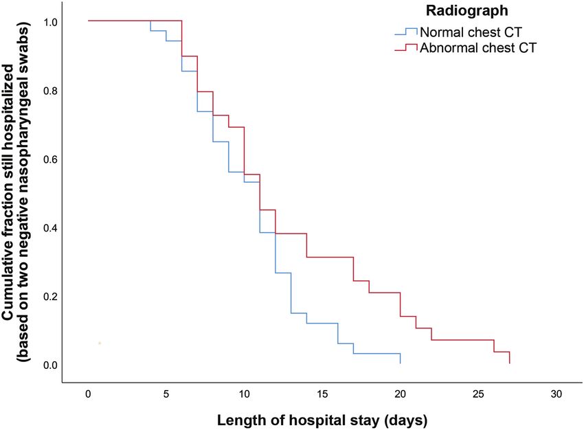
patients have no actual clinical manifestations during the en-        are in accordance with the interim guidance from the World
tire course of the disease [18]. Asymptomatic cases, however,         Health Organization. Following a positive SARS-CoV-2 nucleic
particularly those with no history of contact with a known            acid test, a chest CT scan was done [26]. Individuals were then
SARS-CoV-2 patient, are hard to identify. There are several case      divided into 2 groups based on chest CT scan: a group with
reports of SARS-CoV-2 transmission from asymptomatic in-              changes visible on the radiographic imaging, called asympto-
dividuals [8, 9, 13, 19], including a report of presumed trans-       matic COVID-19 patients, and a group without any detectable
mission by asymptomatic carriers with normal chest computed           imaging changes, called asymptomatic with normal chest CT.
tomography (CT) scans [20]. Together, these traits make con-          During hospitalization, all participants, regardless of group,
trol of SARS-CoV-2 very difficult.                                    had follow-up chest CT scans every 4 to 5 days until discharge.
   The exact profile of these asymptomatic cases and their            Individuals initially allocated to the asymptomatic with normal
role within the wider epidemic are unclear. It is important           chest CT group were immediately reallocated to the asympto-
to understand the proportion of asymptomatic or mildly                matic COVID-19 patient group if a new abnormal finding was
ill cases and their role in driving the epidemic [21]. Since          detected on any chest CT during hospitalization.

                                                                                                                                         Downloaded from https://academic.oup.com/cid/article/71/16/2132/5842166 by guest on 25 November 2020
many SARS-CoV-2 infections are asymptomatic, subclin-                    With the approval of the ethics committee, we collected both
ical, or very mild, the hallmarks and course of asympto-              epidemiological data and medical reports for these 2 groups.
matic infection warrant further investigation [22–24]. Our            Epidemiological data collection was achieved by interviewing
goal was to document the characteristics of asymptomatic              each patient and their family members, including the dates and
infections and identify factors associated with asympto-              times of close contact with (working, living, or gathering to-
matic infection, enabling the formulation of corresponding            gether) or to exposure individuals from the affected area (not
strategies and control measures.                                      only Wuhan) with confirmed or suspected SARS-CoV-2 in-
                                                                      fection. All the data were checked by 2 researchers. If anything
METHODS                                                               was ambiguous, we consulted the attending physician as soon
Data Sources                                                          as possible.
We conducted a retrospective study that focused on the charac-
                                                                      Laboratory Tests
teristics and length of real-time reverse transcription polymerase
                                                                      Diagnosis was made by nasopharyngeal swab and RT-PCR
chain reaction (RT-PCR) positivity and associated hospitaliza-
                                                                      to confirm SARS-CoV-2 infection. The virus detection
tion of confirmed asymptomatic cases of SARS-CoV-2 in the
                                                                      protocol was repeated on subsequent days until a negative
northeast region of Chongqing, China, in the Chongqing Three
                                                                      nucleic acid test was obtained on 2 consecutive occasions.
Gorges Center Hospital and Wanzhou District Hospital since
                                                                      Other laboratory tests were conducted at admission.
January 2020.
   Active contact tracing in China includes nasopharyngeal
                                                                      Treatment
swab diagnostic testing of all contacts regardless of symptoms.
In accordance with Chinese guidelines for the investigation and       Per standard Chinese protocol, all patients were treated
management of close contacts of COVID-19 cases, close con-            with putative antiviral agents on admission [25]. Fifty-
tacts were defined as people who had not taken effective protec-      eight of the 63 in the asymptomatic COVID-19 cohort
tion while in close contact with suspected or confirmed cases         received antiviral treatment with α-interferon inhalation
2 days prior to symptom onset or, if the case was asymptomatic,       (5 million units twice daily) and lopinavir/ritonavir oral
2 days before sampling [25]. We screened close contacts from          (400 mg/100 mg twice daily) with 10 mg thymosin injec-
January 2020 through March 2020 in northeast Chongqing                tion every day to improve immunity. Of the remaining 5
by RT-PCR of nasopharyngeal swabs. Only patients with an              patients, 1 received lopinavir/ritonavir (400 mg/100 mg
RT-PCR–confirmed infection and without any apparent symp-             twice daily), Arbidol (200 mg 3 times daily), α-interferon
toms, including (but not limited to) cough, fever, shortness of       inhalation (5 million units twice daily), thymosin, and
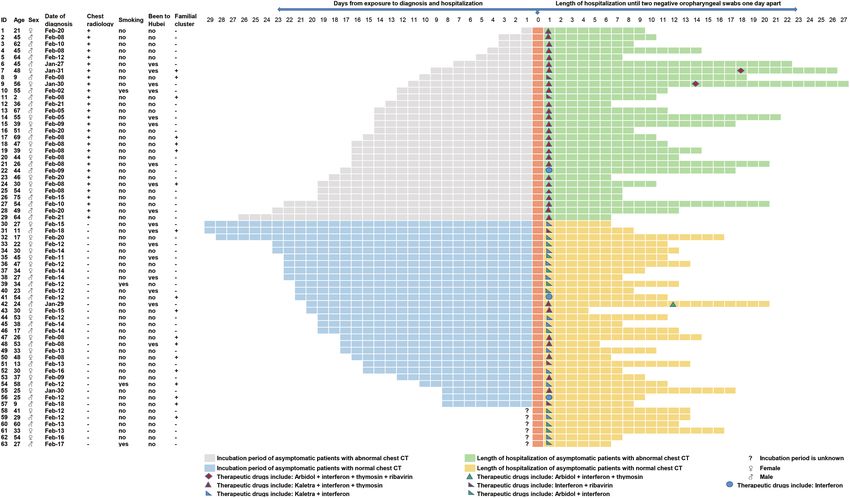
breathless, and muscle soreness, were enrolled in the study.          ribavirin (500 mg twice daily; Figure 1). RT-PCR testing
   These individuals were identified through close symptomatic        was repeated 5 to 7 days after treatment. Patients were dis-
contacts in both the clinic and the community. In accordance          charged after receiving 2 negative results at least 24 hours
with China’s prevention and control policy, all RT-PCR–positive       apart and a normal chest CT scan [24].
cases were hospitalized and treated, whether they had symptoms
or not. We monitored asymptomatic individuals daily during            Statistical Analyses
hospitalization to document the development of any signs and          Continuous variables were expressed as the means and standard
symptoms. We recategorized patients who were asymptomatic             deviations or medians with interquartile ranges. Categorical
at baseline to symptomatic if they reported any symptoms. Case        variables were summarized as the counts and percentages in
definitions of confirmed human infection with SARS-CoV-2              each category. The t test or Mann-Whitney U test was applied

                                                             Asymptomatic Infections With SARS-COV-2 • cid 2020:71 (15 October) • 2133
Downloaded from https://academic.oup.com/cid/article/71/16/2132/5842166 by guest on 25 November 2020
Figure 1. Clinical course and treatment of the 63 asymptomatic patients with severe acute respiratory syndrome coronavirus 2. Abbreviation: CT, computed tomography.
to continuous variables, and the Fisher exact test or Pearson χ2 the exact date of close contact with someone with confirmed or
test was used for categorical variables. All analyses were con- suspected SARS-CoV-2 infection. Of the remaining 57 cases, the
ducted with SPSS 26.0. mean time from exposure to illness onset was 16.0 days (SD, 6.6).
In asymptomatic COVID-19 patients, this period was 13.4 days
RESULTS (SD, 6.3), while in asymptomatic cases with normal chest CT scans,
it was 18.7 days (SD, 5.8) (P < .05). Among the 63 cases, there were
General Condition
18 cases of familial clustering in which 9 cases (14.3%) transmitted
Using active contact tracing of index cases with RT-PCR screening,
the virus to others outside of their families, 4 from the asympto-
279 cases were diagnosed with SARS-CoV-2 infection. Among
matic COVID-19 patients and 5 from the asymptomatic cases with
them, 63 had no symptoms either at the time of diagnosis or
normal chest CT scans (Figure 1).
throughout hospitalization. However, 29 of the 63 cases had ab-
normal chest CT findings, while the remaining 34 had normal
chest CT findings. In total, the mean age was 39.3 years (standard Clinical Laboratory and Immunologic Findings
deviation [SD], 16.5). The mean age of cases with abnormal chest On admission, only 4 of the 63 (6.3%) cases showed leucopenia
CT scans was 46.3 years (SD, 16.8) compared with 33.4 years (SD, (white blood cell count 10 × 109/L). All the cases had a
than half of the 63 cases were male (34, 54%); however, 62.1% of neutrophil count within the normal range. Eight (12.7%) cases
patients with abnormal CT findings were male. Comorbid condi- had a low lymphocyte count (Table 1. Personal and Clinical Characteristics of 63 Asymptomatic Patients With Coronavirus Disease 2019
Chest Radiology
Characteristic All Asymptomatic Patients (N = 63) Normal Chest CT (n = 34) Abnormal Chest CT (n = 29) P Value
Age (y) 39.30 ± 16.45 33.36 ± 13.81 46.26 ± 16.79 .001
Age groups (y) .003
≤18 7 (11.1%) 5 (14.7%) 2 (6.9%)
19–40 25 (39.7%) 19 (55.9%) 6 (20.7%)
41–65 28 (44.4%) 10 (29.4%) 18 (62.1%)
≥66 3 (4.8%) 0 3 (10.3%)
Sex .233
Male 34 (54%) 16 (47.1%) 18 (62.1%)
Female 29 (46%) 18 (52.9%) 11 (37.9%)
Coexisting conditions .103
Downloaded from https://academic.oup.com/cid/article/71/16/2132/5842166 by guest on 25 November 2020
Any 53 (84.1%) 31 (91.2%) 22 (75.9%)
Hypertension 3 (4.8%) 0 3 (10.3%)
Cardiovascular disease 1 (1.6%) 0 1 (3.4%)
Diabetes 1 (1.6%) 0 1 (3.4%)
Chronic obstructive pulmonary disease 1 (1.6%) 1 (2.9%) 0
Systemic lupus erythematosus 1 (1.6%) 0 1 (3.4%)
Gout 1 (1.6%) 1 (2.9%) 0
Chronic gastritis 2 (3.2%) 1 (2.9%) 1 (3.4%)
Exposure history in Hubei .504
Yes 17 (27%) 8 (23.5%) 9 (31%)
No 46 (73%) 26 (76.5%) 20 (69%)
Familial cluster 18 (28.6%) 11 (32.4%) 7 (24.1%) .472
Days from exposure to diagnosis 15.98 ± 6.55 (n = 57) 18.68 ± 5.79 (n = 28) 13.38 ± 6.25 (n = 29) .002
Number of others infected 1.000
No 54 (85.7%) 29 (85.3%) 25 (86.2%)
Yes 9 (14.3%) 5 (14.7%) 4 (13.8%)
Abbreviation: CT, computed tomography.
Clinical Outcome source of infection. A recent study reported that the viral load
Fitness for discharge was based on 2 consecutive negative in asymptomatic patients is similar to that in symptomatic pa-
RT-PCR tests of oropharyngeal swabs and a normal chest CT tients, further underscoring the transmission potential of these
scan. All patients were discharged, and no one died. The median asymptomatic patients [27]. Together, these results suggest that
time between the initial positive RT-PCR test and discharge was strict isolation and screening should be carried out in all asymp-
significantly longer in asymptomatic COVID-19 patients than tomatic close contacts. CT scans can assist in the detection of
in asymptomatic patients with normal chest CT scans, 13.0 and asymptomatic pneumonia but cannot identify asymptomatic
10.4 days, respectively (P < .05; Figure 2). cases with normal chest CT scans. If nucleic acid testing were
reserved only for suspected patients with obvious symptoms,
many contacts with mild symptoms or those who are asympto-
DISCUSSION
matic would be missed, as would their contacts.
The number of SARS-CoV-2 infections is still rising rapidly in Since there are no obvious symptoms, asymptomatic cases
many parts of the world, and asymptomatic infections likely typically remain undiagnosed for a relatively long time. In our
play a large role in transmission [22]. To our surprise, 23% of study, the mean latency between close contact and diagnosis
those identified with SARS-CoV-2 infection, including those was 16.0 days, with a maximum of 29 days. This suggests that
with abnormal chest CT scans on admission, were completely RT-PCR screening should be undertaken even if an individual
asymptomatic throughout their infections—a much higher shows no symptoms more than 14 days after close contact with
proportion than previously reported [18]. The high propor- an infected person. In asymptomatic patients, those whose CT
tion may be related to the extensive and strict close-contact scans show signs of pneumonia are typically diagnosed earlier
screening policy adopted locally that resulted in early detec- than those without such signs.
tion and treatment of cases. This finding suggests that there are Because the pathogenesis of COVID-19 is not well under-
actually more asymptomatic COVID-19 cases than previously stood, all nucleic acid–positive cases received antiviral treat-
thought, highlighting asymptomatic cases as a noteworthy ment to prevent progression of the disease. Although the
Asymptomatic Infections With SARS-COV-2 • cid 2020:71 (15 October) • 2135Table 2. Laboratory Findings in 63 Asymptomatic Patients With Coronavirus Disease 2019
Chest Radiology
Variable All Asymptomatic Patients (N = 63) Normal Chest CT (n = 34) Abnormal Chest CT (n = 29) P Value
White blood cell count (×109/L) 6.81 ± 1.98 7.10 ± 2.04 6.47 ± 1.88 .207
White blood cell count (×109/L) .854
10 4 (6.3%) 3 (8.8%) 1 (3.4%)
Neutrophil count (×109/L) 4.59 ± 1.78 4.91 ± 1.89 4.21 ± 1.59 .122
Lymphocyte count (×109/L) 1.61 (1.24–1.92) 1.66 (1.35–1.95) 1.59 (1.19–1.93) .490
Lymphocyte count (×109/L) .129infection [29]. As a group, the asymptomatic cases in our reading and editing of the manuscript. With the permission of the cor-
responding authors, we can provide participant data without names and
study were younger and had fewer comorbidities compared
identifiers. Data can be provided after the article is published. Once the
with severe cases [11, 12]. Research shows that the adaptive data is made public, the research team will provide an email address for
immune response against the virus in people with asympto- communication. The corresponding authors have the right to decide
matic infection is stronger than in those with symptomatic whether to share the data or not based on the research objectives and
plan provided. This study was reviewed and approved by the Medical
infection [30, 31]. Asymptomatic individuals without im- Ethical Committee of Daping Hospital and Chongqing Three Gorges
aging abnormalities are younger than those with pneumonia. Central Hospital.
From our results, it seems that young individuals with normal Financial support. This study was supported by Excellent Talent
Pool grants for Y. W. and Y. H. and Training Plan of Clinical Medical
immune function and without comorbidities are more likely
Scientific Research Talents for Y. H. from the Army medical university
to become asymptomatically infected. (2018XLC1015).
All patients in the study, with and without abnormal chest Potential conflicts of interest. The authors: No reported conflicts of
CT scans, steadily improved and were discharged smoothly interest. All authors have submitted the ICMJE Form for Disclosure of
Potential Conflicts of Interest.
after testing negative by RT-PCR. However, we do not know
Downloaded from https://academic.oup.com/cid/article/71/16/2132/5842166 by guest on 25 November 2020
whether these asymptomatic cases, particularly those who had References
developed an abnormal chest CT scan, would have progressed 1. World Health Organization. Novel coronavirus (COVID-19) situation. 2020.
to more severe disease without early antiviral treatment. The re- Available at: https://experience.arcgis.com/experience/685d0ace521648f8a5beee
ee1b9125cd. Accessed 21 February 2020.
sults also suggest the need for rigorous randomized, controlled 2. Lipsitch M, Cohen T, Cooper B, et al. Transmission dynamics and control of se-
studies to determine what antiviral therapies are safe and ef- vere acute respiratory syndrome. Science 2003; 300:1966–70.
3. Wallinga J, Teunis P. Different epidemic curves for severe acute respiratory
fective and whether early antiviral treatment for asymptomatic syndrome reveal similar impacts of control measures. Am J Epidemiol 2004;
RT-PCR+ individuals can prevent progression of disease and/or 160:509–16.
4. Lin Q, Chiu AP, Zhao S, He D. Modeling the spread of Middle East respiratory
the length of time cases remain RT-PCR+. Finally, SARS-CoV-2
syndrome coronavirus in Saudi Arabia. Stat Methods Med Res 2018; 27:1968–78.
vaccine study designs should not simply use symptomatic 5. Wang C, Horby PW, Hayden FG, Gao GF. A novel coronavirus outbreak of global
COVID-19 as an end point but should include asymptomatic health concern. Lancet 2020; 395:470–3.
6. van Doremalen N, Bushmaker T, Morris DH, et al. Aerosol and surface stability of
infections as well. SARS-CoV-2 as compared with SARS-CoV-1. N Engl J Med 2020; 382:1564–7.
This study has several limitations. First, only 63 asympto- 7. New coronavirus pneumonia prevention and control program. 5th ed. (in
Chinese). 2020. Available at: http://www.nhc.gov.cn/xcs/zhengcwj/202002/a
matic cases were included; as such it may be that we found no 5d6f7b8c48c451c87dba14889b30147.shtml. Accessed 21 February 2020.
other associated epidemiological factors due to sample size. 8. Pan X, Chen D, Xia Y, et al. Asymptomatic cases in a family cluster with SARS-
CoV-2 infection. Lancet Infect Dis 2020; doi: S1473-3099(20)30114-6
Second, we used a qualitative RT-PCR test and could not quan-
9. Yu P, Zhu J, Zhang Z, et al. A familial cluster of infection associated with the 2019
titate viral load; thus, asymptomatic cases with low viral load novel coronavirus indicating potential person-to-person transmission during the
may have been missed. Third, a precise incubation period and incubation period. J Infect Dis 2020. doi: 10.1093/infdis/jiaa077
10. Li Q, Guan X, Wu P, et al. Early transmission dynamics in Wuhan, China, of novel
transmissibility are difficult to document due to the concealed coronavirus-infected pneumonia. N Engl J Med 2020; 382:1199–207.
nature of asymptomatic infection. Finally, our study does not 11. Huang C, Wang Y, Li X, et al. Clinical features of patients infected with 2019 novel
coronavirus in Wuhan, China. Lancet 2020; 395:497–506.
provide a mechanistic explanation for asymptomatic status. 12. Chen N, Zhou M, Dong X, et al. Epidemiological and clinical characteristics of 99
A study that links asymptomatic status with differences in in- cases of 2019 novel coronavirus pneumonia in Wuhan, China: a descriptive study.
Lancet 2020; 395:507–13.
dividual immunity, virus serotypes, viral load or other factors
13. Chan JF, Yuan S, Kok KH, et al. A familial cluster of pneumonia associated with
would contribute useful insights. the 2019 novel coronavirus indicating person-to-person transmission: a study of
In conclusion, we provide an initial assessment of epi- a family cluster. Lancet 2020; 395:514–23.
14. Li JY, You Z, Wang Q, et al. The epidemic of 2019-novel-coronavirus (2019-
demiological characteristics of asymptomatic infections of nCoV) pneumonia and insights for emerging infectious diseases in the future.
SARS-CoV-2. Early identification of SARS-CoV-2 cases with Microbes Infect 2020; doi: S1286-4579(20)30030-7
15. Lai CC, Shih TP, Ko WC, et al. Severe acute respiratory syndrome coronavirus 2
subsequent isolation and treatment may contribute to decreased (SARS-CoV-2) and coronavirus disease-2019 (COVID-19): the epidemic and the
transmission and mortality. challenges. Int J Antimicrob Agents 2020; 55:105924.
16. Zhu N, Zhang D, Wang W, et al. A novel coronavirus from patients with pneu-
monia in China, 2019. N Engl J Med 2020; 382:727–33.
Notes 17. Zhou P, Yang XL, Wang XG, et al. A pneumonia outbreak associated with a new
Author contributions. Y. H. and J. lX. conceived of and designed the coronavirus of probable bat origin. Nature 2020; 579:270–3.
study, had full access to all data, and take responsibility for the integrity of 18. The Novel Coronavirus Pneumonia Emergency Response Epidemiology
the data and the accuracy of the data analysis. Y. W., J. T., Y. Q., T. X., J. hX., Team. The epidemiological characteristics of an outbreak of 2019 novel coro-
Y. C., J. rL., and J. lX. contributed to data acquisition. Y. H., Y. W., E. S. H., navirus diseases (COVID-19) in China. Zhonghua Liu Xing Bing Xue Za Zhi
2020; 41:145–51.
and J. T. contributed to writing the report. J. hL. contributed to the statistical
19. Hu Z, Song C, Xu C, et al. Clinical characteristics of 24 asymptomatic infections
analysis. All authors contributed to data analysis or data interpretation and
with COVID-19 screened among close contacts in Nanjing, China. Sci China Life
reviewed and approved the final version. Sci 2020; 63:706–11.
Acknowledgments. The authors thank Dr Saffron Willis-Owen and Dr 20. Bai Y, Yao L, Wei T, et al. Presumed asymptomatic carrier transmission of
Youming Zhang from the National Heart and Lung Institute, Imperial COVID-19. JAMA 2020. doi: 10.1001/jama.2020.2565
College London, and the Dr Robert Taylor from the National Institute of 21. Qiu J. Covert coronavirus infections could be seeding new outbreaks. Nature
Allergy and Infectious Diseases, National Institutes of Health, for critical 2020. doi: 10.1038/d41586-020-00822-x
Asymptomatic Infections With SARS-COV-2 • cid 2020:71 (15 October) • 213722. Al-Tawfiq JA. Asymptomatic coronavirus infection: MERS-CoV and SARS-CoV-2 27. Zou L, Ruan F, Huang M, et al. SARS-CoV-2 viral load in upper respiratory spe-
(COVID-19). Travel Med Infect Dis 2020; 101608. doi: 10.1016/j.tmaid.2020.101608 cimens of infected patients. N Engl J Med 2020; 382:1177–9.
23. Gao WJ, Li LM. Advances on presymptomatic or asymptomatic carrier transmis- 28. MO X, Qin W, Qihua F, Ming G. Understanding the influence factors in viral nu-
sion of COVID-19 . Zhonghua Liu Xing Bing Xue Za Zhi 2020; 41:485–8. cleic acid test of 2019 novel coronavirus (2019-nCoV). Chin J Lab Med 2020.
24. Wang Y, Wang Y, Chen Y, et al. Unique epidemiological and clinical features of 43:E002.
the emerging 2019 novel coronavirus pneumonia (COVID-19) implicate special 29. Zhang W, Du RH, Li B, et al. Molecular and serological investigation of 2019-
control measures. J Med Virol 2020. doi: 10.1002/jmv.25748 nCoV infected patients: implication of multiple shedding routes. Emerg Microbes
25. Chinese Center For Disease Control and Prevention. The guideline for the inves- Infect 2020; 9:386–9.
tigation and management of close contacts of COVID cases. 2020. Available at: 30. Simon-Loriere E, Duong V, Tawfik A, et al. Increased adaptive immune responses
http://www.gov.cn/xinwen/2020-01/26/content_5472235.htm. and proper feedback regulation protect against clinical dengue. Sci Transl Med
26. Office of the National Health Commission. The guideline on diagnosis and treat- 2017; 9. pii: eaal5088.
ment of COVID (The Trial, fourth edition). 2020. Available at: http://swjj.gzlps. 31. Guan WJ, Ni ZY, Hu Y, et al. Clinical characteristics of coronavirus disease 2019
gov.cn/gzdt/gzdt_45694/202002/t20200221_50515109.html. in China. N Engl J Med 2020. doi: 10.1056/NEJMoa2002032
Downloaded from https://academic.oup.com/cid/article/71/16/2132/5842166 by guest on 25 November 2020
2138 • cid 2020:71 (15 October) • Wang et alYou can also read


