Controllable cardiac synthesis via disentangled anatomy arithmetic
←
→
Page content transcription
If your browser does not render page correctly, please read the page content below
Controllable cardiac synthesis via disentangled
 anatomy arithmetic

 Spyridon Thermos1 , Xiao Liu1 , Alison O’Neil1,3 , and Sotirios A. Tsaftaris1,2
 1
 School of Engineering, University of Edinburgh, Edinburgh EH9 3FB, UK
 2
 The Alan Turing Institute, London NW1 2DB, UK
 3
 Canon Medical Research Europe, Edinburgh EH6 5NP, UK
arXiv:2107.01748v1 [eess.IV] 4 Jul 2021

 {SThermos,Xiao.Liu,S.Tsaftaris}@ed.ac.uk Alison.ONeil@mre.medical.canon

 Abstract. Acquiring annotated data at scale with rare diseases or con-
 ditions remains a challenge. It would be extremely useful to have a
 method that controllably synthesizes images that can correct such un-
 derrepresentation. Assuming a proper latent representation, the idea of
 a “latent vector arithmetic” could offer the means of achieving such
 synthesis. A proper representation must encode the fidelity of the in-
 put data, preserve invariance and equivariance, and permit arithmetic
 operations. Motivated by the ability to disentangle images into spatial
 anatomy (tensor) factors and accompanying imaging (vector) represen-
 tations, we propose a framework termed “disentangled anatomy arith-
 metic”, in which a generative model learns to combine anatomical factors
 of different input images such that when they are re-entangled with the
 desired imaging modality (e.g. MRI), plausible new cardiac images are
 created with the target characteristics. To encourage a realistic combina-
 tion of anatomy factors after the arithmetic step, we propose a localized
 noise injection network that precedes the generator. Our model is used
 to generate realistic images, pathology labels, and segmentation masks
 that are used to augment the existing datasets and subsequently improve
 post-hoc classification and segmentation tasks. Code is publicly available
 at https://github.com/vios-s/DAA-GAN.

 Keywords: Disentangled anatomy arithmetic · semantic image synthe-
 sis · cardiac data augmentation.

 1 Introduction
 Whilst large scale public datasets are available for traditional vision tasks, med-
 ical data are difficult to acquire. Even in a large-scale medical training dataset,
 examples of rare diseases and anatomies are scarce. As a result, generalisation to
 observations that are not seen during training will be reduced. To increase the
 diversity of training data and for instance, to increase the incidence of rare char-
 acteristics, we would like the ability to mix and match factors that encode these
 variations [4] in a controllable way i.e. perform controllable image synthesis.
 The idea of generating realistic images to augment existing limited data is
 not new in medical image analysis. Generative Adversarial Networks (GANs) [16]
2 S. Thermos et al. Generator Input 1 1 1 መ1 2 2 2 መ2 3 3 3 መ3 Set of Cardiac Images (I) and Their Anatomy (C) Factors Disentangled Anatomy Arithmetic Subject A Subject B Subject A’ ± = Normal (Healthy) Hypertrophic Cardiomyopathy Hypertrophic Cardiomyopathy Fig. 1. Top: overview of the “disentangled anatomy arithmetic” concept illustrated with 3 factors that represent 3 different anatomical parts of the heart (e.g. left/right ventricle and myocardium). Bottom: DAA-GAN generated example. Given a healthy Subject A, we aim to generate an image A’ which exhibits hypertrophic cardiomyopathy (HCM). We select a Subject B with HCM and remove the anatomical factors from A (i.e. the ones that encode the myocardium and left ventricular cavity) and add the corresponding factors of B (inner part of the red circle). Arrows in A’ point to local deformations showing the non-linear abilities of our arithmetic. Arithmetic operations are denoted with ±. have been used to generate variants for a given input image based on sampling from a random noise vector. In fact, more recent GAN architectures pursue con- trollability by disentangling existing factors of variation, conditioning the gener- ation process using semantic priors (e.g. segmentation masks) [10,17,18,22,25,28, 35] and class labels [13,20], or learning cross-modality translations [3,8,11,24,29]. An alternative to relying on sampling from a noise vector for new medical images, is the idea of mixing existing information from different populations (e.g. pa- tients with different anatomical characteristics) to learn the intermediate latent space and generate more realistic data in a more controllable way. A concept that approximates this idea is “vector arithmetic” [33], where existing vector- based latent representations are combined using simple arithmetic operations to produce new images. However, vector representations do not exploit the spatial equivariance of the image content and the respective task (e.g. segmentation) and have shown poor reconstruction quality. An alternative is to use to disentan- gled representations that use both spatial (tensor) and vector representations to
Controllable cardiac synthesis via disentangled anatomy arithmetic 3

capture factors of variation and permit decomposition of the input in spatially
equivariant (and closely to be semantic) and imaging information [7, 9, 38].
 Herein using disentangled representations we propose the concept of “dis-
entangled anatomy arithmetic” (DAA), visualized in Fig. 1, that enables con-
trollable image synthesis of plausible images with a target pathology, which we
show to be useful for augmenting existing medical training data. We design
the DAA-GAN model that learns to combine and transform spatial anatomi-
cal factors –we provide a visual example of anatomy factors in Fig. 1 of the
supplemental– from input images captured by different vendors or from differ-
ent populations, and then re-entangle them with the chosen imaging factors
(e.g. MRI) to generate unseen intermediate representations. Inspired by recent
findings regarding the advantages of introducing spatial stochasticity in the gen-
eration process [1,2,14,23], we propose a convolutional module for the combined
anatomical factor representation transformation, in which structured noise is
injected at the spatial locations where the arithmetic operations take place.
 Our contributions are to:

 – Introduce the concept of “disentangled anatomy arithmetic” on spatial rep-
 resentations of the anatomy.
 – Propose DAA-GAN, a generative model that to the best of our knowledge,
 is the first to condition image generation using spatial anatomy factors as
 semantic priors.
 – Propose the noise injection module that encourages local deformations to
 realistically blend the new factors after the arithmetic step.
 – Evaluate the impact of using DAA-GAN for cardiac data augmentation in
 the context of a classification and a semantic segmentation post-hoc task.

2 Generative Model Architecture

Our model assumes disentangled anatomy and imaging representations as inputs.
To obtain them, we use SDNet [7] as this model provides binary spatial factors
that correspond to the whole anatomy and can be used as semantic priors.
 Model overview. As depicted in Fig. 2, DAA-GAN has 4 distinct steps: 1)
We combine anatomy factors, using disentangled anatomy arithmetic, to obtain
a new mixed anatomical representation Ĉ. 2) A noise injection network J takes
Ĉ and aims to create a plausible and more refined anatomical representation C̃.
3) A generator G reconstructs an image corresponding to C̃ and a given imaging
representation. 4) Two critics, namely a discriminator D and a pathology classi-
fier F ensure good image fidelity, but also that the reconstructed image contains
the right target characteristics. We proceed detailing these steps.
 Disentangled anatomy arithmetic. As shown in Fig. 1 (top), we consider
an example with 3 cardiac anatomy populations Ca , Cb , Cc (e.g. patients from
3 imaging populations a, b, and c) with 3 anatomical factors per population,
which –when combined– form a nested structure that corresponds to the heart
region. These factors are extracted from I a , I b , I c medical images of dataset
4 S. Thermos et al.
Gaussian Noise Patch ℒ
Mixed Factors ( ) Pathology Classifier
ℒ ( )
ℒ ℎ
Anatomy Factors ⊕ ⊕ ⊕ Generator ( )
( ) Discriminator ( )
Localized Noise Injection ෩
G ℒ
Network ( ) ሚ
AdaIN
Anatomy Factors Imaging Factors
( ) ( )
Fig. 2. DAA-GAN overview (from left to right): arithmetic operations are performed
between anatomical factors Ca and Cb to produce a mixed representation Ĉ which is
then refined by the noise injection network J . The generator G receives this refined
representation C̃ and re-entangles it with the the input imaging factors to generate the
new image I.˜ Finally, a discriminator is responsible to judge if I˜ is real or fake, whilst
a pathology classifier assesses if I˜ has the desired cardiac pathology.
Z∼{I a ,I b ,I c } . Based on this setup, we define factor arithmetic between popula-
tions any swapping operation between the corresponding factors. Following this
example, to create a new Ca anatomy by mixing factors, we first swap Ca1 with
Cc1 , and then swap Ca2 with Cb2 . The result is an intermediate Ĉ that is used as
input to the next module of DAA-GAN. Note that before swapping two factors,
assuming upright anatomies, we perform a registration step (warping) to align
the swapped-in factor with the center of mass location of the swapped-out one.
Noise injection. Since cardiac anatomy is a nested structure, the output
of the arithmetic step might be non-realistic, e.g. have factors that overlap with
each other. This can lead to generated images with ambiguous pathology. We
tackle this problem with module J , which receives Ĉ and produces a refined
representation C̃. Inspired by recent work of Karras et al. [23], we introduce
stochastic variation (as Gaussian noise) at specific spatial locations of each con-
volutional (CONV) layer’s activations. This variation is exploited by the network
to cause local deformations around the added (swapped-in) factor(s) in order
to preserve the non-overlapping nested structure of the heart (see Fig. 1(bot-
tom)). J consists of 4 CONV layers, whilst the noise is injected in the form of
noise patches (see Fig. 3) added to each CONV layer’s activation features in an
element-wise fashion. The last CONV layer is followed by a Gumbel-Softmax
operator that bounds C̃ to [0, 1].
Generator. The generator is responsible for the re-entanglement of anatomy
and imaging information, and by extension for the generation of a new image I.˜
G consists of 4 CONV-ReLU layers followed by a hyperbolic tangent activation
function. The first CONV layer receives C̃ as input, while after each CONV-
ReLU block, there is an Adaptive Instance Normalization (AdaIN) layer [21]
that scales and shifts the activations based on the input imaging factors. Since
C̃ has the same dimensions as I and I, ˜ there is no need for any upsampling.Controllable cardiac synthesis via disentangled anatomy arithmetic 5 Critics. Since for I˜ we have no ground truth we use a discriminator to guide reconstruction. We adopt the architecture of the LSGAN discriminator [26] for faster convergence, better stability and ultimately better image quality (com- pared to a variant with spectral normalization layers adopted from [27]). Addi- tionally, since we want I˜ to have desired properties, we use a VGG16 model [36] F to classify the pathology represented in I. ˜ F has 7 CONV-BN-ReLU blocks and 3 fully-connected layers followed by a softmax function. Note that F is pre- trained on the original data, and is then used as a pathology predictor during training of the generative model. Having presented each module in detail, we now proceed to describe the 4 losses used to train our model as a whole: Adversarial loss (Ladv ). We use an adversarial loss to encourage our model to generate realistic images. We choose to minimize the LSGAN loss [26] as it is more stable during training and leads to higher quality image generation compared to the traditional GAN loss. Our adversarial loss is defined as: Ladv = LD + LG , 1 1 LD = EI 0 ∼p(Z) [(D(M · I 0 ) − 1)2 ] + EC∼p(CZ ) [(D(M · G(Ĉ)))2 ], (1) 2 2 1 LG = EC∼p(CZ ) [(D(G(Ĉ)) − 1)2 ], 2 where I 0 is the image that contributes only the anatomy factor(s) C0 which are used to form Ĉ (e.g. I b in Fig. 2). M is a binary mask produced by the union of the anatomical factors that contain information about the heart. Note that M (j) = 0 for the remaining (i.e. non-heart) pixels. Pathology classification (Lpath ). Since we know that we add the anatom- ical factor Ck to the mixed representation, we expect to be able to recognize the pathology corresponding to Ck in the generated image ˜ PΩ I. To achieve this we minimize the cross entropy loss, defined as Lpath = − i=1 yi log(p(xi )), where yi and p(xi ) are the ground truth and predicted pathology labels, and Ω is the number of pathology classes. Anatomical and background consistency (Lcons and Lbg ). Lcons en- courages the anatomical factors which are not related with the heart to remain unaltered after the arithmetic and noise injection steps. To find which pixels should not be changed, we use the blurred mask produced during the noise patch Anatomy Blurred Gaussian Localized Gaussian Factor Gaussian Factor Mask Φ Noise Noise Patch Kernel ⊗ ⊙ Fig. 3. Visualization of the localized Gaussian noise patch generation process. ⊗ and denote convolution and element-wise multiplication, respectively.
6 S. Thermos et al.

generation, denoted as Φ(j), with Φ(j) = 0 for each P pixel location j that is not
part of the anatomy mixing. We define Lcons = N1 j (1 − Φ(j))||Ĉ(j) − C̃(j)||1 .
Lbg is the same as Lcons , but on images. Namely, Lbg = N1 j (1−M (j))||(I a (j)−
 P
 ˜
I(j))||1 where M is defined previously, and N is a the total number of pixels.
 Total loss (Ltotal ). These losses discussed above, are combined as:
 Ltotal = Ladv + Lpath + λ1 (Lcons + Lbg ), (2)
where λ1 = 10 is a weighting hyperparameter for the consistency losses.

3 Experiments
In this section we present key information on datasets and metrics, and discuss
the results of our experiments (see training details in Sec. 1 of the supplemental).
 Data. We use the ACDC [5] dataset and data from the M&Ms challenge [6].
ACDC consists of cardiac images acquired from MRI scanners across 100 sub-
jects, and provides pathology annotations (5 classes) for all images and pixel-level
segmentation masks for end-systole (ES) and end-diastole (ED) per subject.
M&Ms has cardiac-MR images acquired from 4 different (known) sites (do-
mains), across 345 subjects. It provides ED/ES pixel-level segmentation masks
annotations for 320 (of 345) subjects and pathology annotations (4 classes) for
all images (for more dataset details see Sec. 2 of the supplemental).
 Metrics. We approach this in several levels. To measure image quality we
compute the Fréchet Inception Distance (FID) [19], which quantifies the distance
between feature vectors from real and generated images. To quantify the utility
of the generated images we study if they help improving the performance of two
post-hoc tasks: a) pathology classification, and b) semantic segmentation. For
the former, we measure the classification accuracy achieved by a VGG16 model,
whilst for the latter, we measure the Dice score [12, 37] achieved by a U-Net [34]
model. The hardest is to assess controllability, i.e. the ability to generate im-
ages that faithfully create combinations of anatomical factors. We approximate
this by examining the existence of the added or removed pathologies. We train
two VGG16 classifiers, one 5-class for ACDC data and one 4-class for M&Ms
data, and measure the pathology classification accuracy on the generated images.
 Setup. We generate data with our DAA-GAN; SPADE [31] a model that
conditions synthesis using segmentation masks; and AC-GAN [30] a model that
conditions synthesis on class rather than semantic priors. Then, we train the
two posthoc task-specific models (i.e. VGG16 and U-Net) with the following
training sets (applies to both datasets): i) original data (OD), ii) OD augmented
with data from DAA-GAN, iii) OD augmented with data from SPADE, and iv)
OD augmented with data from AC-GAN. For (i)-(iv) we further augment the
training set using traditional intensity (blur, gamma correction) and geometric
(crop, rotate, flip, elastic transformation) augmentations for fairness (validation
and test sets remain unaltered). Note that for each generated image, we extract
and binarize only the heart-related C̃ factors (output of J ) and use them as near-
ground truth masks for retraining U-Net in the context of post-hoc segmentation.
Controllable cardiac synthesis via disentangled anatomy arithmetic 7

 Balanced Un/ed Class Un/ed Vendor Image Qual.
 ACDC M&Ms M&Ms
 Generation ACDC M&Ms
 Acc. Dice Acc. Dice Acc. Dice
 Original data 88.31.6 84.95.0 82.70.6 83.53.7 82.70.6 83.63.6 n/a n/a
 Ours 91.4∗1.4 86.5∗∗
 4.7 86.0∗0.8 85.2∗∗
 4.1 85.8∗0.7 84.7∗∗
 4.0 17.2 24.8
 /without F 89.01.6 85.35.5 84.10.8 83.94.0 84.20.5 83.93.7 18.8 26.3
 /without J 89.31.5 n/a 84.30.6 n/a 84.50.7 n/a 21.5 27.8
 /without F, J 88.71.6 n/a 83.10.8 n/a 82.90.7 n/a 22.5 31.2
 AC-GAN [30] 89.21.2 n/a 83.90.7 n/a 83.00.8 n/a 17.1 24.7
 SPADE [31] 88.31.5 n/a 83.50.5 n/a 83.30.7 n/a 20.2 28.7

Table 1. Comparing data augmentation methods in the context of the 4 questions
defined in the Results section (see text for details). We report average (standard de-
viation as subscript) classification accuracy (Acc.) and segmentation performance as
average Dice score. “n/a” denotes not applicable experiment. * and ** denote signif-
icant improvement over the 2nd best method with p < 0.05 and p < 0.1 (Wilcoxon
non-parametric test), respectively.

 Results. To demonstrate the effectiveness of our method, we answer several
key questions referring to quantitative results presented in Table 1.
 Does DAA-GAN augmentation improve learning on a balanced dataset? For
this experiment we use ACDC, which is balanced in terms of subjects per pathol-
ogy class. We split the OD into 70%, 15%, 15% subjects for training, validation,
and testing, and use DAA-GAN to generate 50 new images for the 5 pathology
classes (this corresponds to a 25% samples increase). These images are picked
based on F confidence. As Table 1 shows (column “Balanced”) data generated
by our model lead to a 3.1% absolute classification accuracy improvement com-
pared to using the OD with traditional augmentations, whilst outperforming
both AC-GAN and SPADE. Regarding segmentation, compared to only using
the OD, our model improves the Dice score by an absolute 1.6%. Since AC-GAN
does not condition on masks and SPADE does not have a mechanism to refine
the fused segmentation masks, they cannot be used in this experiment.
 Does DAA-GAN augmentation improve learning of underrepresented classes?
The M&Ms dataset is imbalanced in terms of the Abnormal Right Ventricle
(ARV) pathology class, which represents only 5% of the dataset. We show-
case here the impact of our generative model in augmenting underrepresented
classes. We split the data in a 70-15-15 percentage fashion (as with ACDC), and
use DAA-GAN to generate 168 –high F confidence– new images (matching the
highest represented class) with the ARV pathology by mixing the factor that cor-
responds to the RV with healthy anatomies from other subjects. We use this new
balanced training set that is used to re-train VGG16 and U-Net. As reported in
Table 1, column “Un/ed Class”, augmentations improve both accuracy by 3.3%
and Dice by 1.7% Dice outperforming AC-GAN and SPADE where applicable.
 Does DAA-GAN augmentation improve learning of underrepresented or new
domains? As stated above, M&Ms comprises data captured from 4 different sites
8 S. Thermos et al.
 i=0 i=3 i=6 i=9 i=0 i=3 i=6 i=9

Fig. 4. Two examples of anatomy factor traversals. For each step i increases by 3: a)
top row depicts the transformed factor C, b) middle row depicts the generated images
˜ and c) bottom row shows the factor difference between the current generated image I˜
I,
and the input image. LV, MYO denote the left ventricular cavity and the myocardium.

(A-D), thus 4 different populations. However, data from site D represent only
14% in the dataset. Here we showcase the augmentation of site D. Thus, in this
experiment we aim to balance the training set of M&Ms by augmenting site D
data. We adopt the same split percentage with the previous experiments, and
augment the original training set by mixing pathological factors from subjects
of vendors A-C with anatomies of vendor D. Results in Table 1, column “Un/ed
Vendor”, show that by augmenting site D with 101 generated images of high F
confidence value, we improve the two tasks’ performance by 3.1% in accuracy
and 1.2% Dice, respectively, while outperforming AC-GAN and SPADE.
 Does DAA-GAN achieve good image quality? Table 1 (rightmost) reports
generated image quality for each model. Our model outperforms SPADE, which
is unsurprising since SPADE has no mechanism to mix semantic priors, thus gen-
erating images with unusual heart representations due to overlapping anatomical
factors (see examples generated using SPADE in Fig. 3 of the supplemental). AC-
GAN, has slightly better FID compared to DAA-GAN in both datasets quality
but is the worst model in terms of controllability and utility.
 Can we control DAA-GAN synthesis? We explore this visually by generat-
ing images through manipulation of the anatomical factors of the same subject
(i.e. without mixing between subjects). We erode and dilate only a single factor
of each subject and generate images based on the altered anatomy. From the ex-
amples in Fig. 4, we observe that the generated cardiac parts do not correspond
linearly to the generative factors when approaching extreme cases, i.e. for large
kernel size. Thus, we argue that our model’s controllability is constrained only
by the dataset bias, i.e. “unseen” extreme (possibly unrealistic) combinations.
 Ablations. To evaluate the impact of J and F on the augmentation results,
we replicate each experiment removing one or both modules. From the results
in Table 1, we conclude that the two modules have similar contribution to post-
hoc classification improvement, F slightly improves segmentation, whilst we also
observe that J plays an important role in the quality of the generated images.
Controllable cardiac synthesis via disentangled anatomy arithmetic 9

4 Conclusions
In this paper we introduced a novel framework for controllable cardiac image
synthesis. In particular, we presented a generative model that learns to produce
unseen images from existing images using a user-selected combination of spatial
anatomy factors. We conducted experiments demonstrating the controllability of
the generation process, whilst showcasing the potential of augmenting existing
medical data with images generated using the concept of “disentangled anatomy
arithmetic”. Future work will focus on extending the capability of our model
beyond simple mixing and morphology to richer anatomy arithmetic operations.

5 Acknowledgement
This work was supported by the University of Edinburgh, the Royal Academy of
Engineering and Canon Medical Research Europe. This work was partially sup-
ported by the Alan Turing Institute under the EPSRC grant EP/N510129/1. We
thank Nvidia for donating a Titan-X GPU. S. A. Tsaftaris acknowledges the sup-
port of Canon Medical and the Royal Academy of Engineering and the Research
Chairs and Senior Research Fellowships scheme (grant RCSRF1819\8\25).

References
 1. Alharbi, Y., Smith, N., Wonka, P.: Latent filter scaling for multimodal unsupervised
 image-to-image translation. In: Proc. CVPR. pp. 1458–1466 (2019)
 2. Alharbi, Y., Wonka, P.: Disentangled image generation through structured noise
 injection. In: Proc. CVPR. pp. 5134–5142 (2020)
 3. Ben-Cohen, A., Klang, E., Raskin, S.P., Amitai, M.M., Greenspan, H.: Virtual pet
 images from ct data using deep convolutional networks: Initial results. In: Tsaftaris,
 S.A., Gooya, A., Frangi, A.F., Prince, J.L. (eds.) Proc. SASHIMI. pp. 49–57 (2017)
 4. Bengio, Y., Courville, A., Vincent, P.: Representation learning: A review and new
 perspectives. IEEE TPAMI 35(8), 1798–1828 (2013)
 5. Bernard, O., Lalande, A., Zotti, C., et al.: Deep learning techniques for automatic
 MRI cardiac multi-structures segmentation and diagnosis: is the problem solved?
 IEEE TMI 37(11), 2514–2525 (2018)
 6. Campello, V.M., et al.: Multi-centre, multi-vendor and multi-disease cardiac seg-
 mentation: The M&Ms challenge. IEEE TMI (Under Review) (2020)
 7. Chartsias, A., Joyce, T., Papanastasiou, G., Semple, S., Williams, M., Newby, D.E.,
 Dharmakumar, R., Tsaftaris, S.A.: Disentangled representation learning in cardiac
 image analysis. MIA 58 (2019)
 8. Chartsias, A., Papanastasiou, G., Wang, C., Stirrat, C., Semple, S., Newby, D.,
 Dharmakumar, R., Tsaftaris, S.A.: Multimodal cardiac segmentation using disen-
 tangled representation learning. In: Pop, M., Sermesant, M., Camara, O., Zhuang,
 X., Li, S., Young, A., Mansi, T., Suinesiaputra, A. (eds.) Proc. STACOM. pp.
 128–137 (2019)
 9. Chen, C., Dou, Q., Jin, Y., Chen, H., Qin, J., Heng, P.A.: Robust multimodal
 brain tumor segmentation via feature disentanglement and gated fusion. In: Shen,
 D., Liu, T., Peters, T.M., Staib, L.H., Essert, C., Zhou, S., Yap, P.T., Khan, A.
 (eds.) Proc. MICCAI. pp. 447–456 (2019)
10 S. Thermos et al.

10. Costa, P., Galdran, A., Meyer, M.I., Niemeijer, M., Abràmoff, M., Mendonça, A.M.,
 Campilho, A.: End-to-end adversarial retinal image synthesis. IEEE TMI 37(3),
 781–791 (2018)
11. Dar, S.U., Yurt, M., Karacan, L., Erdem, A., Erdem, E., Çukur, T.: Image synthesis
 in multi-contrast MRI with conditional generative adversarial networks. IEEE TMI
 38(10), 2375–2388 (2019)
12. Dice, L.R.: Measures of the amount of ecologic association between species. Ecology
 26(3), 297–302 (1945)
13. Frid-Adar, M., Diamant, I., Klang, E., Amitai, M., Goldberger, J., Greenspan, H.:
 Gan-based synthetic medical image augmentation for increased cnn performance
 in liver lesion classification. Neurocomputing 321, 321–331 (2018)
14. Gabbay, A., Hoshen, Y.: Demystifying inter-class disentanglement. In: ICLR (2020)
15. Glorot, X., Bengio, Y.: Understanding the difficulty of training deep feedforward
 neural networks. In: Proc. AISTATS (2010)
16. Goodfellow, I., Pouget-Abadie, J., Mirza, M., Xu, B., Warde-Farley, D., Ozair,
 S., Courville, A., Bengio, Y.: Generative adversarial nets. In: Proc. NeurIPS. pp.
 2672–2680 (2014)
17. Guibas, J.T., Virdi, T.S., Li, P.S.: Synthetic medical images from dual generative
 adversarial networks. Advances in Neural Information Processing Systems Work-
 shop (2017)
18. Havaei, M., Mao, X., Wang, Y., Lao, Q.: Conditional generation of medical images
 via disentangled adversarial inference (2020)
19. Heusel, M., Ramsauer, H., Unterthiner, T., Nessler, B., Hochreiter, S.: GANs
 trained by a two time-scale update rule converge to a local Nash equilibrium.
 In: Proc. NeurIPS. pp. 6626–6637 (2017)
20. Hu, X., Chung, A.G., Fieguth, P., Khalvati, F., Haider, M.A., Wong, A.: Prostate-
 GAN: Mitigating data bias via prostate diffusion imaging synthesis with generative
 adversarial networks. NeurIPS Workshop (2018)
21. Huang, X., Belongie, S.: Arbitrary style transfer in real-time with adaptive instance
 normalization. In: Proc. ICCV. pp. 1501–1510 (2017)
22. Jin, D., Xu, Z., Tang, Y., Harrison, A.P., Mollura, D.J.: CT-Realistic lung nodule
 simulation from 3D conditional generative adversarial networks for robust lung
 segmentation. In: Frangi, A.F., Schnabel, J.A., Davatzikos, C., Alberola-López,
 C., Fichtinger, G. (eds.) Proc. MICCAI. pp. 732–740 (2018)
23. Karras, T., Laine, S., Aila, T.: A style-based generator architecture for generative
 adversarial networks. In: Proc. CVPR. pp. 4396–4405 (2019)
24. Li, K., Yu, L., Wang, S., Heng, P.A.: Unsupervised retina image synthesis via
 disentangled representation learning. In: Burgos, N., Gooya, A., Svoboda, D. (eds.)
 Proc. SASHIMI. pp. 32–41 (2019)
25. Li, Q., Yu, Z., Wang, Y., Zheng, H.: Tumorgan: A multi-modal data augmentation
 framework for brain tumor segmentation. Sensors 20(15) (2020)
26. Mao, X., Li, Q., Xie, H., Lau, R.Y.K., Wang, Z., Smolley, S.P.: Least squares
 generative adversarial networks. In: Proc. ICCV. pp. 2813–2821 (2017)
27. Miyato, T., Kataoka, T., Koyama, M., Yoshida, Y.: Spectral normalization for
 generative adversarial networks. In: ICLR (2018)
28. Mok, T.C., Chung, A.C.: Learning data augmentation for brain tumor segmenta-
 tion with coarse-to-fine generative adversarial networks. In: Proc. MICCAI Brain
 Lesion Workshop. pp. 70–80 (2018)
29. Nie, D., Trullo, R., Lian, J., Petitjean, C., Ruan, S., Wang, Q., Shen, D.: Med-
 ical image synthesis with context-aware generative adversarial networks. In: De-
Controllable cardiac synthesis via disentangled anatomy arithmetic 11

 scoteaux, M., Maier-Hein, L., Franz, A., Jannin, P., Collins, D.L., Duchesne, S.
 (eds.) Proc. MICCAI. pp. 417–425 (2017)
30. Odena, A., Olah, C., Shlens, J.: Conditional image synthesis with auxiliary classifier
 GANs. In: Proc. ICML. pp. 2642–2651 (2017)
31. Park, T., Liu, M.Y., Wang, T.C., Zhu, J.Y.: Semantic image synthesis with
 spatially-adaptive normalization. In: Proc. CVPR. pp. 2337–2346 (2019)
32. Paszke, A., Gross, S., Chintala, S., Chanan, G., Yang, E., DeVito, Z., Lin, Z.,
 Desmaison, A., Antiga, L., Lerer, A.: Automatic differentiation in PyTorch (2017)
33. Radford, A., Metz, L., Chintala, S.: Unsupervised representation learning with
 deep convolutional generative adversarial networks. In: ICLR (2016)
34. Ronneberger, O., Fischer, P., Brox, T.: U-Net: Convolutional networks for biomed-
 ical image segmentation. In: Navab, N., Hornegger, J., Wells, W.M., Frangi, A.F.
 (eds.) Proc. MICCAI. pp. 234–241 (2015)
35. Shin, H.C., Tenenholtz, N.A., Rogers, J.K., Schwarz, C.G., Senjem, M.L., Gunter,
 J.L., Andriole, K.P., Michalski, M.: Medical image synthesis for data augmentation
 and anonymization using generative adversarial networks. In: Gooya, A., Goksel,
 O., Oguz, I., Burgos, N. (eds.) Proc. SASHIMI. pp. 1–11 (2018)
36. Simonyan, K., Zisserman, A.: Very deep convolutional networks for large-scale
 image recognition. In: ICLR (2015)
37. Sørensen, T.: A method of establishing groups of equal amplitude in plant soci-
 ology based on similarity of species content and its application to analyses of the
 vegetation on danish commons. Royal Danish Academy of Sciences and Letters
 5(4), 1–34 (1948)
38. Yang, J., Dvornek, N.C., Zhang, F., Chapiro, J., Lin, M., Duncan, J.S.: Unsuper-
 vised domain adaptation via disentangled representations: Application to cross-
 modality liver segmentation. In: Shen, D., Liu, T., Peters, T.M., Staib, L.H., Essert,
 C., Zhou, S., Yap, P.T., Khan, A. (eds.) Proc. MICCAI. pp. 255–263 (2019)

A Experimental Setting
Both J and D were trained for 90 epochs, using the Adam optimizer with β1 = 0,
β2 = 0.999, and a learning rate of 0.0001. Our pathology classification model F
was pre-trained separately using the Adam optimizer with β1 = 0.9, β2 = 0.999,
and a learning rate of 0.0001, with classification accuracy in the corresponding
validation set as the early stopping criterion. We use the the pre-trained decoder
of SDNet as our generator G. During training of the generative model, the weights
of F and G are frozen4 . Finally, the weights of D were initialized using the
Xavier method [15]. DAA-GAN is implemented using PyTorch [32], while all
experiments were conducted on an Nvidia GTX 1080 Ti graphics processor.
 For all experiments, when performing anatomy arithmetic we allow only one
pathology per subject. That is, we may add a factor exhibiting a certain pathol-
ogy into an otherwise healthy subject, or swap one factor for another factor of
the same pathology, but we do not combine two factors with different patholo-
gies in the same subject. During inference, our model uses J and G to generate
an image with the targeted pathology and F to generate the pathology label,
while D is discarded.
4
 For the ablation study, when we remove J , we instead fine-tune G during generative
 model training, with learning rate set to 0.0001.
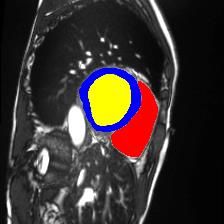
12 S. Thermos et al.

 MRI sample Segmentation Masks

 Disentangled Anatomical Factors

Fig. 5. Example of MRI sample from ACDC cardiac dataset (top left), the predicted
segmentation masks of the ROI (top right), and the 5 (out of 12) most semantic dis-
entangled anatomical factors. Blue, yellow, and red show the segmentation prediction
for the myocardium (MYO), left ventricle (LV) and right ventricle (RV), respectively.

B Dataset Details

ACDC pathology class annotations: a) normal (NOR), b) myocardial infarction
(MINF), c) dilated cardiomyopathy (DCM), d) hypertrophic cardiomyopathy
(HCM), and e) abnormal right ventricle (ARV). ACDC segmentation annota-
tions (3 semantic classes): left ventricular cavity (LV), myocardium (MYO) of
the LV, and right ventricle (RV). All images are resampled to 1.37mm2 /pixel
resolution and cropped to 224 × 224 pixels.
M&Ms (see also https://www.ub.edu/mnms/) comprises class annotations for
19 pathologies. However, 15 of them represent the 12% of the subjects that
corresponds to 1-3 subjects per class, thus we choose to experiment on the 4
most dominant classes that are: a) NOR, b) DCM, c) HCM, and d) ARV. Note
that ARV in our experiments is the underrepresented class, but with 26 subjects.
M&M segmentation annotations are identical with ACDC ones. All images are
resampled to 1.2mm2 /pixel resolution and cropped to 224 × 224 pixels.
 For both datasets during both model training and evaluation we combine
pairs of MR images from the same heart axis slice level.
Controllable cardiac synthesis via disentangled anatomy arithmetic 13

Fig. 6. Four SPADE-generated images using overlapped anatomical factors as input.
Arrows point to the areas where the performed arithmetic operations lead to such
overlaps.
You can also read