LensID: A CNN-RNN-Based Framework Towards Lens Irregularity Detection
←
→
Page content transcription
If your browser does not render page correctly, please read the page content below
LensID: A CNN-RNN-Based Framework
                                                          Towards Lens Irregularity Detection
                                                             in Cataract Surgery Videos?

                                              Negin Ghamsarian1 , Mario Taschwer1 , Doris Putzgruber-Adamitsch2 , Stephanie
                                                        Sarny2 , Yosuf El-Shabrawi2 , and Klaus Schoeffmann1
                                                   1
arXiv:2107.00875v1 [eess.IV] 2 Jul 2021

                                                       Department of Information Technology, Alpen-Adria-Universität Klagenfurt
                                                               2
                                                                 Department of Ophthalmology, Klinikum Klagenfurt
                                                                               negin@itec.aau.at

                                                  Abstract. A critical complication after cataract surgery is the dislocation of the
                                                  lens implant leading to vision deterioration and eye trauma. In order to reduce the
                                                  risk of this complication, it is vital to discover the risk factors during the surgery.
                                                  However, studying the relationship between lens dislocation and its suspicious
                                                  risk factors using numerous videos is a time-extensive procedure. Hence, the sur-
                                                  geons demand an automatic approach to enable a larger-scale and, accordingly,
                                                  more reliable study. In this paper, we propose a novel framework as the major step
                                                  towards lens irregularity detection. In particular, we propose (I) an end-to-end re-
                                                  current neural network to recognize the lens-implantation phase and (II) a novel
                                                  semantic segmentation network to segment the lens and pupil after the implanta-
                                                  tion phase. The phase recognition results reveal the effectiveness of the proposed
                                                  surgical phase recognition approach. Moreover, the segmentation results confirm
                                                  the proposed segmentation network’s effectiveness compared to state-of-the-art
                                                  rival approaches.

                                                  Keywords: Semantic Segmentation · Surgical Phase Recognition · Cataract Surgery.

                                          1     Introduction
                                          Cataract refers to the eye’s natural lens, having become cloudy and causing vision de-
                                          terioration. Cataract surgery is the procedure of restoring clear eye vision via cataract
                                          removal followed by artificial lens implantation. This operation is conducted with the
                                          aid of a binocular microscope providing a 3D magnified and illuminated image of the
                                          eye. The camera mounted on the microscope records and stores the whole surgery for
                                          postoperative objectives.
                                              Over several years, there have been numerous advances in surgical techniques, tools,
                                          and instruments in ophthalmic surgeries. Such advances resulted in decreasing the risk
                                          of severe intraoperative and postoperative complications. Still, there are many ongoing
                                          research efforts to prevent the current implications during and after surgery. A criti-
                                          cal issue in cataract surgery that has not yet been addressed is intraocular lens (IOL)
                                          dislocation. This complication leads to various human sight issues such as vision blur,
                                          ?
                                              This work was funded by the FWF Austrian Science Fund under grant P 31486-N31.
2          N. Ghamsarian et al.

double vision, or vision inference as observing the lens implant edges. Intraocular in-
flammation, corneal edema, and retinal detachment are some other consequences of
lens relocation. Since patient monitoring after the surgery or discharge is not always
possible, the surgeons seek ways to diagnose evidence of potential irregularities that
can be investigated during the surgery.
    Recent studies show that particular intraocular lens characteristics can contribute to
lens dislocation after the surgery [15]. Moreover, the expert surgeons argue that there
can be a direct relationship between the overall time of lens unfolding and the risk of
lens relocation after the surgery. Some surgeons also hypothesize that severe lens in-
stability during the surgery is a symptom of lens relocation. To discover the potential
correlations between lens relocation and its possibly contributing factors, surgeons re-
quire a tool for systematic feature extraction. Indeed, an automatic approach is required
for (i) detecting the lens implantation phase to determine the starting time for lens statis-
tics’ computation and (ii) segmenting the lens and pupil to compute the lens statistics
over time. The irregularity-related statistics can afterward be extracted by tracking the
lens’s relative size (normalized by the pupil’s size) and relative movements (by calibrat-
ing the pupil). Due to the dearth of computing power in the operation rooms, automatic
phase detection and lens/pupil segmentation on the fly is not currently achievable. Al-
ternatively, this analysis can be performed in a post hoc manner using recorded cataract
surgery videos. The contributions of this paper are:

 1. We propose a novel CNN-RNN-based framework for evaluating lens unfolding
    delay and lens instability in cataract surgery videos.
 2. We propose and evaluate a recurrent convolutional neural network architecture to
    detect the “implantation phase” in cataract surgery videos.
 3. We further propose a novel semantic segmentation network architecture termed as
    AdaptNet1 , that can considerably improve the segmentation performance for the
    intraocular lens (and pupil) compared to ten rival state-of-the-art approaches.
 4. We introduce three datasets for phase recognition, pupil segmentation, and lens
    segmentation that are publicly released with this paper to support reproducibility
    and allow further investigations for lens irregularity detection2 .

2      Related Work

Since this work involves phase recognition and semantic segmentation, we briefly re-
view the state-of-the-art approaches related to the mentioned subjects.
    Phase recognition in surgical videos has experienced three stages of development.
In the first stage, hand-engineered features such as color, shape, and instrument pres-
ence information [14,19,21] are exploited as the input to classical machine learning
approaches. Using Conditional Random Fields (CRF), Random Forests, and Hidden
Markov Models (HMM), the corresponding phase of each feature vector is classified
 1
     The PyTorch implementation of AdaptNet is publicly available at https://github.com/Negin-
     Ghamsarian/AdaptNet-MICCAI2021.
 2
     http://ftp.itec.aau.at/datasets/ovid/LensID/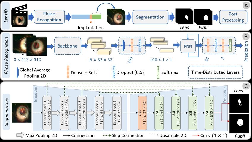
LensID: A CNN-RNN-Based Framework Towards Lens Irregularity Detection          3

Fig. 1: The block diagram of LensID and the architecture of Phase Recognition and
Semantic Segmentation networks.

[23,19,21]. Because of pruning to suboptimal results, the classical approaches are re-
placed with convolutional neural networks in the second stage [22]. To improve the
classification accuracy by taking advantage of the temporal information, the third gen-
eration of phase recognition approaches adopt LSTM [11] or GRU [4] or bidirectional
recurrent layers [27,13,9].
    Since semantic segmentation plays a prominent role in medical image analysis,
considerable attention has been devoted to this subject in recent years. Specifically,
U-Net [20], which takes advantage of skip connections between symmetric layers in
encoder and decoder, demonstrated pioneering results in medical image segmentation.
Many approaches based on U-Net are proposed to improve its segmentation accuracy or
address its weaknesses [10,12,24,7,26]. UNet++ [26] ensembles varying-depth U-Nets
to deal with network depth optimization. CPFNet [7] adopts a scale-aware module by
fusing the output feature maps of atrous convolutions [2] with different dilation rates.
MultiResUNet [12] fuses the feature maps coming from sequential convolutions as an
effective alternative to convolutional layers with large filters and atrous convolutions.
Many other attention modules are proposed to boost segmentation accuracy for differ-
ent medically-relevant content such as surgical instruments [18,17,16], liver lesion [3],
and general medical image segmentation [10].

3   Methodology

Fig. 1 demonstrates the block diagram of LensID and the network architecture of the
phase recognition and segmentation steps. As the first step towards lens irregularity
detection, we adopt a recurrent convolutional network (Fig. 1-B) to detect the lens im-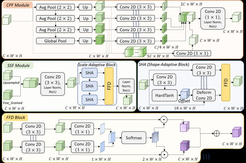
4       N. Ghamsarian et al.

     Fig. 2: The detailed architecture of the CPF and SFF modules of AdaptNet.

plantation phase (the temporal segment in which the lens implantation instrument is
visible). We start segmenting the artificial lens and pupil exactly after the lens implan-
tation phase using the proposed semantic segmentation network (Fig. 1-C). The pupil
and lens segmentation results undergo some post-processing approaches to compute
lens instability and lens unfolding delay. More precisely, we draw the smallest convex
polygon surrounding pupil’s and lens’ masks using binary morphological operations.
For lens instability, we use the normalized distance between the lens and pupil centers.
For lens unfolding, we track the lens’ area over time, considering its relative position.

Phase Recognition. As shown in Fig. 1-B, we use a pre-trained backbone followed by
global average pooling to obtain a feature vector per each input frame. These features
undergo a sequence of Dense, Dropout, and ReLU layers to extract higher-order seman-
tic features. A recurrent layer with five units is then employed to improve the feature
representation by taking advantage of temporal dependencies. These features are then
fed into a sequence of layers to finally output the predicted class for each input frame.

Lens & Pupil Segmentation. In cataract surgery, a folded artificial lens is implanted
inside the eye. The lens is transparent and inherits the pupil’s color after implanta-
tion. Moreover, it is usually being unfolded very fast (sometimes with the help of
an instrument). The transparency and unpredictable formation of this object, as well
as occlusion, defocus blur, and motion blur [8], make lens segmentation and track-
ing more challenging. Hence, we require a semantic segmentation network that can be
adapted to the changes in the artificial lens’s shape and scale. We adopt a U-Net-based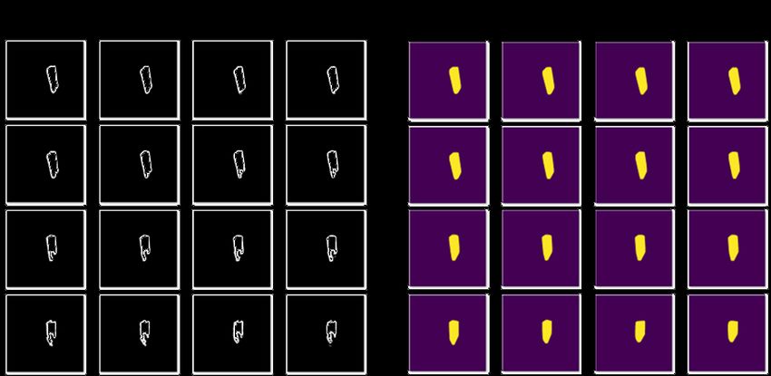
LensID: A CNN-RNN-Based Framework Towards Lens Irregularity Detection          5

encoder-decoder architecture for the proposed semantic segmentation network termed
as AdaptNet. AdaptNet consists of three main components: encoder, cascade pooling
fusion (CPF) module, and shape/scale-adaptive feature fusion (SSF) module. We use
the VGG16 network as the encoder network. The encoder’s output feature map is fed
into the CPF module to enhance the feature representation using pyramid features. This
feature map is then fed into a sequence of SSF modules, which decode low-resolution
semantic features.

    As shown in Fig. 2, the CPF module applies a sequence of three average pooling
layers (with a stride of two pixels) followed by a global average pooling layer to the
input features. The obtained feature maps are upsampled to the original size of the input
and concatenated together with the input feature map in a depth-wise manner. Each
group of five channels in the generated feature map undergoes a distinct convolution
for intra-channel feature refinement (which is performed using a convolutional layer
with C groups). Besides, the upsampled features are mapped into a smaller channel
space while extracting higher-order semantic features using convolutional layers with
shared weights. The obtained features are concatenated with the intra-channel refined
features and undergo a convolutional layer for inter-channel feature refinement.

    The SSF module starts with concatenating the upsampled semantic feature map
with the fine-grained feature map coming from the encoder. The concatenated feature
map is fed into a sequence of convolutional, layer normalization, and ReLU layers
for feature enhancement and dimensionality reduction. The resulting features are fed
into the scale-adaptive block, which aims to fuse the features coming from cascade
convolutional blocks. This succession of convolutional layers with small filter sizes
can factorize the large and computationally expensive receptive fields [12]. Moreover,
the fusion of these successive feature maps can play the role of scale-awareness for
the network. The shape-adaptive (SHA) block is responsible for fusing the resulting
feature maps of deformable and structured convolutions. At first, a convolutional layer
followed by a hard tangent hyperbolic function is employed to produce the offsets for
the deformable convolutional layer [5]. The input features are also fed into a regular
convolutional layer that shares the weights with the deformable layer for structured-
feature extraction. These features are then fused to induce the awareness of shape and
deformation to the network.

    The feature fusion decision (FFD) block inspired by CPFNet [7] accounts for de-
termining the importance of each input feature map in improving semantic features.
Fig. 2 shows the FFD Block in the case of two input branches. At first, shared con-
volutional layers are applied to the input feature maps to extract the shared semantic
features. The resulting feature maps undergo shared pixel-wise convolutions to produce
the pixel-wise attention maps. The concatenated attention maps are fed into a softmax
activation layer for normalization. The obtained features are used as pixel-wise weights
of the shared-semantic feature maps. The shape/scale adaptive features are computed as
the sum of pixel-wise multiplications ( ) between the normalized attention maps and
their corresponding semantic feature maps.
6 N. Ghamsarian et al.
4 Experimental Setup
We use three datasets for this study: (i) a large dataset containing the annotations for the
lens implantation phase versus the rest of phases from 100 videos/operations of cataract
surgery, (ii) a dataset containing the lens segmentation of 401 frames from 27 videos
(292 images from 21 videos for training, and 109 images from six videos for testing),
and (iii) a dataset containing the pupil segmentation of 189 frames from 16 videos
(141 frames from 13 videos for training, and 48 frames from three videos for testing).
Regarding the phase recognition dataset, since lens implantation is a very short phase
(around four seconds) compared to the whole surgery (seven minutes on average), cre-
ating a balanced dataset that can cover the entire content of videos from the “Rest” class
is quite challenging. Hence, we propose a video clip generator that can provide diverse
training sequences for the recurrent neural network by employing stochastic functions.
At first, 12 three-second video clips with overlapping frames are extracted from the
implantation phase of each cataract surgery video. Besides, the video segments before
and after the implantation phase are divided into eight and four video clips, respectively
(these clips have different lengths depending on the length of the input video). Ac-
cordingly, we have a balanced dataset containing 2040 video clips from 85 videos for
training and 360 video clips from the other 15 videos for testing. For each training ex-
ample, the video generator uses a stochastic variable to randomly select a three-second
clip from the input clip. We divide this clip into N sub-clips, and N stochastic variables
are used to randomly select one frame per sub-clip (in our experiments, N is set to five
to reduce computational complexity and avoid network overfitting).
We compare the segmentation accuracy of the proposed approach (AdaptNet) with
ten state-of-the-art approaches including UNet++ (and UNet++/DS) [26], MultiRe-
sUNet [12], CPFNet [7], dU-Net [24], CE-Net [10], FEDNet [3], PSPNet [25], Seg-
Net [1], and U-Net [20]. It should be mentioned that the rival approaches employ dif-
ferent backbone networks, loss functions (cross entropy or cross entropy log Dice), and
upsampling methods (bilinear, transposed convolution, pixel-shuffling, or max unpool-
ing).
For phase recognition, all networks are trained for 20 epochs. The initial learning
rate for these networks is set to 0.0002 and 0.0004 for the networks with VGG19 and
Resnet50 backbones, respectively, and halved after ten epochs. Since the segmentation
networks used for evaluations have different depths, backbones, and the number of
trainable parameters, all networks are trained with three different initial learning rates
(lr0 ∈ {0.0005, 0.001, 0.002}). For each network, the results with the highest Dice
coefficient are listed. All segmentation networks are trained for 30 epochs, and the
learning rate is decreased by a factor of 0.8 in every other epoch. To prevent overfitting
and improve generalization performance, we have used motion blur, Gaussian blur,
random contrast, random brightness, shift, scale, and rotation for data augmentation.
The backbones of all networks evaluated for phase recognition and lens/pupil semantic
segmentation are initialized with ImageNet [6] weights. The size of input images to all
networks is set to 512 × 512 × 3. The loss function for the phase recognition network
is set to Binary Cross Entropy. For the semantic segmentation task, we adopt a loss
function consisting of categorical cross entropy and logarithm of soft Dice coefficient
as follows (in Eq. (1), CE stands for Cross Entropy, and XP red and XT rue denote theLensID: A CNN-RNN-Based Framework Towards Lens Irregularity Detection 7
Table 1: Phase recognition results of the end-to-end recurrent convolutional networks.
Backbone: VGG19 Backbone: ResNet50
RNN Precision Recall F1-Score Accuracy Precision Recall F1-Score Accuracy
GRU 0.97 0.96 0.96 0.96 0.9 0.94 0.94 0.94
LSTM 0.98 0.98 0.98 0.98 0.96 0.96 0.96 0.96
BiGRU 0.97 0.96 0.96 0.96 0.95 0.95 0.95 0.95
BiLSTM 1.00 1.00 1.00 1.00 0.98 0.98 0.98 0.98
predicted and ground-truth segmentation images, respectively. Besides, we use a Dice
smoothing factor equal to 1, and set λ = 0.8 in our experiments):
L = λ × CE(XP red , XT rue ) − (1 − λ) × log2 Dice(XP red , XT rue ) (1)
To evaluate the performance of phase recognition networks, we use Precision, Recall,
F1-Score, and Accuracy, which are the common classification metrics. The semantic
segmentation performance is evaluated using Dice coefficient and Intersection over
Union (IoU).
5 Experimental Results and Discussion
Table 1 compares the classification reports of the proposed architecture for phase recog-
nition considering two different backbone networks and four different recurrent layers.
Thanks to the large training set and taking advantage of recurrent layers, all networks
have shown superior performance in classifying the implantation phase versus other
phases. However, the LSTM and bidirectional LSTM (BiLSTM) layers have shown
better performance compared to GRU and BiGRU layers, respectively. Surprisingly,
the network with a VGG19 backbone and BiLSTM layer has achieved 100% accuracy
in classifying the test clips extracted from the videos which are not used during train-
ing. Fig. 3 compares the segmentation results (mean and standard deviation of IoU and
Dice coefficient) of AdaptNet and ten rival state-of-the-art approaches. Overall, it can
be perceived that AdaptNet, UNet++, UNet++/DS, and FEDNet have achieved the top
four segmentation results. However, AdaptNet has achieved the highest mean IoU and
Dice coefficient compared to the rival approaches. In particular, the proposed approach
achieves 3.48% improvement in mean IoU and 2.22% improvement in mean Dice for
lens segmentation compared to the best rival approach (UNet++). Moreover, the smaller
standard deviation of IoU (10.56% vs. 12.34%) and Dice (8.56% vs. 9.65%) for Adapt-
Net compared to UNet++ confirms the reliability and effectiveness of the proposed ar-
chitecture. For pupil segmentation, AdaptNet shows subtle improvement over the best
rival approach (UNet++) regarding mean IoU and Dice while showing significant im-
provement regarding the standard deviation of IoU (1.91 vs. 4.05). Table 2 provides
an ablation study of AdaptNet. We have listed the Dice and IoU percentage with two
different learning rates by gradually adding the proposed modules and blocks (for lens
segmentation). It can be perceived from the results that regardless of the learning rate,8 N. Ghamsarian et al.
IoU(%) Dice(%)
92.62
90.44
100
89.95
88.68
87.09
Lens Segmentation
85.26
83.75
83.61
82.32
82.22
81.53
80.59
75.38
73.88
73.86
73.17
70.56
80
71.4
65.81
61.89
61.42
56.86
60
97.98
97.96
97.53
97.12
96.06
96.02
95.99
95.28
94.43
94.18
93.32
Pupil Segmentation
92.33
100 91.31
89.55
89.36
87.66
84.24
83.51
82.4
75.34
80
69.59
63.16
60
et Net Net -Net E-Net U-Net PFNet sUNet +/DS Net++ aptNe
t
U-N Seg PSP FED C d C R e e t+ U d
lti UN A
Mu
Fig. 3: Quantitative comparison of segmentation results for the proposed approach
(AdaptNet) and rival approaches.
Table 2: Impact of different modules on the segmentation results of AdaptNet.
Components lr = 0.001 lr = 0.002
Baseline SSF SHA CPF IoU(%) Dice(%) IoU(%) Dice(%)
X 7 7 7 82.79 89.94 84.33 90.90
X X 7 7 83.54 90.33 84.99 91.22
X X X 7 84.76 91.12 86.34 92.17
X X X X 85.03 91.28 87.09 92.62
each distinctive module and block has a positive impact on segmentation performance.
We cannot test the FFD block separately since it is bound with the SSF module.
Fig. 4 shows the post-processed lens segments (pink) and pupil segments (cyan)
from a representative video in different time slots (a), the relative lens area over time
(b), and relative lens movements over time (c). Due to lens instability, a part of the
lens is sometimes placed behind the iris, as shown in the segmentation results in the
35th second. Accordingly, the visible area of the lens can change independently of
the unfolding state. Hence, the relative position of the lens should also be taken into
account for lens unfolding delay computations. As can be perceived, the visible area of
the lens is near maximum at 20 seconds after the implantation phase, and the lens is
located nearly at the center of the pupil at this time. Therefore, the lens unfolding delay
is 20 seconds in this case. However, the lens is quite unstable until 70 seconds after
implantation.LensID: A CNN-RNN-Based Framework Towards Lens Irregularity Detection 9
Fig. 4: The lens statistics for one representative cataract surgery video.
6 Conclusion
Lens irregularity detection is a highly relevant problem in ophthalmology, which can
play a prominent role in predicting and preventing lens relocation after surgery. This
paper focuses on two significant steps towards lens irregularity detection: (i) “lens im-
plantation phase” detection and (ii) lens/pupil segmentation. In particular, We propose
an end-to-end recurrent convolutional network that can extract spatio-temporal depen-
dencies to detect the lens implantation phase accurately. Moreover, we propose a novel
semantic segmentation network termed as AdaptNet. The proposed approach can deal
with severe deformations and scale variations in the intraocular lens by adaptively fus-
ing sequential and parallel feature maps. Experimental results reveal the effectiveness
of the proposed phase recognition and semantic segmentation networks.
References
1. Badrinarayanan, V., Kendall, A., Cipolla, R.: Segnet: A deep convolutional encoder-decoder
architecture for image segmentation. IEEE Transactions on Pattern Analysis and Machine
Intelligence 39(12), 2481–2495 (2017). https://doi.org/10.1109/TPAMI.2016.2644615
2. Chen, L., Papandreou, G., Kokkinos, I., Murphy, K., Yuille, A.L.: Deeplab: Semantic image
segmentation with deep convolutional nets, atrous convolution, and fully connected crfs.
IEEE Transactions on Pattern Analysis and Machine Intelligence 40(4), 834–848 (2018).
https://doi.org/10.1109/TPAMI.2017.2699184
3. Chen, X., Zhang, R., Yan, P.: Feature fusion encoder decoder network for automatic liver
lesion segmentation. In: 2019 IEEE 16th International Symposium on Biomedical Imaging
(ISBI 2019). pp. 430–433 (2019). https://doi.org/10.1109/ISBI.2019.8759555
4. Cho, K., van Merrienboer, B., Gülçehre, Ç., Bougares, F., Schwenk, H., Bengio, Y.: Learning
phrase representations using RNN encoder-decoder for statistical machine translation. CoRR
abs/1406.1078 (2014), http://arxiv.org/abs/1406.1078
5. Dai, J., Qi, H., Xiong, Y., Li, Y., Zhang, G., Hu, H., Wei, Y.: Deformable convolutional
networks. In: 2017 IEEE International Conference on Computer Vision (ICCV). pp. 764–
773 (2017). https://doi.org/10.1109/ICCV.2017.8910 N. Ghamsarian et al.
6. Deng, J., Dong, W., Socher, R., Li, L.J., Li, K., Fei-Fei, L.: ImageNet: A Large-Scale Hier-
archical Image Database. In: CVPR09 (2009)
7. Feng, S., Zhao, H., Shi, F., Cheng, X., Wang, M., Ma, Y., Xiang, D., Zhu, W., Chen, X.:
Cpfnet: Context pyramid fusion network for medical image segmentation. IEEE Transactions
on Medical Imaging 39(10), 3008–3018 (2020). https://doi.org/10.1109/TMI.2020.2983721
8. Ghamsarian, N., Taschwer, M., Schoeffmann, K.: Deblurring cataract surgery videos using
a multi-scale deconvolutional neural network. In: 2020 IEEE 17th International Symposium
on Biomedical Imaging (ISBI). pp. 872–876 (2020)
9. Ghamsarian, N., Taschwer, M., Putzgruber-Adamitsch, D., Sarny, S., Schoeffmann, K.: Rel-
evance detection in cataract surgery videos by spatio- temporal action localization. In: 2020
25th International Conference on Pattern Recognition (ICPR). pp. 10720–10727 (2021).
https://doi.org/10.1109/ICPR48806.2021.9412525
10. Gu, Z., Cheng, J., Fu, H., Zhou, K., Hao, H., Zhao, Y., Zhang, T., Gao, S., Liu, J.: Ce-net:
Context encoder network for 2d medical image segmentation. IEEE Transactions on Medical
Imaging 38(10), 2281–2292 (2019). https://doi.org/10.1109/TMI.2019.2903562
11. Hochreiter, S., Schmidhuber, J.: Long Short-Term Memory. Neural Computation 9(8), 1735–
1780 (1997). https://doi.org/10.1162/neco.1997.9.8.1735, https://doi.org/10.1162/
neco.1997.9.8.1735
12. Ibtehaz, N., Rahman, M.S.: Multiresunet : Rethinking the u-net architecture
for multimodal biomedical image segmentation. Neural Networks 121, 74–
87 (2020). https://doi.org/https://doi.org/10.1016/j.neunet.2019.08.025, https:
//www.sciencedirect.com/science/article/pii/S0893608019302503
13. Jin, Y., Dou, Q., Chen, H., Yu, L., Qin, J., Fu, C., Heng, P.: SV-RCNet: Workflow Recogni-
tion From Surgical Videos Using Recurrent Convolutional Network. IEEE Transactions on
Medical Imaging 37(5), 1114–1126 (May 2018). https://doi.org/10.1109/TMI.2017.2787657
14. Lalys, F., Riffaud, L., Bouget, D., Jannin, P.: A framework for the recognition of high-level
surgical tasks from video images for cataract surgeries. IEEE Transactions on Biomedical
Engineering 59(4), 966–976 (April 2012). https://doi.org/10.1109/TBME.2011.2181168
15. Mayer-Xanthaki, C.F., Pregartner, G., Hirnschall, N., Falb, T., Sommer, M.,
Findl, O., Wedrich, A.: Impact of intraocular lens characteristics on intraocular
lens dislocation after cataract surgery. British Journal of Ophthalmology (2020).
https://doi.org/10.1136/bjophthalmol-2020-317124, https://bjo.bmj.com/content/
early/2020/09/18/bjophthalmol-2020-317124
16. Ni, Z.L., Bian, G.B., Wang, G.A., Zhou, X.H., Hou, Z.G., Chen, H.B., Xie, X.L.: Pyra-
mid attention aggregation network for semantic segmentation of surgical instruments.
Proceedings of the AAAI Conference on Artificial Intelligence 34(07), 11782–11790
(Apr 2020). https://doi.org/10.1609/aaai.v34i07.6850, https://ojs.aaai.org/index.
php/AAAI/article/view/6850
17. Ni, Z.L., Bian, G.B., Wang, G.A., Zhou, X.H., Hou, Z.G., Xie, X.L., Li, Z., Wang, Y.H.:
Barnet: Bilinear attention network with adaptive receptive fields for surgical instrument seg-
mentation. In: Bessiere, C. (ed.) Proceedings of the Twenty-Ninth International Joint Con-
ference on Artificial Intelligence, IJCAI-20. pp. 832–838. International Joint Conferences
on Artificial Intelligence Organization (7 2020). https://doi.org/10.24963/ijcai.2020/116,
https://doi.org/10.24963/ijcai.2020/116, main track
18. Ni, Z.L., Bian, G.B., Zhou, X.H., Hou, Z.G., Xie, X.L., Wang, C., Zhou, Y.J., Li, R.Q., Li, Z.:
Raunet: Residual attention u-net for semantic segmentation of cataract surgical instruments.
In: Gedeon, T., Wong, K.W., Lee, M. (eds.) Neural Information Processing. pp. 139–149.
Springer International Publishing, Cham (2019)
19. Quellec, G., Lamard, M., Cochener, B., Cazuguel, G.: Real-time segmentation and recogni-
tion of surgical tasks in cataract surgery videos. IEEE Transactions on Medical Imaging 33,
2352–60 (12 2014). https://doi.org/10.1109/TMI.2014.2340473LensID: A CNN-RNN-Based Framework Towards Lens Irregularity Detection 11
20. Ronneberger, O., Fischer, P., Brox, T.: U-net: Convolutional networks for biomedical image
segmentation. In: Navab, N., Hornegger, J., Wells, W.M., Frangi, A.F. (eds.) Medical Image
Computing and Computer-Assisted Intervention – MICCAI 2015. pp. 234–241. Springer
International Publishing, Cham (2015)
21. Stauder, R., Okur, A., Peter, L., Schneider, A., Kranzfelder, M., Feussner, H., Navab, N.:
Random forests for phase detection in surgical workflow analysis. In: Stoyanov, D., Collins,
D.L., Sakuma, I., Abolmaesumi, P., Jannin, P. (eds.) Information Processing in Computer-
Assisted Interventions. pp. 148–157. Springer International Publishing, Cham (2014)
22. Twinanda, A.P., Shehata, S., Mutter, D., Marescaux, J., de Mathelin, M., Padoy, N.: EndoNet:
A Deep Architecture for Recognition Tasks on Laparoscopic Videos. IEEE Transactions on
Medical Imaging 36(1), 86–97 (Jan 2017). https://doi.org/10.1109/TMI.2016.2593957
23. Zappella, L., Béjar, B., Hager, G., Vidal, R.: Surgical gesture classification
from video and kinematic data. Medical Image Analysis 17(7), 732 – 745
(2013). https://doi.org/https://doi.org/10.1016/j.media.2013.04.007, http://www.
sciencedirect.com/science/article/pii/S1361841513000522, special Issue
on the 2012 Conference on Medical Image Computing and Computer Assisted Intervention
24. Zhang, M., Li, X., Xu, M., Li, Q.: Automated semantic segmentation of red blood cells for
sickle cell disease. IEEE Journal of Biomedical and Health Informatics 24(11), 3095–3102
(2020). https://doi.org/10.1109/JBHI.2020.3000484
25. Zhao, H., Shi, J., Qi, X., Wang, X., Jia, J.: Pyramid scene parsing network. In: Proceedings
of the IEEE Conference on Computer Vision and Pattern Recognition (CVPR) (July 2017)
26. Zhou, Z., Siddiquee, M.M.R., Tajbakhsh, N., Liang, J.: Unet++: Redesigning skip connec-
tions to exploit multiscale features in image segmentation. IEEE Transactions on Medical
Imaging 39(6), 1856–1867 (2020). https://doi.org/10.1109/TMI.2019.2959609
27. Zisimopoulos, O., Flouty, E., Luengo, I., Giataganas, P., Nehme, J., Chow, A., Stoyanov, D.:
DeepPhase: Surgical Phase Recognition in CATARACTS Videos. CoRR abs/1807.10565
(2018), http://arxiv.org/abs/1807.1056512 N. Ghamsarian et al.
7 Supplementary Material
Table S1 lists the specifications of the rival state-of-the-art approaches used in our eval-
uations. In “Upsampling” column, “Trans Conv” stands for Transposed Convolution.
Table S1: Specifications of the proposed and rival segmentation approaches.
Model Backbone Params Upsampling Reference Year
UNet++ (/DS) VGG16 24.24 M Bilinear [26] 2020
MultiResUNet 7 9.34 M Trans Conv [12] 2020
CPFNet ResNet34 34.66 M Bilinear [7] 2020
dU-Net 7 31.98 M Trans Conv [24] 2020
CE-Net ResNet34 29.90 M Trans Conv [10] 2019
FED-Net ResNet50 59.52 M Trans Conv & PixelShuffle [3] 2019
PSPNet ResNet50 22.26 M Bilinear [25] 2017
SegNet VGG16 14.71 M Max Unpooling [1] 2017
U-Net 7 17.26 M Bilinear [20] 2015
AdaptNet VGG16 23.61 M Bilinear Proposed
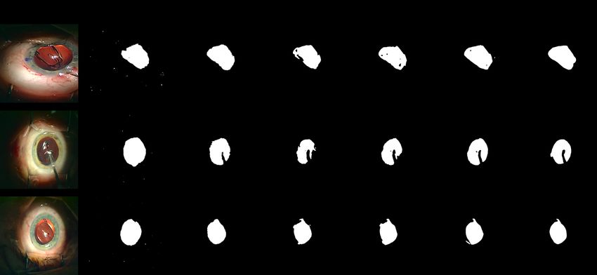
Fig. S1 presents qualitative comparisons among the top five approaches for lens
segmentation in three representative frames. It can be perceived from the figure that
AdaptNet can provide the most visually close segmentation results to the ground truth.
Moreover, AdaptNet is more robust against lens deformations as it provides the most
delineated predictions compared to the rival approaches.
Fig. S1: Qualitative comparisons among the top five segmentation approaches.
Fig. S2 demonstrates the effect of post-processing on segmentation results. We use
three morphological operations to improve the semantic segmentation results: (i) open-
ing (with the kernel size of 10 × 10) to attach the separated regions due to instrumentLensID: A CNN-RNN-Based Framework Towards Lens Irregularity Detection 13
Fig. S2: Qualitative comparisons among the top five segmentation approaches.
covering, (ii) closing (with the kernel size of 15 × 15) to remove the distant wrong de-
tections, and (iii) convex polygon. Since instruments usually cover a part of the pupil
and intraocular lens during surgery, the segmentation results may contain some holes in
the location of instruments. However, the occluded parts should be included in the lens
and pupil area. Since the pupil is inherently a convex object, and the intraocular lens
is usually convex during unfolding, we draw the smallest convex polygon around these
objects to retrieve the occluded segments. For convex polygons, we used the “Scipy
ConvexHull” function.You can also read