Case Report Spontaneous Splenic Artery Rupture as the First Symptom of Systemic Amyloidosis
←
→
Page content transcription
If your browser does not render page correctly, please read the page content below
Hindawi
Case Reports in Critical Care
Volume 2021, Article ID 6676407, 6 pages
https://doi.org/10.1155/2021/6676407

Case Report
Spontaneous Splenic Artery Rupture as the First Symptom of
Systemic Amyloidosis

 Øyvind Bruserud,1 Tor Henrik Anderson Tvedt,2 Aymen Bushra Ahmed,2
 Olav Karsten Vintermyr,3,4 Tor Hervig,4,5 Anne Berit Guttormsen,1,6
 and Håkon Reikvam 2,4
 1
 Department of Anaesthesia and Intensive Care, Haukeland University Hospital, Bergen, Norway
 2
 Department of Medicine, Haukeland University Hospital, Bergen, Norway
 3
 Department of Pathology, Haukeland University Hospital, Bergen, Norway
 4
 Department of Clinical Science, University of Bergen, Bergen, Norway
 5
 Department of Immunology and Transfusion Medicine, Haugesund Hospital, Norway
 6
 Department of Clinical Medicine, University of Bergen, Bergen, Norway

 Correspondence should be addressed to Håkon Reikvam; hakon.reikvam@uib.no

 Received 14 December 2020; Revised 20 January 2021; Accepted 24 February 2021; Published 9 March 2021

 Academic Editor: Chiara Lazzeri

 Copyright © 2021 Øyvind Bruserud et al. This is an open access article distributed under the Creative Commons Attribution
 License, which permits unrestricted use, distribution, and reproduction in any medium, provided the original work is
 properly cited.

 Spontaneous splenic rupture is a life-threatening condition leading to a rapidly progressing hypovolemic shock due to intra-
 abdominal blood loss, with a mortality rate of about 10%. Spontaneous splenic rupture can be caused by widely different
 disorders including acute and chronic infections, neoplastic disorders, and inflammatory noninfectious disorders. In this case
 report, we present a 67-year-old male patient with hemorrhagic shock caused by an acute bleeding from the splenic artery. The
 patient was massively transfused with blood products and fluids and underwent laparotomy for hemostatic control and clinical
 stabilization. Multiorgan involvement by amyloid light-chain amyloidosis (AL-amyloidosis) caused by plasma cell dyscrasia,
 specifically with infiltration of the spleen artery, was found to be the underlying cause of his life-threatening bleeding. Based on
 this case, we discuss the features of serious spleen bleeding, massive transfusion therapy in the intensive care setting, and AL-
 amyloidosis pathophysiology and treatment.

1. Introduction bleeding from the splenic artery. The patient required mas-
 sive transfusion therapy and underwent laparotomy for
Spontaneous splenic rupture can be caused by several differ- hemostatic control and clinical stabilization. Multiorgan
ent disorders including acute and chronic infections, neo- involvement by amyloid light-chain (AL-) amyloidosis was
plastic disorders, and inflammatory noninfectious disorders found to be the underlying cause of bleeding.
and has the potential to cause a hemorrhagic life-
threatening form of hypovolemic shock [1]. Injuries that 2. Case Report
penetrate the capsule result in an acute bleeding, while dam-
age to the parenchymal tissue leads to subcapsular hematoma A 67-year-old man was admitted to the emergency depart-
and a delayed rupture. Although splenic rupture is a well- ment (ED) with acute abdominal pain and hypotension.
known entity, it is relatively rarely occurring, and only a very Physical examination revealed blood pressure at
few studies have systematically evaluated its pathophysiology 60/30 mmHg, pulse 110 per minute, and a respiratory rate
and outcome. In this case report, we present a 67-year-old at 40 per minute. He was pale and the abdomen was dis-
male patient with hemorrhagic shock caused by an acute tended and painful, although without clear peritoneal signs.
2 Case Reports in Critical Care

Table 1: The table gives the laboratory tests at the emergency Table 2: Blood gas analysis in the emergency department.
department.
 Normal range Value
 Normal range Value pH 7.36-7.44 7.10
Hemoglobin 13.4–17.0 g/dL 9.2 pCO2 4.5-6.1 kPa 3.8
Leucocyte count 4:3 – 10:7 × 10⁹ /L 17.5 pO2 >9.2 kPa 31.4
 9
Platelet count 145 – 348 × 10 /L 50 Anion gap 7-12 mmol/L 25.8
Creatinine 45-90 μmol/L 170 Hemoglobin 13.4–17.0 g/dL 9.9
Albumin 39-48 g/L 34 Lactate 0.4-1.3 mmol/L 13.3
Ionized calcium 1.13-1.28 mmol/L 1.20 Glucose 4.0-6.0 mmol/L 9.0
Lactate dehydrogenase 105-205 U/L 441 Sodium 137-145 mmol/L 140
C-reactive protein
Case Reports in Critical Care 3

 Emergency Radiologic Intensive Operating Intensive Operating Intensive
 department department care unit room care unit Room care unit
 Stabilized
 - Clinical - CT imaging Laparotomy Re-laparotomy Diagnostics
 Medical examination - Coiling procedure Extubated
 procedure - Intravenous
 access
 - Arterial
 cannulation
 - Bedside US

 Transfusion Crystalloides 3 plasma Crystalloides 4 plasma 4 RCC
 4 whole blood 2 albunorm 7 RCC 4 plasma
 3 RCC 1 platelets 10 plasma
 3 plasma 1 platelets

 Vasopressor
 Noradr 0.08-0.30 ug/kg/min

 Timeline
 Day 1 Day 2
 Accumulated numbers of

 30
 blood products given

 20

 10

 0

Figure 1: The figure gives an overview over the medical procedures and massive transfusion therapy the patient received for the first two days
in hospital. He was initially hemodynamically stabilized by transfusion therapy in the emergency department before the CT imaging and
coiling procedure. However, vasopressor was then needed to keep the mean arterial pressure > 60 mmHg and he was massively transfused
during laparotomy. The patient finally stabilized the second day in hospital after a relaparotomy to secure hemostasis. The bar chart gives
the accumulated number of blood products given; blue bar, plasma; orange bar, red cell concentrates (RCC); and grey bar, platelet
concentrates.

and improved cardiac function (NYHA stage 0-I). However, hemorrhage, the patient is often better served by rapid
no change in serum lambda levels was observed during three diagnostic and therapeutic interventions, such as operative
courses of chemotherapy. Treatment was therefore changed exploration, angiography with embolization, or gastrointes-
to daratumumab, a monoclonal antibody that targets CD38 tinal endoscopy.
expressed in plasma cells. Three weeks after the first injection The initial management of patients with hemorrhagic
of daratumumab, normalization of serum lambda levels was shock should focus on restoring the intravascular volume
observed. The patient stayed in hospital for 30 days and is and rapidly localize and control of hemorrhage. Massive-
currently doing well. transfusion protocols provide a survival benefit for patients
 with acute bleeding [4] and mobilize universal donor blood
3. Discussion products (e.g., RBCs, plasma, and platelet concentrates) to
 the patient’s bedside in prespecified ratios, together with
In this case, spontaneous rupture of the spleen artery caused pharmaceutical adjuncts such as calcium and tranexamic
by systemic amyloidosis was found to be the underlying acid. A ratio of red cell concentrates, plasma, and platelet
cause of hemorrhagic shock. However, the potential sources concentrates close to 1 : 1 : 1 is suggested to avoid dilution of
of hemorrhage should be identified during the initial evalua- coagulation factors and thrombocytopenia [4–8]. Transfu-
tion of patients with hemorrhagic shock. Bedside US can be sion of whole blood can also be administrated in massive
helpful to identify occult sources of bleeding such as a rup- hemorrhage [9]. Laboratory measurements including base
tured abdominal aortic aneurysm or a ruptured spleen. Nota- deficit, lactate, hemoglobin, international normalized ratio,
bly, CT imaging should only be performed if the source of platelet count, fibrinogen, electrolytes, and thromboelasto-
bleeding remains uncertain and the patient’s condition has graphy (TEG) are crucial for guiding the blood product
been stabilized with initial resuscitation. In cases with severe resuscitation [1].
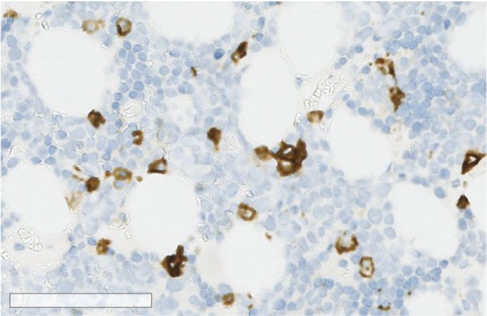
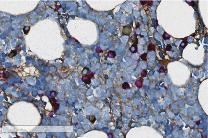
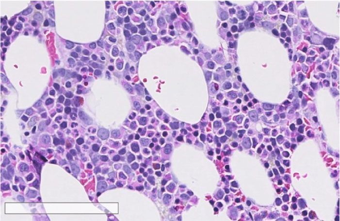
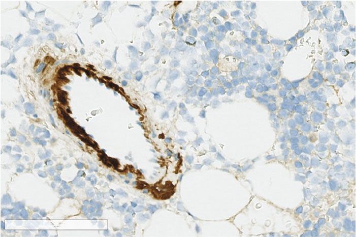
4 Case Reports in Critical Care

 100 m 100 m

 (a) (b)

 100 m 100 m

 (c) (d)

Figure 2: (a) Illustration of representative bone marrow morphology on a formalin-fixed paraffin-embedded hematoxylin-eosin-stained
section. (b–d) Immune histochemistry on formalin-fixed paraffin-embedded cut sections using CD138 (b), light chain kappa and lambda
(c), and amyloid P (d). There were less than 10% plasma cells (b), and the plasma cells did selective express light-chain lambda (c), and
focal deposits of amyloid (d) were noted in vessel walls.

 (a) (b)

Figure 3: Formalin-fixed paraffin-embedded tissue section from the spleen. (a) Amyloid deposits in an arterial wall using Congo staining.
Bar: 100 mm. (b) Green fluorescence due to amyloid deposits in an arterial wall using polarized light on a Congo-stained formalin-fixed
paraffin-embedded tissue section from the spleen. Bar: 100 mm.

 Amyloidosis is a term referring to a group of complex Although rare, these diseases can also be associated with
diseases caused by protein misfolding and aggregation into potentially life-threatening hemorrhage [11, 12]. The patho-
highly ordered amyloid fibrils depositing in tissues, result- physiological mechanisms predisposing for abnormal hem-
ing in progressive organ damage [10]. AL-amyloidosis is a orrhage in patients with systemic amyloidosis are
complication of plasma cell dyscrasia, a monoclonal plasma heterogeneous [11]. Amyloid is often associated with
cell population producing a monoclonal antibody or light acquired coagulation-factor deficiencies [13, 14]. Amyloid
chains with potential to deposit in several organs. Organ fibrils bind and remove factor X from plasma thus inducing
dysfunction is caused by several mechanisms including a a factor X deficiency that is resistant to replacement therapy.
direct cytotoxic effect of light chain and bystander proteins, Spontaneously elevated international normalized ratio (INR)
as well as spatial disruption by the amyloid deposits. Car- and activated partial thromboplastin time (aPTT) may indi-
diac involvement is by far the most significant negative cate factor X deficiency. This acquired factor X deficiency is
prognostic marker, and the diagnosis can be made using found in about 1/3 of AL-amyloidosis patients, and splenec-
markers of myocardial involvement, cNT-proBNP, and tro- tomy results in normalization of factor X levels [11]. Isolated
ponin [2]. deficiency of other coagulation factors, e.g., factor II, V, VII,
Case Reports in Critical Care 5

and IX, has also been described. In addition, abnormal fibrin Consent
polymerization and hyperfibrinolysis are also described to be
involved in AL-amyloidosis coagulopathy [15, 16]. Platelet The patient gave written consent for publication of this case
dysfunction reflected by decreased aggregability of platelets report.
upon stimulation is found in patients with systemic amyloid-
osis and may contribute to abnormal hemorrhage [11]. Finally, Conflicts of Interest
amyloid deposition in blood vessels and perivascular tissue
may lead to amyloid angiopathy with increased vessel fragility The authors declare that they have no conflicts of interest.
and impaired vasoconstriction [17, 18], which was found to be
the cause of the splenic artery rupture in our patient. References
 Spontaneous rupture of the spleen or splenic artery is a
rare condition. Although a large number of single case [1] J. W. Cannon, “Hemorrhagic shock,” The New England jour-
reports have been published, only a very limited number of nal of medicine, vol. 378, no. 4, pp. 370–379, 2018.
articles have systematically analyzed cause and treatment [2] A. D. Wechalekar, S. O. Schonland, E. Kastritis et al., “A Euro-
outcome after spontaneous spleen rupture. Renzulli et al. pean collaborative study of treatment outcomes in 346 patients
[19] reviewed 632 publications reporting a total of 845 with cardiac stage III AL amyloidosis,” Blood, vol. 121, no. 17,
patients. In this material, approximately 30% of spontaneous pp. 3420–3427, 2013.
spleen rupture were due to neoplastic disorders, 27% due to [3] A. Dispenzieri, M. A. Gertz, R. A. Kyle et al., “Serum cardiac
infectious disorders, 20% to inflammatory noninfectious dis- troponins and N-terminal pro-brain natriuretic peptide: a
orders, and the remaining cases to mechanical trauma, staging system for primary systemic amyloidosis,” Journal of
related to medical treatment, or of unknown etiology. clinical oncology, vol. 22, no. 18, pp. 3751–3757, 2004.
Among neoplastic disorders, hematological malignancies, [4] J. W. Cannon, M. A. Khan, A. S. Raja et al., “Damage control
i.e., non-Hodgkin’s lymphomas, acute myeloid leukemia, resuscitation in patients with severe traumatic hemorrhage,”
and myeloproliferative disorders, were the most common The journal of trauma and acute care surgery, vol. 82, no. 3,
 pp. 605–617, 2017.
causes, although also pancreatic tail tumors with spleen
 [5] P. I. Johansson, J. Stensballe, R. Oliveri, C. E. Wade, S. R.
involvement can cause spleen rupture [20]. Mononucleosis
 Ostrowski, and J. B. Holcomb, “How I treat patients with mas-
was the most common cause among patients with an infec- sive hemorrhage,” Blood, vol. 124, no. 20, pp. 3052–3058, 2014.
tious disorder, reported in 102 of 137 cases (74%). Approxi-
 [6] J. B. Holcomb, D. J. del Junco, E. E. Fox et al., “The prospective,
mately 85% of patients required surgical treatment, and the observational, multicenter, major trauma transfusion
reported mortality was 12%. Age and neoplastic disorders (PROMMTT) study: comparative effectiveness of a time-
conferred a higher probability of death. varying treatment with competing risks,” JAMA surgery,
 However, the primary treatment goal of AL-amyloidosis vol. 148, no. 2, pp. 127–136, 2013.
is to eliminate the monoclonal plasma cell clone producing [7] J. B. Holcomb, B. C. Tilley, S. Baraniuk et al., “Transfusion of
the pathological light chain. Hence, AL-amyloidosis applies plasma, platelets, and red blood cells in a 1: 1:1 vs a 1: 1: 2 ratio
similar treatment protocols as for multiple myeloma, with and mortality in patients with severe trauma: the PROPPR
the goal of a rapid suppression of the amyloidogenic organ randomized clinical trial,” Journal of the American Medical
toxic light chains. Particularly, the proteasome inhibitor bor- Association, vol. 313, no. 5, pp. 471–482, 2015.
tezomib that ensures a rapid reduction in plasma cell light- [8] J. Hallet, F. Lauzier, O. Mailloux et al., “The use of higher plate-
chain production is often incorporated into treatment proto- let: RBC transfusion ratio in the acute phase of trauma resusci-
cols [10]. Nevertheless, patients with amyloidosis are often tation: a systematic review,” Critical care medicine, vol. 41,
too fragile, due to age and to organ involvement, to tolerate no. 12, pp. 2800–2811, 2013.
the most intensive approach which includes high-dose che- [9] A. P. Cap, A. Beckett, A. Benov et al., “Whole blood transfu-
motherapy and autologous hematopoietic stem cell trans- sion,” Military medicine, vol. 183, suppl_2, pp. 44–51, 2018.
plantation [10], and patient-adapted risk assessment is [10] G. Merlini, A. Dispenzieri, V. Sanchorawala et al., “Systemic
required to reduce the treatment-related mortality. immunoglobulin light chain amyloidosis,” Nature reviews Dis-
 ease primers, vol. 4, no. 1, p. 38, 2018.
 [11] C. Sucker, G. R. Hetzel, B. Grabensee, M. Stockschlaeder, and
4. Conclusion R. E. Scharf, “Amyloidosis and bleeding: pathophysiology,
 diagnosis, and therapy,” American journal of kidney diseases,
Spontaneous splenic rupture is a rare condition leading to a vol. 47, no. 6, pp. 947–955, 2006.
rapidly progressing life-threatening hypovolemic shock due
 [12] P. Renzulli, A. Schoepfer, E. Mueller, and D. Candinas, “Atrau-
to intra-abdominal blood loss and can be caused by disorders matic splenic rupture in amyloidosis,” Amyloid, vol. 16, no. 1,
such as acute and chronic infections, neoplastic disorders, pp. 47–53, 2009.
and inflammatory noninfectious disorders. Although rare, [13] E. B. Choufani, V. Sanchorawala, T. Ernst et al., “Acquired fac-
plasma cell dyscrasia causing amyloid light-chain amyloid- tor X deficiency in patients with amyloid light-chain amyloid-
osis also has the potential to cause spontaneous splenic osis: incidence, bleeding manifestations, and response to high-
rupture and life-threatening bleeding. However, early recog- dose chemotherapy,” Blood, vol. 97, no. 6, pp. 1885–1887,
nition of hemorrhagic shock and prompt action to stop the 2001.
bleeding and restore the patient’s intravascular volume and [14] B. Furie, L. Voo, K. P. McAdam, and B. C. Furie, “Mechanism
oxygen-carrying capacity are lifesaving. of factor X deficiency in systemic amyloidosis,” The New
6 Case Reports in Critical Care

 England journal of medicine, vol. 304, no. 14, pp. 827–830,
 1981.
[15] G. Gamba, N. Montani, E. Anesi et al., “Clotting alterations in
 primary systemic amyloidosis,” Haematologica, vol. 85, no. 3,
 pp. 289–292, 2000.
[16] D. A. Gastineau, M. A. Gertz, T. M. Daniels, R. A. Kyle, and
 E. J. Bowie, “Inhibitor of the thrombin time in systemic amy-
 loidosis: a common coagulation abnormality,” Blood, vol. 77,
 no. 12, pp. 2637–2640, 1991.
[17] M. O. McCarron and J. A. Nicoll, “Cerebral amyloid angiopa-
 thy and thrombolysis-related intracerebral haemorrhage,” The
 Lancet Neurology, vol. 3, no. 8, pp. 484–492, 2004.
[18] R. A. Yood, M. Skinner, A. Rubinow, L. Talarico, and A. S.
 Cohen, “Bleeding manifestations in 100 patients with amyloid-
 osis,” Journal of the American Medical Association, vol. 249,
 no. 10, pp. 1322–1324, 1983.
[19] P. Renzulli, A. Hostettler, A. M. Schoepfer, B. Gloor, and
 D. Candinas, “Systematic review of atraumatic splenic rup-
 ture,” The British journal of surgery, vol. 96, no. 10,
 pp. 1114–1121, 2009.
[20] S. Gurzu, T. Bara, C. Molnar et al., “The epithelial-
 mesenchymal transition induces aggressivity of mucinous cys-
 tic neoplasm of the pancreas with neuroendocrine component:
 an immunohistochemistry study,” Pathology, research and
 practice, vol. 215, no. 1, pp. 82–89, 2019.
You can also read