Clinical Study on Evaluation of Autonomic Nervous Dysfunction Based on Imaging Urodynamic Examination with Slow Filling and Synchronous Blood ...
←
→
Page content transcription
If your browser does not render page correctly, please read the page content below
Open Journal of Urology, 2021, 11, 112-123
                                                                                            https://www.scirp.org/journal/oju
                                                                                                       ISSN Online: 2160-5629
                                                                                                         ISSN Print: 2160-5440

Clinical Study on Evaluation of Autonomic
Nervous Dysfunction Based on Imaging
Urodynamic Examination with Slow Filling and
Synchronous Blood Pressure Monitoring in the
Patients with Cervicothoracic Spinal Cord
Injury

Qingqing Li*, Hui Chen, Xihui Xiao, Weibin Zeng, Shuqing Wu, Maping Huang, Xinghua Yang

Department of Neurological Rehabilitation, Guangdong Provincial Work Injury Rehabilitation Hospital, Guangzhou, China

How to cite this paper: Li, Q.Q., Chen, H.,   Abstract
Xiao, X.H., Zeng, W.B., Wu, S.Q., Huang,
M.P. and Yang, X.H. (2021) Clinical Study     Objective: Explore the rule of autonomic nervous dysfunction in the patients
on Evaluation of Autonomic Nervous Dys-       with urination disorder after high level spinal cord injury, and seek a safe,
function Based on Imaging Urodynamic          objective and accurate method to evaluate autonomic nervous function. Pa-
Examination with Slow Filling and Syn-
chronous Blood Pressure Monitoring in the
                                              tients and Method: 48 patients with dysuria after cervicothoracic SCI were
Patients with Cervicothoracic Spinal Cord     selected. Before, during and after imaging urodynamic examination with slow
Injury. Open Journal of Urology, 11,          filling in supine position, blood pressure and ECG were monitored simulta-
112-123.                                      neously. The symptoms of sweating, shivering, headache, flushing and chills
https://doi.org/10.4236/oju.2021.114012
                                              were observed and recorded. The study of the relationship among the
Received: March 5, 2021                       changes of blood pressure, heart rate and urodynamic indexes and the above
Accepted: April 24, 2021                      symptoms was analyzed. Results: They were divided into three groups: group
Published: April 27, 2021                     A (no obvious abnormality), group B (hyperactivity) and group C (hypoac-
                                              tivity) according to their BP, HR and existing the symptoms or not. Conclu-
Copyright © 2021 by author(s) and
Scientific Research Publishing Inc.           sion: The incidence of autonomic dysfunction in the high level SCI patients
This work is licensed under the Creative      with dysuria was very high (79.17%), most of them were hyperactivity, and a
Commons Attribution International             few were low function. The changes of SBP and DBP in the hypoactivity
License (CC BY 4.0).
                                              group all appeared an increasing and then declining trend, while the change
http://creativecommons.org/licenses/by/4.0/
                                              of HR in the low function one was lower than normal and decreased conti-
                Open Access
                                              nuously. The main inducements of AD are neurogenic detrusor overactivity,
                                              detrusor sphincter dyssynergia, elevated abdominal pressure and abnormal
                                              bladder sensitivity. The asymptomatic patients had a higher occurrence rate
                                              (43.75%). Only by imaging urodynamic examination with slow filling and
                                              synchronous blood pressure monitoring, can autonomic nervous function of

DOI: 10.4236/oju.2021.114012 Apr. 27, 2021                     112                                    Open Journal of Urology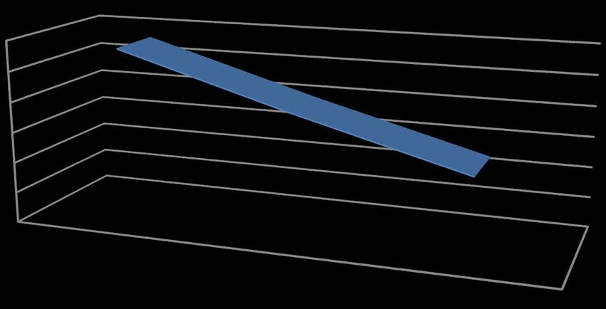
Q. Q. Li et al.

                                 the patients be evaluated safely, objectively, early and accurately.

                                 Keywords
                                 High Level Spinal Cord Injury, Autonomic Nervous Function, Imaging
                                 Urodynamic Examination, Slow Filling, Synchronous Blood Pressure
                                 Monitoring

                               1. Introduction
                               The Patients with dysuria of high level spinal cord injury (SCI) above T6 often
                               have autonomic nervous dysfunction, such as sudden significant increasement of
                               blood pressure, tachycardia, or transient hypotension, bradycardia, and or ac-
                               companied by sweating, shivering, headache, blushing, chills, hyperspasmia of
                               muscles below the injury level and other symptoms [1] [2]. Severe cases have the
                               risk of cerebral hemorrhage [3], retinal hemorrhage [4], or seizures, and even
                               cardiac arrest [5], which is one of the most serious complications after SCI. It
                               may even aggravate the neurological symptoms of the patients [6] and affect the
                               rehabilitation process. How can the blood pressure and heart rate change dy-
                               namically when autonomic nerve dysfunction occurs in such patients? What is
                               the relationship between it and bladder filling (intestinal irritation)? What are
                               the corresponding mechanisms and main incentives? How to evaluate auto-
                               nomic nervous function safely and scientifically? The above issues urgently need
                               to be studied and discussed. Therefore, it is very important to clarify the change
                               rule of autonomic nerve dysfunction in the patients with dysuria after high level
                               SCI, to explore its causes, to quantify autonomic nerve function of the patients
                               scientifically, to find a safe, objective, early and accurate method to evaluate au-
                               tonomic nerve function of them in clinic.

                               2. Patients and Method
                               From July 2017 to July 2020, 48 in patients with dysuria after cervicothoracic SCI
                               in our hospital were selected.
                                  The inclusion criteria: 1) 18 ≤ age ≤ 60 years old; 2) Dysuria caused by ASIA
                               classification (A-D grade) of cervical and thoracic spinal cord injury; 3) Course
                               of disease ≥ 1 month; 4) No urinary calculi; 5) No urinary tract tumor; 6) No
                               obvious stress urinary incontinence; 7) No history of urinary tract, bladder and
                               prostate surgery; 8) No serious urinary tract infection and there is a stable con-
                               dition within a week; 9) Signed the informed consent.
                                  The exclusion criteria: 1) Age < 18 or age > 60 years old; 2) Dysuria caused by
                               injury of lumbar spinal cord and below level; 3) Urination disorder caused by
                               cuaniocerebral diseases; 4) Course of disease < 1 month; 5) Have urinary calculi;
                               6) Have urinary tract tumor; 7) Have obvious stress urinary incontinence; 8)
                               Have history of urinary tract, bladder and prostate surgery; 9) The symptomatic
                               urinary tract infection occurred within the past week; 10) Those who have been

DOI: 10.4236/oju.2021.114012                        113                                     Open Journal of Urology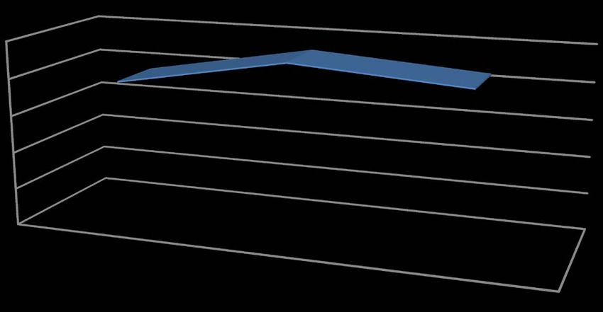
Q. Q. Li et al.

                               unstable condition within a week; 11) People who are allergic to iohexol; 12)
                               People with allergies; 13) People who refuse to participate in the study.
                                 The research method: Firstly, the patients were given adequate bowel prepara-
                               tion: They were given cleaning enema once at the night before the examination
                               and once at four hours before the examination on the same day. The enema so-
                               lution was a mixture of 100 ml normal saline and 40 ml glycerin. Then after their
                               skin preparation in the perineum area, all patients underwent imaging urody-
                               namic examination with slow filling in zero degree supine position. 10% iohexol
                               sodium chloride solution was continuously pumped into the bladder manometer
                               tube. The bladder filling rate was 8 - 10 ml/min. Before, during and after exami-
                               nation, blood pressure and ECG were monitored. Among them, the blood pres-
                               sure and heart rate during examination are the corresponding values of the lea-
                               kage point of the patients, or the immediate values when the patients had related
                               discomfort symptoms, or the values at the time of stopping bladder filling. Ob-
                               served and recorded whether the patients had sweating, shivering, headache,
                               blushing, chills and other symptoms.
                                 Inspection instrument: a set of German Siemens imaging urodynamic exami-
                               nation equipment, a wireless Bluetooth urodynamic instrument from Canada
                               Labry, an ECG monitor of China Shenzhen Jinlu UT4000B.
                                 Statistical methods: SPSS 21.0 software was used for data analysis. The mea-
                               surement data were represented by mean ± SD, and the count data were indi-
                               cated by two or three categories. T-test was used for the measurement data, P <
                               0.05 was statistically significant.

                               3. Results
                               The patients were divided into three groups according to their BP, HR and ex-
                               isting the symptoms of autonomic nervous dysfunction or not: group A (no ob-
                               vious abnormality of autonomic nervous function): before, during and after
                               examination, their BP and HR were normal, without accompanying symptoms;
                               group B (hyperactivity of it): both their SBP and DBP rised more than 20% dur-
                               ing examination than before, and at least they were accompanied with one of the
                               five symptoms; and group C (hypoactivity of it): they had lower HR (BP) than
                               normal, and or they were accompanied with one of the five symptoms. The val-
                               ues of BP and HR of the patients were shown in Table 1. For the sociodemo-
                               graphic and clinical characteristics, see Table 2. The data of age, disease course,
                               bladder safety capacity and bladder compliance was listed in Table 3. T-test of
                               age, disease course, bladder safe volume and compliance was used to compare
                               between three groups in Table 4. T-test of BP, HR was used to contrast between
                               three groups in Table 5. The incidence of three groups’ patients was evinced in
                               Table 6. The results of urodynamic examination were manifested in Table 7.
                               The results of a typical case’ blood pressure synchronous monitoring were
                               demonstrated in Table 8. Group B’ SBP trend of change was appeared in Figure
                               1; Group B’ DBP trend of change was indicated in Figure 2. Group C’ HR trend

DOI: 10.4236/oju.2021.114012                         114                                   Open Journal of Urology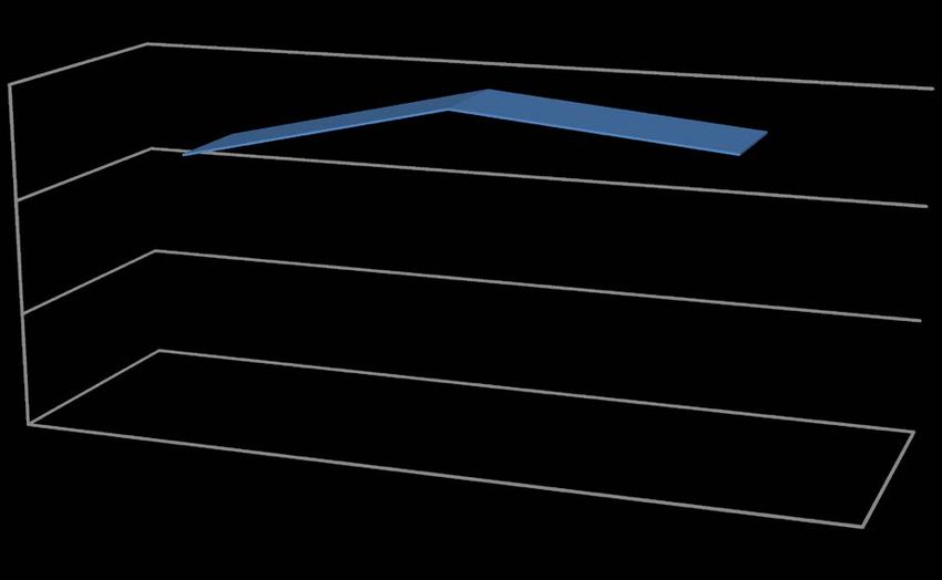
Q. Q. Li et al.

Table 1. Values of BP and HR of the patients (mean ± SD).

                     SBP (mmHg)                                         DBP (mmHg)                                           HR (Times/min)
          SBP1          SBP2           SBP3                DBP1              DBP2              DBP3                HR1              HR2                HR3

  A   112.25 ± 4.86 113.00 ± 11.81 116.38 ± 8.99        69.63 ± 5.34    71.25 ± 10.55       74.75 ± 6.67      66.00 ± 3.82     64.75 ± 4.37      66.38 ± 4.69
  B   117.13 ± 16.78 144.57 ± 29.67 134.10 ± 18.68 76.40 ± 14.85 92.80 ± 22.50              86.27 ± 14.36     66.03 ± 14.90 66.30 ± 18.70 66.27 ± 12.51
  C   103.38 ± 9.59 120.63 ± 5.66 110.13 ± 11.91        60.50 ± 7.87    74.63 ± 6.89        70.75 ± 10.86     59.63 ± 8.85     58.00 ± 8.78      56.50 ± 7.23

                                          Table 2. Sociodemographic and clinical characteristics of the patients.

                                                                       Group                                   A                 B                     C

                                                                                       M                       5                 23                    7
                                                        Gender
                                                                                       F                       3                    7                  1

                                                   Hypertension                       No                       8                 26                    8
                                                     history                          Yes                      0                    3                  0

                                              Hypotension history                     No                       8                 29                    7
                                                intermittently                        Yes                      0                    1                  1
                                                                               Cervical segment            5 (62.50%)        28 (93.33%)        7 (87.50%)
                                                                                Upper thoracic
                                                                                                           3 (37.50%)         1 (3.33%)          0 (0.00%)
                                               Plane of SCI (%)                 segment (≤T6)
                                                                                Lower thoracic
                                                                                                            0 (0.00%)         1 (3.33%)         1 (12.50%)
                                                                                segment (>T6)
                                                                                  Complete                 3 (37.50%)        13 (43.33%)        1 (12.50%)
                                            ASIA grades of SCI (%)
                                                                                 Incomplete                5 (62.50%)        17 (56.67%)        7 (87.50%)

                                                    Concomitant                       No                    8 (100%)         15 (50.00%)        3 (37.50%)
                                                   symptoms (%)                       Yes                   0 (0.00%)        15 (50.00%)        5 (62.50%)
                                                                                      No                   7 (87.50%)        28 (93.33%)        7 (87.50%)
                                              Hydronephrosis (%)
                                                                                      Yes                  1 (12.50%)         2 (6.67%)         1 (12.50%)

                                          Table 3. Patients’ age, disease course, bladder safety capacity and bladder compliance
                                          (mean ± SD).

                                                                  Group                                 A                       B                      C

                                                               Age (years)                         42.25 ± 7.92           37.07 ± 10.73        40.13 ± 14.84
                                                        Disease course (months)                   12.50 ± 13.30           26.03 ± 13.91        31.75 ± 36.10
                                                       Bladder safety capacity (ml)              134.13 ± 178.90         211.17 ± 137.83      200.63 ± 116.89
                                               Bladder compliance (ml/cmH2O)                      19.66 ± 48.65           18.52 ± 26.57        27.51 ± 45.22

                                          Table 4. Inter-group comparison of the patients’ age, disease course, bladder safe volume
                                          and bladder compliance (t-test).

                                                   p           Age           Disease course         Bladder safety capacity             Bladder compliance

                                               A-B            0.212*             0.355*                        0.195*                         0.929*
                                               A-C            0.726*             0.179*                        0.394*                         0.743*
                                               B-C            0.513*             0.716*                        0.844*                         0.472*

                                          *: p > 0.05.

DOI: 10.4236/oju.2021.114012                                              115                                                    Open Journal of Urology
Q. Q. Li et al.
Table 5. Inter-group comparison of the patients’ BP and HR (t-test).
p SBP (mmHg) DBP (mmHg) HR (Times/min)
SBP1 0.425* DBP1 0.217* HR1 0.995*
A-B SBP2 0.006*** DBP2 0.013** HR2 0.819*
SBP3 0.014** DBP3 0.035** HR3 0.981*
SBP1 0.035** DBP1 0.017** HR1 0.082*
A-C SBP2 0.122* DBP2 0.461* HR2 0.072*
SBP3 0.256* DBP3 0.390* HR3 0.006***
SBP1 0.034** DBP1 0.006*** HR1 0.226*
B-C SBP2 0.031** DBP2 0.032** HR2 0.234*
SBP3 0.002*** DBP3 0.007*** HR3 0.043**
*: p > 0.05; **: p < 0.05; ***: p < 0.01.
Table 6. Incidence of three groups’ patients.
Group A B C
Number 8 30 8
Percentage (%) 16.67 62.50 16.67
Total (B + C) 79.17
Table 7. Results of the patients’ urodynamic examination.
Group A B C
Overactivity 5 (62.50%) 23 (76.67%) 7 (87.50%)
Detrusor type (%)
Bladder atony 3 (37.50%) 7 (23.33%) 1 (12.50%)
Decreasement 7 (87.50%) 23 (76.67%) 5 (62.50%)
Bladder compliance (%) Increasement 1 (12.50%) 5 (16.67%) 1 (12.50%)
Normal 0 (0.00%) 2 (6.67%) 2 (25.00%)
External urethral Incoordination 6 (75.00%) 22 (73.33%) 5 (62.50%)
sphincter-detrusor (%) Achalasia 2 (25.00%) 8 (26.67%) 3 (37.50%)
Sensitive 4 (50.00%) 17 (56.67%) 5 (62.50%)
Bladder sensation (%) lost 4 (50.00%) 9 (30.00%) 2 (25.00%)
Declining 0 (0.00%) 4 (13.33%) 1 (12.50%)
No 6 (75.0%) 14 (46.67%) 1 (12.50%)
Higher abdominal pressure (%)
Yes 2 (25.00%) 16 (53.33%) 7 (87.50%)
Table 8. Results of a typical case’ blood pressure synchronous monitoring.
SBP (mmHg) DBP (mmHg) HR (Times/min)
Start filling 113 70 77
Stop filling 137 87 65
End examination 130 82 66
DOI: 10.4236/oju.2021.114012 116 Open Journal of UrologyQ. Q. Li et al.
B
150
100
50 B
0
B
SBP1
SBP2
SBP3
Figure 1. Trend chart of SBP in group B.
B
100
80
60
40
20 B
0
B
DBP1
DBP2
DBP3
Figure 2. Trend chart of DBP in group B.
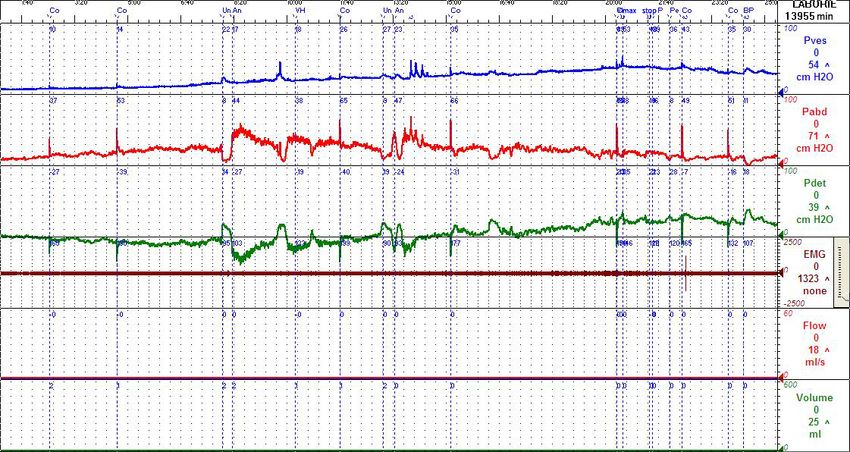
of change was revealed in Figure 3. The urodynamic graph of a typical case was
disclosed in Figure 4.
Table 4 exposed that there was no significant difference in age, disease course,
bladder safety capacity and compliance between three groups (T-test) (P > 0.05).
However, from Table 5, compared of BP and HR between three groups (T-test),
it can be seen that SBP2 of group A-B had extremely prominent discrepancy (P
< 0.01); SBP3, DBP2 and DBP3 of it had marked distinction (P < 0.05). HR3 of
group A-C was greatly remarkable different (P < 0.01), SBP1 and DBP1 of it had
obvious dissimilarity (P < 0.05). The diversity between group B-C was more evi-
dent. Figure 1, Figure 2 and Table 1 revealed that the changes of SBP and DBP
of group B were increased earlier and then decreased. Figure 3 and Table 1 ex-
hibited that the changes of HR of group C was lower than normal and declined
continuously. Figure 4 suggested that the abdominal pressure was significantly
higher than the bladder pressure, from the whole course of inspection. Table 6
displayed the incidence of autonomic dysfunction of the patients with dysuria
after high level SCI was very high (about 79.17%, adding B and C), most of them
were hyperactivity (62.50%), and a few were hypoactivity (16.67%). Table 7 ma-
nifested: The incidence of NDO [7], low compliant bladder (62.50% - 87.50%),
DOI: 10.4236/oju.2021.114012 117 Open Journal of UrologyQ. Q. Li et al.
C
60
59
58
57
56
C
55
54
C
HR1
HR2
HR3
Figure 3. Trend chart of HR in group C.
Figure 4. Urodynamic graph of a typical case.
NDSD [8] of group B and C was very high, that of high abdominal pressure of
group B, C (53.33%, 87.50%) was evidently more quantity than that of group A
(25.00%), the ratio of bladder disappearance of group B, C (30.00%, 25.00%) was
fewer than that of group A (50.00%). From Table 8, compared with the baseline,
it can be demonstrated that the values of SBP and DBP of a typical case at he
time of stopping filling increased more than 20 mmHg. From Table 2, we found
out that the incidence of symptoms without autonomic nervous dysfunction of
group B and C was not low (50.00%, 37.50%), and the average incidence was
43.75%.
4. Discussion
Paraplegic patients with high level SCI may have abnormal autonomic reflex or
autonomic dysreflexia (AD), especially in the patients with T6 or higher plane,
the incidence is nearly 1/2 - 3/4, the latest research shows that: as high as 93%
[9], the patients with cervical SCI are more common. Its severity is mainly re-
lated to the sudden and distinct increase of BP: such as conscious change, visual
impairment, seizures, intracranial hemorrhage, and even death. Headache, facial
DOI: 10.4236/oju.2021.114012 118 Open Journal of UrologyQ. Q. Li et al.
flushing, chest tightness, palpitation, sweating and other symptoms are often
used as a warning sign of elevated BP. Nevertheless, sometimes patients have
elevated BP without any symptoms, which is called “asymptomatic AD” [10]. In
this case, patients and doctors may have missed the best time to manage the po-
tential powerful afferent stimuli, which may lead to severe sequelae. In fact, this
“silent killer” is more harmful to patients. The Patients with high level SCI can
also present hypotension, orthostatic hypotension (OH) [11] [12], reflex brady-
cardia and cardiac arrest. Available data indicate that the incidence of bradycar-
dia and cardiac arrest of SCI patients can be as high as 30%, and the average HR
is 54 times/min, including some patients with severe bradycardia less than 50
times/min [13]. Therefore, we should pay enough attention to the occurrence of
serious cardiovascular complications, such as bradycardia and cardiac arrest,
especially in the patients with high level paraplegia. At present, circulatory dis-
turbance has become the second leading cause of death in the patients with
chronic SCI. Therefore, it is necessary and urgent to study the autonomic dys-
function in the patients with SCI.
The study demonstrated that the incidence of autonomic dysfunction in the
high level SCI patients with dysuria was very high (almost 80.00%), most of
them were hyperactivity (62.50%), and a few were low function (16.67%).
Among them, the increasement of SBP and DBP in the hyperfunction group was
greater than 20% of the baseline value when there was no attack, and they all
showed a rise firstly and then a fall; the decline of HR (BP) in the hypoactivity
one was lower than normal and decreased continuously, before, during and after
examination, and or two groups of patients were accompanied with one of the
five symptoms: sweating, shivering, headache, facial congestion, chills [14].
Indeed, In addition to bladder overinflation caused by NDO and NDSD is the
main causes of AD, the results in Table 7 suggest that the increase of abdominal
pressure is also the main cause of AD. Previous studies have shown that the most
common cause of AD is bladder dilation, accounting for 75% - 85%; the second
common cause is intestinal irritation, such as fecal impaction, accounting for
20% [15] [16]. Most patients with dysuria of SCI are accompanied by neurogenic
intestinal dysfunction. Therefore, with the increament of abdominal pressure
caused by bladder filling and intestinal irritation, the conduction through viscer-
al sensory nerve: pelvic nerve and pudendal nerve to the sacral spinal cord can
also cause the above AD performance [17]. In this study, the patients of group B
and C had more transient paroxysmal increasement of abdominal pressure than
ones of group A, and or they were accompanied by transient enhancement of the
myoelectric amplitude of the external urethral sphincter. Most of patients’ ab-
dominal pressure fluctuated within 10 - 30 cm H2O for a short period of time,
even as high as about 70 cm H2O, as shown in the urodynamic map of a typical
case of SCI (T3 AIS grade B). Although good bowel preparations (cleaning ene-
ma twice) had been done before the examination, the patient’ bladder was filled
slowly in the supine position, and detrusor was recorded uninhibitory contrac-
tion during the urine storage period, the maximum detrusor pressure is 35 cm
DOI: 10.4236/oju.2021.114012 119 Open Journal of UrologyQ. Q. Li et al.
H2O, without significant increase, the peak value of abdominal pressure was 71
cm H2O. Simultaneous cystography showed that when the bladder was filled to
100 ml, it presented the X-ray appearance of neurogenic bladder protruded to
the right. The results of synchronous blood pressure monitoring in Table 8 in-
dicated that the values of SBP and DBP at the time of stopping filling raised by
more than 20 mmHg compared to the values at the beginning of filling, even
though the patient had no symptoms of AD. Imaging urodynamic diagnosis:
bladder sensation disappeared; detrusor was overactivity in the storage period,
detrusor contraction was weakness in the voiding period; bladder compliance
decreased; bladder safety capacity, urinary bladder capacity was about 356 ml;
partial incordination was existed between external urethral sphincter and detru-
sor; rectal pressure increased significantly; there was no vesicoureteral reflux;
autonomic hyperfunction. In Table 7, the incidence of high abdominal pressure
of group B and C was noticeably higher than that of group A, illustrating that
elevated abdominal pressure was actually very common in AD patients.
The results in Table 7 also imply that the patients with abnormal bladder sen-
sitivity are more likely to induce AD. Under normal circumstances, bladder fill-
ing or intestinal stimulation can cause sympathetic nerve excitation, increase
blood pressure, stimulate aorta and carotid baroreceptor to regulate and nor-
malize blood pressure; however, the patients with SCI can not reduce blood
pressure through visceral vasodilation, the brainstem also can not transmit inhi-
bitory impulses to the sympathetic nerves, If they were accompanied by hyper-
sensitivity of the bladder below the injury level, AD is more likely to occur, and
the sympathetic nerve mechanism is dominant, Elevated blood pressure is a
clinical manifestation [18]; After the aortic and carotid baroreceptors are stimu-
lated, the impulse is uploaded to the cardiovascular motor center of the brain
stem, which can also cause parasympathetic dominance at the level above the
injury, excitation of the vagus nerve, thus slowing down the heart rate, but the
blood pressure is not necessarily lower, the situation of decline in heart rate is
rare [19]. In Table 1 and Table 6 of this study, the results of group B and C are
consistent with this.
It can be seen from Table 2 that the average incidence of asymptomatic AD is
higher (43.75%), which indicates that asymptomatic AD has great potential
harm to the patients with chronic SCI [20]. Therefore, by accurately and objec-
tively evaluating whether the autonomic nervous function of the patients is ab-
normal, early detection and identification of such asymptomatic patients, timely,
scientific and standardized management of bladder and intestinal tract, and
comprehensive treatment can effectively, maximally avoid acute attack of AD or
deterioration of symptoms in the patients with SCI, reduce or ward off the oc-
currence of clinical malignant emergencies.
For the SCI patients with urination disorder, the autonomic nerve function is
mostly hyperactive and blood pressure tends to rise. Therefore, during the whole
process of image urodynamic examination, it is necessary to pay attention to the
speed of bladder perfusion should be slow rather than fast, that is, slow filling,
DOI: 10.4236/oju.2021.114012 120 Open Journal of UrologyQ. Q. Li et al.
also known as physiological filling refers to the speed of bladder filling is less
than 10 ml/min [21], and synchronized blood pressure monitoring is performed
to observe and record the patients’ blood pressure and heart rate dynamic
changes, before, during and after the examination, as well as whether there are
five main symptoms of sweating, chills, headache, flushing and chills. Only in
this way can the autonomic nervous function of patients be evaluated safely, ob-
jectively, early and accurately.
5. Conclusion
The incidence of autonomic dysfunction in the high level SCI patients with dy-
suria was very high (nearly 80.00%), most of them were hyperactivity, and a few
were low function. The changes of SBP and DBP in the hypoactivity group all
appeared an increasing and then declining trend, while the change of HR in the
low function one was lower than normal and decreased continuously. The main
inducements of AD are neurogenic detrusor overactivity, detrusor sphincter
dyssynergia, elevated abdominal pressure and abnormal bladder sensitivity. The
asymptomatic patients had a higher occurrence rate (43.75%). Only by imaging
urodynamic examination with slow filling and synchronous blood pressure
monitoring, can autonomic nervous function of the patients be evaluated safely,
objectively, early and accurately.
Acknowledgements
This study was supported by Medical Scientific Research Foundation of Guang-
dong Province, China (grant number: A2017288).
I sincerely thank Dr. Hui Chen and other medical workers for their strong
support and assistance in this study.
Conflicts of Interest
The authors declare no conflicts of interest regarding the publication of this pa-
per.
References
[1] Teasell, R.W., Arnold, J.M., Krassioukov, A., et al. (2000) Cardiovascular Conse-
quences of Loss of Supraspinal Control of the Sympathetic Nervous System after
Spinal Cord Injury. Archives of Physical Medicine and Rehabilitation, 81, 506-516.
https://doi.org/10.1053/mr.2000.3848
[2] Claydon, V.E., Elliott, S.L., Sheel, A.W. and Krassioukov, A. (2006) Cardio Vascular
Responses to Vibrostimulation for Sperm Retrieval in Men with Spinal Cord Injury.
Journal of Spinal CordMedicine, 29, 207-216.
https://doi.org/10.1080/10790268.2006.11753876
[3] Eker, A., Yigitoglu, P.H., Ipekdal, H.I. and Tosun, A. (2014) Acute Onset of Intra-
cerebral Hemorrhage Due to Autonomic Dysreflexia. Journal of Korean Neurosur-
gical Society, 55, 277-279. https://doi.org/10.3340/jkns.2014.55.5.277
[4] Rosenthal, J. and Colachis, S. (2011) Cortical Blindness Associated with Autonomic
DOI: 10.4236/oju.2021.114012 121 Open Journal of UrologyQ. Q. Li et al.
Dysreflexia in a Man with Tetraplegia: A Rare but Serious Complication. Journal of
Spinal Cord Medicine, 34, 527-529.
https://doi.org/10.1179/2045772311Y.0000000021
[5] Amit, J., Babita, G., Kajal, J., Jeetinder, K.M., Kishore, M. and Supriya, S. (2013) Se-
vere Autonomic Dysreflexia Induced Cardiac Arrest under Isoflurane Anesthesia
Spine Injury in a Patient with Lower Thoracic Spine Injury. Journal of Anaesthesi-
ology Clinical Pharmacology, 29, 241-243.
https://doi.org/10.4103/0970-9185.111652
[6] Harrop, J.S., Sharan, A.D., Vaccaro, A.R. and Przybylski, G.J. (2001) The Cause of
Neurologic Deterioration after Acute Cervical Spinal Cord Injury. Spine, 26,
340-346. https://doi.org/10.1097/00007632-200102150-00008
[7] Walter, M., Knüpfer, S.C., Cragg, J.J., Leitner, L., Schneider, M.P., Mehnert, U., et
al. (2018) Prediction of Autonomic Dysreflexia during Urodynamics: A Prospective
Cohort Study. BMC Medicine, 16, 53. https://doi.org/10.1186/s12916-018-1040-8
[8] Liu, N., Zhou, M.W., Biering-Sorensen, F. and Krassioukov, A.V. (2016) Cardi-
ovascular Response during Urodynamics in Individuals with Spinal Cord Injury.
Spinal Cord, 55, 279-284. https://doi.org/10.1038/sc.2016.110
[9] Lee, E.S. and Joo, M.C. (2017) Prevalence of Autonomic Dysreflexia in Patients with
Spinal Cord Injury above T6. Biomed Research International, 2017, Article ID:
2027594. https://doi.org/10.1155/2017/2027594
[10] Huang, Y.H., Bih, L.I., Liao, J.M., Chen, S.L., Chou, L.W. and Lin, P.H. (2013)
Blood Pressure and Age Associated with Silent Autonomic Dysreflexia during Uro-
dynamic Examinations in Patients with Spinal Cord Injury. Spinal Cord, 51,
401-405. https://doi.org/10.1038/sc.2012.155
[11] Noreau, L., Proulx, P., Gagnon, L., Drolet, M. and Laramée, M. (2000) Secondary
Impairments after Spinal Cord Injury: A Population-Based Study. American Journal
of Physical Medicine & Rehabilitation, 79, 526-535.
https://doi.org/10.1097/00002060-200011000-00009
[12] Cariga, P., Ahmed, S., Mathias, C.J. and Gardner, B.P. (2002) The Prevalence and
Association of Neck (Coat-Hanger) Pain and Orthostatic (Postural) Hypotension in
Human Spinal Cord Injury. Spinal Cord, 40, 77-82.
https://doi.org/10.1038/sj.sc.3101259
[13] Furlan, J.C. and Fehlings, M.G. (2008) Cardiovascular Complications after Acute
Spinal Cord Injury: Pathophysiology, Diagnosis, and Management. Neurosurgical
Focus, 25, E13. https://doi.org/10.3171/FOC.2008.25.11.E13
[14] Krassioukov, A., Biering-Sorensen, F., Donovan, W., Kennelly, M., Kirshblum, S.,
Krogh, K., et al. (2012) International Standards to Document Remaining Autonom-
ic Function after Spinal Cord Injury (ISAFSCI). Journal of Spinal Cord Medicine,
35, 201. https://doi.org/10.1179/1079026812Z.00000000053
[15] Chen, C.Y., Chuang, T.Y., Tsai, Y.A., Tai, H.C., Lu, C.L., Kang, L.J., et al. (2004)
Loss of Sympathetic Coordination Appears to Delay Gastrointestinal Transit in Pa-
tients with Spinal Cord Injury. Digestive Diseases and Sciences, 49, 738-743.
https://doi.org/10.1023/B:DDAS.0000030082.05773.c9
[16] Cotterill, N., Madersbacher, H., Wyndaele, J.J., Apostolidis, A., Drake, M.J., Ga-
jewski, J., et al. (2018) Neurogenic Bowel Dysfunction: Clinical Management Rec-
ommendations of the Neurologic Incontinence Committee of the Fifth Internation-
al Consultation on Incontinence 2013. Neurourology and Urodynamics, 37, 46-53.
https://doi.org/10.1002/nau.23289
[17] Blackmer, J. (2003) Rehabilitation Medicine: 1. Autonomic Dysreflexia. Canadian
DOI: 10.4236/oju.2021.114012 122 Open Journal of UrologyQ. Q. Li et al.
Medical Association Journal, 169, 931-935.
[18] Weaver, L.C. (2002) What Causes Autonomic Dysreflexia after Spinal Cord Injury?
Clinical Autonomic Research, 12, 424-426.
https://doi.org/10.1007/s10286-002-0076-0
[19] Legramante, J.M., Raimondi, G., Massaro, M. and Iellamo, F. (2001) Positive and
Negative Feedback Mechanisms in the Neural Regulation of Cardiovascular Func-
tion in Healthy and Spinal Cord-Injured Humans. Circulation, 103, 1250-1255.
https://doi.org/10.1161/01.CIR.103.9.1250
[20] Grossman, R.G., Frankowski, R.F., Burau, K.D., Toups, E.G., Crommett, J.W.,
Johnson, M.M., et al. (2012) Incidence and Severity of Acute Complications after
Spinal Cord Injury. Journal of Neurosurgery Spine, 17, 119-128.
https://doi.org/10.3171/2012.5.AOSPINE12127
[21] Liao, L.M. (2012) Urodynamics. People’s Military Medical Press, Beijing, 119.
DOI: 10.4236/oju.2021.114012 123 Open Journal of UrologyYou can also read