Prevention of lymphedema via axillary reverse mapping for arm lymph-node preservation following breast cancer surgery: a randomized controlled trial
←
→
Page content transcription
If your browser does not render page correctly, please read the page content below
Faisal et al. Patient Safety in Surgery (2019) 13:35
https://doi.org/10.1186/s13037-019-0217-1

 RESEARCH                                                                                                                                        Open Access

Prevention of lymphedema via axillary
reverse mapping for arm lymph-node
preservation following breast cancer
surgery: a randomized controlled trial
Mohammed Faisal* , Mohamed Gamal Sayed, Kerolos Antonious, Ahmmed Abo Bakr and Sherif Hussein Farag

  Abstract
  Background: Breast cancer, with an incidence of 32%, is the most frequent cancer among Egyptian women.
  The frequency of arm lymphedema after axillary surgery for breast cancer ranges from 7 to 77%. Axillary reverse
  mapping is a technique aimed to distinguish and conserve upper-limb lymphatics and lymph nodes during the
  course of axillary surgery and could help to prevent arm lymphedema.
  Methods: Patients (n = 48) were prepared for axillary lymph-node dissection. The study group and the control
  group each contained 24 individuals. In the study group, following dye injection, stained arm lymph nodes
  and lymphatics were conserved during axillary dissection, whereas control-group participants underwent the
  conventional procedure. All participants were re-evaluated after 6 months, and the incidence of lymphedema was
  recorded by measuring arm circumference at a level 10 cm proximal to the medial epicondyle. Arm lymphedema
  was defined as a change in the circumference of the ipsilateral upper extremity > 2 cm during the follow-up period.
  Results: Age, tumor size and N stage were not significantly different between the study and control groups.
  Lymph-node visualization was achieved in 20 participants (83.3%) in the study group. Suspicious stained lymph
  nodes were surgically removed from four individuals but showed no metastatic involvement. In 20 individuals in
  the study group, no stained lymph nodes were removed. The incidence of lymphedema in the control group was
  16.7%, and the incidence in the study group was 4.2%.
  Conclusions: Axillary reverse mapping is a minimally invasive technique that can be performed during axillary
  lymph-node dissection, helping to prevent the subsequent development of arm lymphedema.
  Trial registration: #SCURCTN3276, retrospectively registered on 11 April 2017 at Research Ethics Committee at the
  Faculty of medicine-Suez Canal University.
  Keywords: Breast cancer, Lymphedema, Blue dye, Axillary reverse mapping

Background                                                                            increases, the importance of limitation of the adverse ef-
Worldwide, ~ 1.67 million women are diagnosed with                                    fects of axillary surgery will also increase. One of these
breast cancer every year. Breast cancer is second on the                              adverse effects is arm lymphedema, which has a fre-
list of the most common cancers in the world and has                                  quency of 7–77% after axillary surgery [3].
the highest cancer incidence among the female popula-                                   Axillary reverse mapping (ARM) is a technique that
tion [1], including that in Egypt, where it represents                                has been devised based on the assumption that arm
32.04% of female cancer incidence [2]. As the number of                               lymphedema results from disruption of arm lymphatics
patients recovering from breast cancer treatment                                      during axillary dissection. The axillary lymphatics and
                                                                                      lymph nodes are anatomically linked to the lymphatic
* Correspondence: M.faisal@med.suez.edu.eg                                            drainage of the arm; these nodes include the lateral or
Surgical Oncology Unit, Department of Surgery, Faculty of Medicine, Suez
Canal University hospital, Circular Road, Ismailia 411522, Egypt
                                                                                      brachial group that lies below the axillary vein [4]. The

                                        © The Author(s). 2019 Open Access This article is distributed under the terms of the Creative Commons Attribution 4.0
                                        International License (http://creativecommons.org/licenses/by/4.0/), which permits unrestricted use, distribution, and
                                        reproduction in any medium, provided you give appropriate credit to the original author(s) and the source, provide a link to
                                        the Creative Commons license, and indicate if changes were made. The Creative Commons Public Domain Dedication waiver
                                        (http://creativecommons.org/publicdomain/zero/1.0/) applies to the data made available in this article, unless otherwise stated.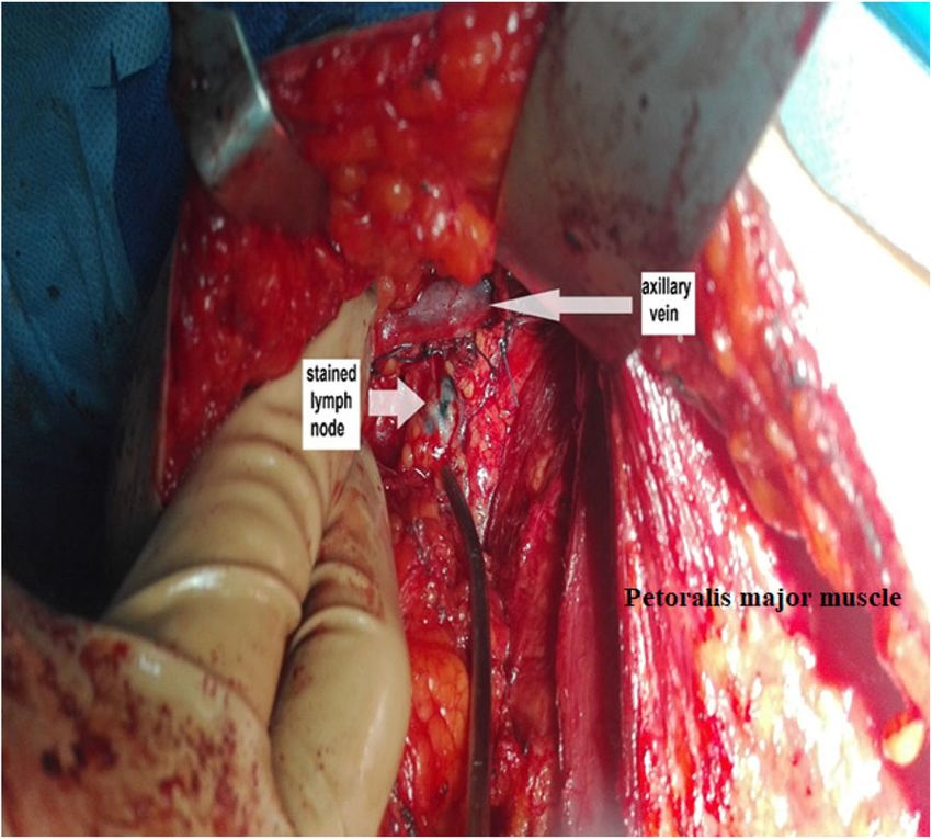
Faisal et al. Patient Safety in Surgery (2019) 13:35 Page 2 of 6
purpose of ARM is to differentiate the lymphatics and
lymph nodes of the arm from those of the breast during
axillary surgery, to enable preservation of the arm lym-
phatics [5]. Currently available methods for ARM are
based on injection and tracking of blue dye, fluorescent
dye or a radioisotope, as reviewed elsewhere [6].
In the current study, our objectives were to assess the
effectiveness of arm-node preservation by ARM for pre-
vention of lymphedema in patients with breast cancer
undergoing axillary lymph-node dissection (ALND), to
determine the rate of arm-node involvement and to in-
vestigate the location of arm nodes. Our overall aim is
to improve the quality of life of patients with breast can-
cer by reducing the adverse effects associated with axil-
lary surgery.
Methods
Fig. 1 Injection of blue dye
A randomized controlled trial was carried out in the
Surgery Department of Suez Canal University Hospital
and Ismailia Teaching Oncology Hospital in Egypt from for a few minutes to aid dye migration toward the axilla.
June 2017 to January 2018. The study design was reviewed Surgery for breast cancer, either modified radical mast-
by the Research Ethics Committee at the Faculty of ectomy or wide local excision of the lesion, was then
medicine-Suez Canal University at its meeting on 11/04/ performed, followed by axillary dissection (~ 20 min after
2017 with reference number (#3276). The study adhered dye injection).
to CONSORT guidelines. Lymphatic arm drainage (LAD) was identified by ob-
The following formula was used to calculate the re- servation of stained lymphatics and stained lymph nodes
quired sample size: draining the arm (within the lateral compartment of the
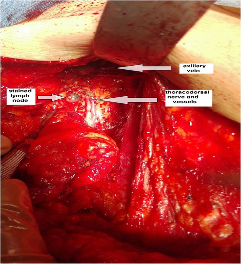
n = (Zα/2 + Zβ)2 * (p1(1 − p1) + p2(1 − p2))/(p1 − p2)2, axilla) (Fig. 2) that were preserved as a part of the ARM
where n is the sample size of each group (with a 1:1 procedure during ALND (Fig. 3). Any variations in
ratio of group sizes), α is the probability of type I error stained arm lymphatics associated with the site and their
(set at 0.01), Zα/2 is the critical value of the normal sizes were noted.
distribution at α/2, β is the probability of type II error Removal of stained lymph nodes was carried out only
(set at 0.2), Zβ is the critical value of the normal distri- in patients with suspicious lymph nodes (Fig. 4), which
bution at β (for β = 0.2, Zβ = 0.84), and p1 and p2 are were identified by the following criteria: 1) multiple
the expected sample proportions (incidences) in the two
groups. Yue et al. previously demonstrated incidences of
lymphedema of 33.1% with ARM and 5.9% without
ARM [7]. These values were used for p1 and p2, result-
ing in a calculation of n = 24 for each group in our study.
A total of 48 patients diagnosed with breast cancer were
investigated.
ALND was indicated for all participants, who were
randomly assigned (via a sequence generated in Micro-
soft Excel) in a 1:1 ratio to either the study or control
groups. The technique was performed by a single sur-
geon, who was given the randomly generated treatment
allocations within sealed, opaque envelopes. When a pa-
tient consented to enter the trial, the envelope was
opened, and the patient underwent the allocated surgery.
In the study group, general anesthesia was employed
then ~ 2.5 ml of 1% (w/v) methylene blue dye was
injected subcutaneously into the medial intramuscular
crease of the upper arm on the same side (Fig. 1). The
Fig. 2 Identification of a stained lymph node below the axillary vein
injection site was massaged, and the arm was elevated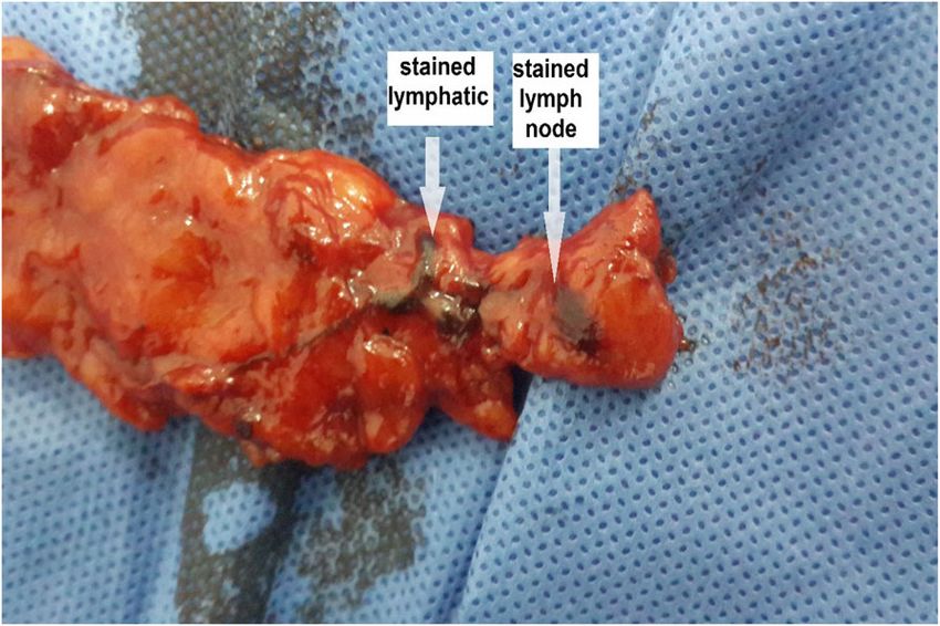
Faisal et al. Patient Safety in Surgery   (2019) 13:35                                                                              Page 3 of 6

                                                                        Arm circumference measurement
                                                                        Arm circumference was measured at a level 10 cm prox-
                                                                        imal to the medial epicondyle before surgery and 6
                                                                        months after surgery. Changes between arm circumfer-
                                                                        ence of the ipsilateral and contralateral upper extremity
                                                                        in each group were compared. In the ipsilateral upper
                                                                        extremity, arm circumferences before surgery and 6
                                                                        months after surgery were also compared. Arm lymph-
                                                                        edema was defined as a change in the circumference of
                                                                        the ipsilateral upper extremity > 2 cm during the follow-
                                                                        up period [8].

                                                                        Results
                                                                        The mean age of the study population was 52 ± 11 years.
                                                                        All participants had invasive ductal carcinoma, as shown
                                                                        by Tru-cut biopsy (Table 1); 22.9% had T1 lesions,
                                                                        56.3% had T2 lesions, and 20.8% had T3 lesions. Patho-
                                                                        logic axillary lymph-node staging demonstrated that
                                                                        29.3% of participants had pN0 disease, 35.4% had pN1
                                                                        disease, 18.8% had pN2 disease, and 16.7% had pN3 dis-
 Fig. 3 Location of a stained lymph node lateral to the thoracodorsal
 trunk 2 cm below the axillary vein
                                                                        ease. Regarding the type of surgery, 27.1% of participants
                                                                        underwent conservative breast surgery (wide local exci-
                                                                        sion of the lesion), whereas 72.9% underwent modified
                                                                        radical mastectomy.
suspicious or amalgamated lymph nodes in the axilla; 2)                   Out of 24 participants with ARM, stained lym-
enlarged node (> 1 cm); 3) firm or hard node; and 4)                    phatics were visualized in 18 (75.0%), and stained
anatomical location other than lateral or above the thor-               lymph nodes were visualized in 20 (83.3%) (Table 2).
acodorsal pedicle.                                                      Of the stained nodes, 95.9% were situated between
  In the control group, classic axillary dissection was                 the second intercostobrachial nerve and the lower
performed without conservation of the upper-limb lym-                   limit of the axillary vein. In four participants, some
phatics and lymph nodes.                                                stained lymphatics and nodes (mean 2.25, range 1–3
  All patients were revaluated after 6 months, and the                  lymph nodes per person) were removed and assessed
incidence of lymphedema was recorded.                                   by histopathological examination. Metastasis was not
                                                                        identified in any of the removed stained lymph
                                                                        nodes (Table 3). Arm lymphedema was reported in
                                                                        one individual in the study group (4.2%), and four in
                                                                        the control group (16.7%) (Table 4).

                                                                        Table 1 Histopathological tumor characteristics of the study
                                                                        population
                                                                        Variable           Parameter    Control group,   Study group,   Total,
                                                                                                        n (%)            n (%)          n (%)
                                                                        Histopathology     IDC          24 (100)         24 (100)       48 (100)
                                                                        T stage            T1           9 (37.5)         2 (8.3)        11 (22.9)
                                                                                           T2           9 (37.5)         18 (75)        27 (56.3)
                                                                                           T3           6 (25)           4 (16.7)       10 (20.8)
                                                                        N stage            N0           7 (29.2)         7 (29.2)       14 (29.2)
 Fig. 4 Example of axillary lymph-node dissection. For this patient,
                                                                                           N1           13 (54.2)        4 (16.7)       17 (35.4)
 the total number of excised lymph nodes was 19. Three of these
 nodes were stained, and there was no evidence of metastasis in the                        N2           2 (8.3)          7 (29.2)       9 (18.8)
 stained nodes. Among the 16 unstained nodes, metastasis was                               N3           2 (8.3)          6 (25)         8 (16.7)
 detected in five nodes
                                                                        IDC invasive ductal carcinoma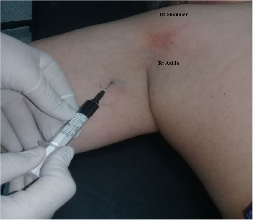
Faisal et al. Patient Safety in Surgery         (2019) 13:35                                                                                    Page 4 of 6

Table 2 Characteristics of axillary lymph-node dissection surgery in the study population
Variable                                  Parameter            Control group, n (%)          Study group, n (%)      Total, n (%)       χ2          p-value
Type of breast cancer surgery             Conservative         8 (33.3)                      5 (20.8)                13 (27.1)          0.949       0.330
                                          MRM                  16 (66.7)                     19 (79.2)               35 (72.9)
Visualization of lymphatics               Yes                  N/A                           18 (75)                 N/A                N/A         N/A
                                          No                   N/A                           6 (25)                  N/A
Visualization of lymph nodes              Yes                  N/A                           20 (83.3)               N/A                N/A         N/A
                                          No                   N/A                           4 (16.7)                N/A
MRM modified radical mastectomy; N/A not applicable

Discussion                                                                           multiple amalgamated nodes, which made visualization
ALND is the main method for final staging of breast                                  of the blue dye difficult. In a previous study conducted
cancer [9]. The morbidities associated with ALND, espe-                              by Nos et al. [12], the LAD mapping rate was 91%, with
cially upper-limb lymphedema, lead to reductions in                                  positive identification in 21 of 23 individuals, with an
quality of life, because affected individuals suffer from                            ARM technique combining injections of radioactive iso-
disfigurement, pain, numbness, restriction of movement                               tope and blue dye. In a larger study, Tummel et al. [13]
and recurrent infections [3].                                                        identified stained arm nodes and lymphatics in 153 of
   The ARM technique was developed on the basis of                                   213 individuals (71.8%) who underwent ARM.
evidence of the existence of separate lymphatic pathways                               In our study, 95.9% of the stained nodes were located
for the arm and the breast. This evidence suggests that                              between the lower aspect of the axillary vein and the
upper-limb lymphedema results from disruption during                                 second intercostobrachial nerve lying lateral to the thor-
axillary dissection of the lymphatic channels and lymph                              acodorsal nerve and vessels. Only one patient (4.1%) had
nodes associated with arm lymph drainage. Identification                             stained nodes that were situated medial to the thoraco-
and preservation of these lymphatics and lymph nodes                                 dorsal nerve and vessels. These results are similar to
should therefore limit the incidence of morbidities asso-                            those of a previous study [8] in which stained lymph
ciated with ALND [5, 10].                                                            nodes inferior to the axillary vein and above the second
   For ARM in this study, the anatomical location of dye                             intercostobrachial were found in 97% of the study popu-
injection in the upper part of the arm along the medial                              lation. Clough et al. [14] identified stained arm nodes 1
intermuscular crease was selected because it is the site                             cm inferior to the axillary vein, lateral to the thoracodor-
where nearly all arm lymphatics aggregate, and injection                             sal nerve and vessels. Kumar et al. [15] found no meta-
in this area enables rapid migration of the dye to the ax-                           static involvement in stained arm nodes located lateral
illa [8]. Moreover, this site is preferred because it con-                           to the thoracodorsal nerve and vessels.
ceals the blue mark resulting from injection, which may                                In our study, the mean number of axillary nodes re-
remain for up to 6 months [11].                                                      moved during ALND was 13.4 per person in the study
   The effectiveness of ARM technique is described by its                            group and 17.4 in the control group; the difference be-
value in identifying arm nodes and reducing arm lymph-                               tween these means was statistically significant. Although
edema also by the safety of the technique. In this study                             the quantity of retrieved lymph nodes in the ARM group
stained lymphatics were visualized in 18 individuals                                 was less than the number of resected nodes with classic
(75.0% of the study group), and stained lymph nodes                                  axillary dissection, there was no statistically significant
were documented in 20 individuals (83.3%). Only two                                  difference between the number of harvested nodes and
participants had no visualization of stained lymph nodes                             the number of nodes that were positive for malignancy.
or lymphatics in the axilla; the axillary dissection showed                          Notably, the mean number of excised nodes in our ARM

Table 3 Histopathological status of surgically removed stained lymph nodes from four patients in the study group
Arbitrary case       N stage         Total number of nodes                 Total number of metastatic      Number of stained        Number of metastatic
number                               removed                               nodes removed                   nodes removed            stained nodes removed
1                    N3              15                                    13                              1                        0
2                    N2              19                                    5                               3                        0
3                    N1              13                                    2                               3                        0
4                    N1              14                                    1                               2                        0
Faisal et al. Patient Safety in Surgery (2019) 13:35 Page 5 of 6
Table 4 Lymphedema incidence and distribution during 6 months follow-up in the study population
Variable Parameter Control group, n (%) Study group, n (%) Total, n (%) χ2 p-value
Incidence of lymphedema Yes 4 (16.7) 1 (4.2) 5 (10.4) 2.009 0.156
No 20 (83.3) 23 (95.8) 43 (89.6)
study group was greater than the 10 nodes previously recommended to validate the results. Another limita-
suggested to be the minimum number resected for axil- tion was that the short-term follow-up of 6 months
lary clearance to be oncologically successful [16]. may not have been long enough to distinguish transi-
In four of the individuals in our study group, stained ent lymphedema resulting from acute surgical edema
lymph nodes were classified as suspicious by virtue of from permanent lymphedema, suggesting that longer
being firm and > 1 cm in diameter or being located med- follow-up is needed.
ial to the thoracodorsal nerve and vessels. These suspi-
cious nodes were surgically removed and separately Conclusions
assessed by histopathological examination; the mean ARM is a minimally invasive technique that can be read-
number of removed blue lymph nodes was 2.25, ranging ily added to ALND and that can help prevent arm
from 1 to 3 nodes. None of the removed stained nodes lymphedema. The use of ARM for LAD mapping and
showed any metastatic spread. avoidance of excision of arm lymphatics and nodes was
In our study, we relied on the variation in arm circum- associated with a lower incidence of arm lymphedema
ference, as previously defined [8], to determine the oc- than classic ALND surgery in our study population.
currence of arm lymphedema during the follow-up However, we recommend implementing future studies
period. Chirag and his colleagues [17] have also indi- on the ARM procedure in a larger number of patients,
cated that self-assessed symptoms of discomfort, numb- to obtain statistically significant results.
ness and pain may be important for assessment of the
incidence of lymphedema. Abbreviations
ARM: Axillary reverse mappingALNDAxillary lymph-node dissectionLADLym-
In our study, the 4.2% incidence of lymphedema in the phatic arm drainage
study group was not significantly different from the
16.7% incidence in the control group. This lack of sig- Acknowledgments
nificance may be the result of the small number of pa- We would like to give special thanks, admiration and respect to all our
department members for their kind help, guidance and valuable support.
tients included in our study. Tausch et al. [18] studied
143 patients with breast cancer, with preservation of Authors’ contributions
arm lymphatic drainage in 52.7% of the cohort. Al- MF carried out the surgical procedures, conceived the study, participated in
though the incidence of lymphedema in that study was study design and sequence alignment, and drafted the manuscript. MG
participated in study design and surgical procedures and helped to draft and
43% in the group without preservation and only 23% in critically revise the manuscript. KA participated in surgery, data collection
the ARM group, as in our study there was no significant and performance of the statistical analysis. AA &SF participated in study
difference in incidence between the groups [18]. How- design, coordination, and critical revision. All authors have read and
approved the final manuscript.
ever, Yue and colleagues enrolled 265 patients with
breast cancer and identified a significant difference in Funding
the incidence of lymphedema (33.7% in the control No funding was received.
group versus 5.93% in the ARM group; P < 0.001) [7].
Availability of data and materials
The methylene blue dye that is used for staining of
The datasets used and/or analyzed during the current study are available
lymph nodes is safe for subcutaneous injection, with from the corresponding author on reasonable request. All data generated or
only rare reports of allergic reactions. One drawback analyzed during this study are included in this published article [and its
supplementary information files].
of the use of this dye, however, is skin tattooing,
which may last from 1 week to 6 months. To counter Ethics approval and consent to participate
this drawback, the inner aspect of the upper arm can This study has been reviewed by the Research Ethics Committee at the
be chosen as the injection site to help mask the tat- Faculty of medicine-Suez Canal University at its meeting on 11/04/2017 with
reference number (#3276). Written and verbal informed consent was ob-
tooed skin [19, 20]. tained from the selected patients.
Study limitations Consent for publication
One limitation of our study was the small sample We obtained consent from all the patients included in our study with
institutional consent forms.
size, which may have prevented us from demonstrat-
ing a significant difference in lymphedema incidence Competing interests
between the groups; large-scale studies are strongly The authors declare that they have no competing interests.Faisal et al. Patient Safety in Surgery (2019) 13:35 Page 6 of 6
Received: 27 June 2019 Accepted: 31 October 2019
References
1. Ferlay J, Soerjomataram I, Dikshit R, et al. Cancer incidence and mortality
worldwide: Sources, methods and major patterns in GLOBOCAN 2012. Int. J.
Cancer. 2015;136:E359–86.
2. Ibrahim AS, Khaled HM, Mikhail NNH, et al. Cancer incidence in Egypt,
results of the national population-based cancer registry program. J Cancer
Epidemiol. 2014;2014:437971.
3. Hack TF, Cohen L, Katz J, et al. Physical and psychological morbidity after
axillary lymph node dissection for breast cancer. J Clin Oncol. 1999;17(1):
143–9.
4. Klimberg VS. A new concept toward the prevention of lymphoedema:
axillary reverse mapping. J Surg Oncol. 2008;97(7):563–4.
5. Thompson M, Korourian S, Henry-Tillman R, et al. Axillary reverse mapping
(ARM): a new concept to identify and enhance lymphatic preservation. Ann
Surg Oncol. 2007;14(6):1890–5.
6. Beek MA, Gobardhan PD, Schoenmaeckers EJ, et al. Axillary reverse
mapping in axillary surgery for breast cancer: An update of the current
status. Breast Cancer Res Treat. 2016;158(3):421–32.
7. Yue T, Zhuang D, Zhou P, et al. A prospective study to assess the feasibility
of axillary reverse mapping and evaluate its effect on preventing
lymphedema in breast cancer patients. Clin Breast Cancer. 2015;15(4):301–6.
8. Han JW, Seo YJ, Choi JE, et al. The Efficacy of Arm Node Preserving Surgery
Using Axillary Reverse Mapping for Preventing Lymphedema in Patients
with Breast Cancer. J. Breast Cancer. 2012;15(1):91–7.
9. National Comprehensive Cancer Network, NCCN, Breast Cancer, version 3.
2017 2017.
10. Klimberg VS. Axillary Reverse Mapping. In: Bland K, Klimberg VS, et al.,
editors. Mastery Techniques in General Surgery: Breast Surgery. Philadelphia:
Lippincott, Williams & Wilkins; 2011. p. 201–8.
11. Simmons R, Thevarajah S, Brennan MB, et al. Methylene blue dye as an
alternative to isosulfan blue dye for sentinel lymph node localization. Ann
Surg Oncol. 2003;10(3):242–7.
12. Nos C, Kaufmann G, Clough KB, et al. Combined axillary mapping (ARM)
technique for breast cancer patients requiring axillary dissection. Ann Surg
Oncol. 2008;15:2550–5.
13. Tummel E, Ochoa D, Korourian S, et al. Does axillary reverse mapping
prevent lymphedema after lymphadenectomy? Ann Surgery. 2016;265(5):
987–92.
14. Clough KB, Nasr R, Nos C, et al. New anatomical classification of the axilla
with implications for sentinel node biopsy. Br. J. Surg. 2010;97(11):1659–65.
PubMed PMID: 20799288.
15. Shiva Kumar K, Hemanth GN, Poonam K, et al. Feasibility of axillary reverse
mapping and Clinicopathological features predicting ARM node metastasis
in breast Cancer—a pilot study. Indian J Surg Oncol. 2017;8(2):119–22.
16. Kirikuta CL, Tausch J. A mathematical model of axillary lymph node
involvement based on 1446 complete axillary dissection in patients with
breast carcinoma. Cancer. 1992;69:2496–501.
17. Shah C, Arthur D, Riutta J, et al. Breast-Cancer related lymphedema: a
review of procedure-specific incidence rates, Clinical Assessment Aids,
Treatment Paradigms, and Risk Reduction. Breast J. 2013;18(4):357–61.
18. Tausch C, Baege A, Dietrich D, et al. Can axillary reverse mapping avoid
lymphedema in node positive breast cancer patients? Eur J Surg Oncol.
2013;39(8):880–6.
19. Simmons RM, Smith SM, Osborne MP. Methylene blue dye as an alternative
to isosulfan blue dye for sentinel lymph node localization. Breast J. 2001;
7(3):181–3.
20. Blessing WD, Stolier AJ, Teng SC, et al. A comparison of methylene blue and
lymphazurin in breast cancer sentinel node mapping. Am J Surg. 2002;
184(4):341–5.
Publisher’s Note
Springer Nature remains neutral with regard to jurisdictional claims in
published maps and institutional affiliations.You can also read