RECURRENCE OF POSITIVE SARS COV 2 VIRAL RNA IN RECOVERED COVID 19 PATIENTS DURING MEDICAL ISOLATION OBSERVATION - NATURE
←
→
Page content transcription
If your browser does not render page correctly, please read the page content below
www.nature.com/scientificreports
OPEN Recurrence of positive SARS‑CoV‑2
viral RNA in recovered COVID‑19
patients during medical isolation
observation
Bo Yuan1,3, Han‑Qing Liu1,3, Zheng‑Rong Yang2, Yong‑Xin Chen1, Zhi‑Yong Liu1, Kai Zhang1,
Cheng Wang1, Wei‑Xin Li1, Ya‑Wen An1, Jian‑Chun Wang1* & Shuo Song1*
Recently, the recurrence of positive SARS-CoV-2 viral RNA in recovered COVID-19 patients is receiving
more attention. Herein we report a cohort study on the follow-up of 182 recovered patients under
medical isolation observation. Twenty (10.99%) patients out of the 182 were detected to be SARS-
CoV-2 RNA positive (re-positives), although none showed any clinical symptomatic recurrence,
indicating that COVID-19 responds well to treatment. Patients aged under 18 years had higher
re-positive rates than average, and none of the severely ill patients re-tested positive. There were
no significant differences in sex between re-positives and non-re-positives. Notably, most of the
re-positives turned negative in the following tests, and all of them carried antibodies against SARS-
CoV-2. This indicates that they might not be infectious, although it is still important to perform
regular SARS-CoV-2 RNA testing and follow-up for assessment of infectivity. The findings of this study
provide information for improving the management of recovered patients, and for differentiating the
follow-up of recovered patients with different risk levels.
The current pneumonia epidemic (COVID-19), caused by the SARS-CoV-2 coronavirus has spread to more than
200 countries. There have been more than 8 million confirmed cases and up to 440,000 deaths (as of June 18,
2020) 1, raising a high level of concern all over the world. Previous studies have mainly focused on the clinical
and epidemiological characteristics of patients infected with SARS-CoV-22–4. With the increase in the number of
recovered patients, follow-up and detection are particularly important. Previous studies have found that patients
who have recovered from COVID-19 are still testing positive for SARS-CoV-25–7. A single center study reported
that 7.41% of COVID-19 patients re-tested positive for SARS-CoV-2 RNA by real-time reverse transcriptase
polymerase chain reaction (RT-PCR) test after d ischarge8, and this finding has challenged the current hospital
discharge criteria for containing the pandemic. The present study analyzed the SARS-CoV-2 viral RNA test
results in all 182 recovered COVID-19 patients in Shenzhen before April 21st during a 14-day medical isolation
observation period, to provide more reference for containing the pandemic more effectively.
Results
Patients under 18 years old, and mild and moderately patients have a higher risk of re‑testing
positive. Among all the recovered and isolated patients, 182 of them satisfied the inclusion criteria of this
study. They were all re-tested at least once. Eighty-four (46.2%) were males and 98 (53.8%) were females, and the
average age was 46.4 ± 17.1 years (median 49 years, range 1–81 years). Thirty-nine (21.4%) had severe symptoms,
and 143 (78.6%) had mild and moderate symptoms (Table 1). A few of them showed different symptoms (mild
flu, allergic rhinitis, smoking-induced sore throat) during medical isolation, although COVID-19 symptoms did
not recur.
Twenty patients out of the 182 re-tested positive (13 females, seven males; 1–72 years old). Differences in
sex, age, basic symptoms, and epidemiological information between those re-testing positive (re-positives) and
1
Science and Education Department, Shenzhen Samii Medical Center, 1 Jinniu West Road, Pingshan District,
Shenzhen, Guangdong Province, China. 2HIV/AIDS Control and Prevention Division, Shenzhen Center for Disease
Control and Prevention, Shenzhen, China. 3These authors contributed equally: Bo Yuan and Han-Qing Liu. *email:
wangjianchun@ssmc‑sz.com; songshuo@ssmc‑sz.com
Scientific Reports | (2020) 10:11887 | https://doi.org/10.1038/s41598-020-68782-w 1
Vol.:(0123456789)www.nature.com/scientificreports/
Re-positive (n = 20) Non-re-positive (n = 162) P value
Epidemiological information
Total (n = 182) 20 (10.99%) 162 /
Severe cases (n = 39) 0** 39 0.014
Wuhan exposure (n = 75) 5 70 0.120
Time from onset to admission 5.1 ± 4.8 4.5 ± 4.0 0.766
Time from admission to discharge 20.8 ± 7.1* 25.6 ± 7.6 0.02
Comorbidity
Hypertension 3 26 0.907
Diabetes 0 12 0.211
Hyperlipemia 0 2 0.627
Cardiovascular disease 2 10 0.520
Malignant tumor 0 5 0.432
Hepatopathy 1 7 0.894
Lung disease 0 3 0.547
Sex
Male (n = 84) 7 (8.3%) 77
0.294
Female (n = 98) 13 (13.3%) 85
Age (years)
Median age (range) 41.5 (1–72) 49 (1–81) /
Average age 39.9 ± 20.1 47.2 ± 16.6 0.073
Under 18 years old (n = 13) 4 (30.8%)* 9
0.018
Over 18 years old (n = 169) 16 (9.5%) 153
Table 1. Basic information of recovered COVID-19 patients. All data were analyzed using the Mann–Whitney
U test. *p < 0.05, **p < 0.01 versus the non-re-positive group.
those not re-testing positive (non-re-positives) were analyzed. The time from admission to discharge of the re-
positives was significantly shorter than for the non-re-positives, indicating that the length of hospital stay might
be important. There were no significant differences between re-positives and non-re-positives in terms of age
median, sex, and comorbidities, although patients aged under 18 years had a higher re-positive rate (Table 1).
Thirteen of them re-tested positive on the 7th day, and another 7 re-tested positive on the 14th day. Fourteen
had positive nasopharyngeal swabs, and six had positive anal swabs. None had both swabs positive (Table 2).
The re-positives were transferred to a designated hospital for quarantine treatment, and RT-PCR testing of
blood, nasopharyngeal swabs, and anal swabs were on the 1st, 4th, and 7th day (some were taken on 2nd and
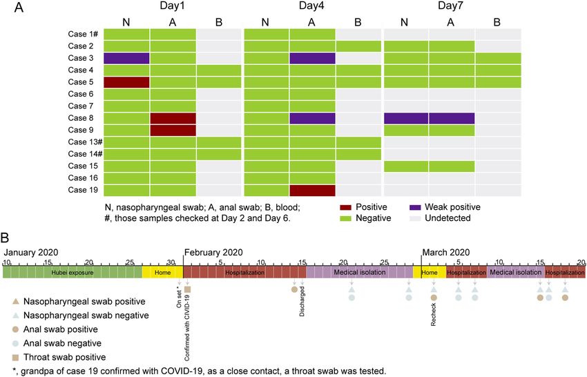
6th day). Among the results of the 14 cases, five were positive, and one of the five (case 8) was positive for tests
on all three testing days. Three (cases 2, 4, and 15) of the 14 were negative for tests on all three testing days, and
none have found positive results in blood tests (Fig. 1A).
Re‑positives and non‑re‑positives have the same level of antibodies. All the COVID-19 recov-
ered patients were advised to undergo antibody detection and laboratory testing of blood. Fourteen out of the
20 re-positives, and 133 out of the 162 non-re-positives took the advice and underwent the tests. These tests
revealed 13 negative results for IgA (13 non-re-positives and zero re-positives), one negative result for IgG (1
non-re-positive and zero re-positives), 42 negative results for IgM (38 non-re-positives and four re-positives),
and positive total antibody (Ab) tests’ results for all 14 re-positives and 133 non-re-positives. Meanwhile, all
14 re-positives were SARS-CoV-2 antibody carriers. There were no significant differences between 133 non-
re-positive recovered COVID-19 patients and 14 re-positives for total Ab, IgA, and IgG. The p-value for IgM
was 0.024, but the median values were similar (2.66 and 3.16) (Figure S1). There were no obvious abnormalities
found in routine laboratory blood testing (Table 3).
Asymptomatic carriers can be re‑positive. We noticed the particular case of an 8-year-old boy (case
19) who had Hubei exposure history during Jan 10–16, 2020, who were re-tested positive for repeated times. He
returned from a journey from Hubei to Shenzhen on Jan 26, 2020. His grandfather was confirmed to be infected
with COVID-19 on Jan 31, 2020. Due to their close contact, throat swab tests were performed for the whole fam-
ily and on Feb 1, 2020, the boy was confirmed as having COVID-19, and hospitalized. No fever or other symp-
toms were detected during his hospitalization. He reached the hospital discharge criteria (according to the 4th
Trail edition) on Feb 15, but was requested to transfer to an isolation hotel for another 14 days (Feb 15–28) due
to the positive results of an anal swab test on Feb 14. During hotel isolation, results on two test days (Feb 21 and
28) were negative, and thus he was allowed to go home. On his return hospital visit on Mar 2, an anal swab test
showed a positive result, and he was hospitalized for a second time. During the second hospitalization, results on
two test days (Mar 5 and 7) were negative, and he was transferred to isolation observation for a second period of
Scientific Reports | (2020) 10:11887 | https://doi.org/10.1038/s41598-020-68782-w 2
Vol:.(1234567890)www.nature.com/scientificreports/
Day 7 check Day 14 check
Case number Sex Age (years) Nasopharyngeal swab Anal swab Nasopharyngeal swab Anal swab
Case 1 Male 38 Negative Negative Negative Positive*
Case 2 Male 53 Negative Negative Positive Negative
Case 3 Female 40 Positive Negative ∕ ∕
Case 4 Female 61 Negative Negative Positive Negative
Case 5 Female 64 Negative Negative Positive Negative
Case 6 Female 53 Negative Negative Positive Negative
Case 7 Female 33 Positive* Negative ∕ ∕
Case 8 Female 1 Negative Positive ∕ ∕
Case 9 Female 34 Negative Positive* ∕ ∕
Case 10 Male 43 Positive Negative ∕ ∕
Case 11 Female 34 Negative Positive ∕ ∕
Case 12 Male 38 Negative Positive ∕ ∕
Case 13 Female 50 Positive Negative ∕ ∕
Case 14 Female 50 Positive* Negative ∕ ∕
Case 15 Female 5 Negative Positive ∕ ∕
Case 16 Female 55 Positive Negative ∕ ∕
Case 17 Female 72 Negative Negative Positive Negative
Case 18 Male 54 Negative Negative Positive* Negative
Case 19 Male 8 Negative Positive ∕ ∕
Case 20 Male 12 Positive Negative / /
Table 2. Recurrence of positive SARS-CoV-2 viral RNA in recovered COVID-19 patients. Bold indicates
positive results. *Results were weakly positive on the first test and Ct values were ≤ 40 when re-tested the next
day. /: Test was not performed.
Figure 1. (A) RT-PCR testing of 15 re-positive cases out of 20. Data shows RT-PCR results of blood,
nasopharyngeal swabs, and anal swabs tested on the 1st, 4th, and 7th day (2nd and 6th day for case 1, 13, and
14). (B) The timeline of case 19.
Scientific Reports | (2020) 10:11887 | https://doi.org/10.1038/s41598-020-68782-w 3
Vol.:(0123456789)www.nature.com/scientificreports/
Thyroid function
Case number Ab(S/CO)# IgA(S/CO)# IgM(S/CO)# IgG(S/CO)# IL-6* Hs-CRP* SAA* PCT* FT3* FT4* TSH* TPO* TG*
Case 1 138.57COI 3.07COI 0.96COI 18.10COI 61.65 pg/mL 0.03www.nature.com/scientificreports/
recent study showed that COVID-19 patients with any comorbidity yielded poorer clinical outcomes than those
without15, but there are no such differences between re-positives and non-re-positives.
Zhao et al. reported that a higher titer of antibody in the plasma of patients with COVID-19 was indepen-
dently associated with disease severity16. We analyzed the total Ab, IgM, IgG, and IgA, but there were no sig-
nificant differences in antibody titers between re-positive recovered COVID-19 patients and non-re-positives
(Figure S1), suggesting that all the 182 recovered patients, including the 20 cases that re-tested positive, are
antibody carriers. Furthermore, we did not find an association between viral load (Table S1) and antibody titer
(Table 2). This may suggest that the re-positives are shedding viral RNA segments, and re-testing positive does
not cause an inflammatory response or antibody level fluctuations.
Taken together, patients aged under 18 years, and mild and moderately ill patients have a higher risk of recur-
rence of positive SARS-CoV-2 viral RNA via an RT-PCR test, although all ages and sexes are at risk of re-testing
positive. No severely ill patients were re-tested positive in our study, although these results are not sufficient
to prove that severely patients are not at risk from re-testing positive. All discharged patients should undergo
medical observation and quarantine for at least 14 days. Longer periods of observation and surveillance might
be necessary.
Methods
As of Feb 21, 2020, COVID-19 patients of Shenzhen city who met all of the hospital discharge criteria were
requested to stay in medical isolation observation for a further 14 days, and the discharge criteria includes:
1. Body temperature below 37 degrees, lasting for at least three consecutive days;
2. Resolved respiratory symptoms;
3. Substantially improved chest lesions on computed tomography (CT) images; and
4. Two consecutive negative RT-PCR test results with at least a 1-day i nterval17.
The clinical classification of COVID-19 is defined clearly in the “Diagnosis and Treatment of Pneumonia
Caused by Novel Coronavirus (Trial Version 7)”. In brief, the mild type has no signs of pneumonia on chest imag-
ing; the moderate type includes fever and respiratory symptoms, and signs of pneumonia on radiologic assess-
ment; the severe type meets any of the following criteria: (1) shortness of breath, RR ≥ 30 times/min; (2) oxygen
saturation ≤ 93% at rest; (3) arterial oxygen partial pressure/fraction of inspiration O2 (PaO2/FiO2 ≤ 300 mmHg);
and (4) pulmonary imaging showing significant progression of lesion > 50% within 24–48 h17.
Cohort information. Patients infected with COVID-19 were divided into severe and non-severe (mild and
moderate) groups according to the guidelines for “Diagnosis and Treatment of Pneumonia Caused by Novel
Coronavirus (Trial Version 7)” 17. All the discharged patients were asked to stay in medical isolation observation
for a further 14 days at the Samii Medical Center, in a single room for each patient. Viral RNA testing of naso-
pharyngeal swabs and anal swabs were carried out on the 7th and 14th day. Antibody detection and laboratory
testing of blood were carried on the 7th day.
RT‑PCR analysis. Nasopharyngeal swabs and anal swabs were taken on the 7th and 14th day of observa-
tion, for RT-PCR tests at the Shenzhen Center for Disease Control and Prevention (CDC), to decide if they
were allowed to go home. The RT-PCR tests were performed by the CDC using the High Pure Viral RNA Kit
(Roche, Mannheim, Germany) and the 2019-nCoV Viral RNA detection kit (Bio-Germ, Shanghai, China), a
similar methods have been described p reviously18. In brief, we put nasopharyngeal swabs/anal swabs into a
collection tube with 1.5 mL of virus preservation solution. Then 200 μL of cell lysate was vortexed for 10 s and
was then allowed to stand at room temperature for 10 min. We then collected the suspension after a 10-min
centrifugation at 1,000 rpm. Two target genes of SARS-CoV-2, including the open reading frame 1ab (ORF1ab)
and the nucleocapsid protein (N), were simultaneously amplified and tested during the RT-PCR assay. Target
1 (ORF1ab): forward primer CCCTGTGGGTTTTACACTTAA; reverse primer ACGATTGTGCATCAGCTG
A; probe: 5′-VIC-CCGTCTGCGGTATGTGGAAAGGTTATGG-BHQ1-3′. Target 2 (N): forward primer GGG
GAACTTCTCCTGCTAGAAT; reverse primer CAGACATTTTGCTCTCAAGCTG; probe 5′-FAM- TTGCTG
CTGCTTGACAGATT-TAMRA-3′. A cycle threshold value (Ct-value) less than 37 was defined as positive, and
a Ct-value no less than 40 was defined as negative. A medium load, 37 ≤ Ct < 40, was defined as weakly positive,
which requires further confirmation by re-testing. If Ct-value ≤ 40 in the re-test on the next day, a positive result
would be reported.
We collected all of the RT-PCR test information from the recovered and isolated for 7 + days COVID-19
patients, and analyzed the re-positive tests results.
Antibody detection and laboratory testing. The main results and indicators of epidemiology, demog-
raphy, clinical manifestation, and laboratory examinations of 182 recovered patients with COVID-19 were col-
lected and analyzed. The Inflammation markers and thyroid functions were tested, including Interleukin-6 (IL-
6), hypersensitive-c-reactive-protein (Hs-CRP), serum amyloid A protein (SAA), procalcitonin (PCT), serum
free triiodothyronine (FT3), free tetraiodothyronine (FT4), thyroid stimulating hormone (TSH), thyroid per-
oxidase (TPO), and thyroglobulin (TG). Total Ab, IgA, IgG and IgM were tested on the 7th day using a SARS-
CoV-2 testing kit (WANTAI BioPharm, Beijing, China) based on the Chemiluminescence method. All tests were
performed according to the manufacturer’s instructions. S/CO < 1 indicated a negative antibody result, and S/
CO ≥ 1 indicated a positive antibody result.
Scientific Reports | (2020) 10:11887 | https://doi.org/10.1038/s41598-020-68782-w 5
Vol.:(0123456789)www.nature.com/scientificreports/
This study was approved by the Shenzhen Samii Medical Center Institutional Review Board (SSMC-
R-20200401) and we declare that all the patients involved in this study have been fully informed and written
informed consents were obtained. Concerning the 20 minors involved in the study, their patients/LARs have
been informed and signed the informed consents on their behalf. These data do not contain any private infor-
mation of the patients. All methods were performed in accordance with the relevant guidelines and regulations.
Statistical analysis. The Mann–Whitney U test was used to analyze differences in basic information
between non-re-positive recovered COVID-19 patients and re-positives. A two-tailed independent sample t-test
was used to test for significant differences in antibody detection between non-re-positives and re-positives. The
Mann–Whitney U tests were performed using ggplot2, and the two-tailed independent sample t-tests were per-
formed using the ggpubr package of R software (version 3.6), respectively.
Data availability
The datasets used and/or analyzed during this study are available from the corresponding author upon reason-
able request.
Received: 2 April 2020; Accepted: 1 July 2020
References
1. Organization, W. H. Novel Coronavirus (2019-nCoV). Situation Report-149, (2020).
2. Chen, N. et al. Epidemiological and clinical characteristics of 99 cases of 2019 novel coronavirus pneumonia in Wuhan, China: a
descriptive study. Lancet 395, 507–513. https://doi.org/10.1016/S0140-6736(20)30211-7 (2020).
3. Novel Coronavirus Pneumonia Emergency Response Epidemiology, T. [The epidemiological characteristics of an outbreak of
2019 novel coronavirus diseases (COVID-19) in China]. Zhonghua Liu Xing Bing Xue Za Zhi 41, 145–151, doi:10.3760/cma.j.i
ssn.0254-6450.2020.02.003 (2020).
4. Xu, X. W. et al. Clinical findings in a group of patients infected with the 2019 novel coronavirus (SARS-Cov-2) outside of Wuhan,
China: retrospective case series. BMJ 368, m606. https://doi.org/10.1136/bmj.m606 (2020).
5. Lan, L. et al. Positive RT-PCR test results in patients recovered from COVID-19. JAMA https://doi.org/10.1001/jama.2020.2783
(2020).
6. 6An, J. et al. Clinical characteristics of the recovered COVID-19 patients with re-detectable positive RNA test. medRxiv,
2020.2003.2026.20044222, doi:10.1101/2020.03.26.20044222 (2020).
7. Qu, Y. M., Kang, E. M. & Cong, H. Y. Positive result of Sars-Cov-2 in sputum from a cured patient with COVID-19. Travel. Med.
Infect. Dis. https://doi.org/10.1016/j.tmaid.2020.101619 (2020).
8. Cao, H., Ruan, L., Liu, J. & Liao, W. The clinical characteristic of eight patients of COVID-19 with positive RT-PCR test after
discharge. J. Med. Virol. https://doi.org/10.1002/jmv.26017 (2020).
9. Bao, L. et al. Reinfection could not occur in SARS-CoV-2 infected rhesus macaques. bioRxiv, 2020.2003.2013.990226.
doi:10.1101/2020.03.13.990226 (2020).
10. Wu, Y. et al. Prolonged presence of SARS-CoV-2 viral RNA in faecal samples. Lancet Gastroenterol. Hepatol. 5, 434–435. https://
doi.org/10.1016/S2468-1253(20)30083-2 (2020).
11. Yao, X. H. et al. Pathological evidence for residual SARS-CoV-2 in pulmonary tissues of a ready-for-discharge patient. Cell. Res.
https://doi.org/10.1038/s41422-020-0318-5 (2020).
12. Young, B. E. et al. Epidemiologic features and clinical course of patients infected with SARS-CoV-2 in Singapore. JAMA https://
doi.org/10.1001/jama.2020.3204 (2020).
13. Ling, Y. et al. Persistence and clearance of viral RNA in 2019 novel coronavirus disease rehabilitation patients. Chin. Med. J. (Engl)
133, 1039–1043. https://doi.org/10.1097/CM9.0000000000000774 (2020).
14. Hu, Z. et al. Clinical characteristics of 24 asymptomatic infections with COVID-19 screened among close contacts in Nanjing
China. Sci. China Life Sci 63, 706–711. https://doi.org/10.1007/s11427-020-1661-4 (2020).
15. Guan, W. J. et al. Comorbidity and its impact on 1590 patients with Covid-19 in China: a nationwide analysis. Eur. Respir. J. https
://doi.org/10.1183/13993003.00547-2020 (2020).
16. Zhao, J. et al. Antibody responses to SARS-CoV-2 in patients of novel coronavirus disease 2019. Clin. Infect. Dis. https://doi.
org/10.1093/cid/ciaa344 (2020).
17. China, N. H. C. O. T. P. S. R. O. Diagnosis and treatment of pneumonia caused by novel coronavirus (trial version 7), (2020).
18. Wang, D. et al. Clinical characteristics of 138 hospitalized patients with 2019 novel coronavirus-infected pneumonia in Wuhan
China. JAMA https://doi.org/10.1001/jama.2020.1585 (2020).
Acknowledgements
This study was financially supported by Grant JCYJ20180302153611416 from the Science and Technology Plan-
ning Project of Shenzhen Municipality (CN). We thank Jia Zhao, Xiao-Peng Hu, and Zhi-Wen Li for help with
data collection, Yi-Fan Zhong and Xiao-Hui Wang (Shenzhen CDC) for help with confirmation of the diagnosis
of viral infection, and Dr. Qi Hu (NEOMICS institute) for statistical analyses.
Author contributions
B.Y. and W.X. L. were responsible for data analyses, chart making, and manuscript writing. Y.W.A. and C.W.
participated in data collection and contributed to the production of Figures. Z.R.Y., K.Z., and Z.Y.L. performed
RT-PCR testing and data collection. Y.X.C. and J.C.W. followed up the patients and collected informed consent.
S.S., J.C.W., and H.Q.L. drafted and edited the manuscript. All authors reviewed the manuscript and approved
the submission.
Competing interests
The authors declare no competing interests.
Scientific Reports | (2020) 10:11887 | https://doi.org/10.1038/s41598-020-68782-w 6
Vol:.(1234567890)www.nature.com/scientificreports/
Additional information
Supplementary information is available for this paper at https://doi.org/10.1038/s41598-020-68782-w.
Correspondence and requests for materials should be addressed to J.-C.W. or S.S.
Reprints and permissions information is available at www.nature.com/reprints.
Publisher’s note Springer Nature remains neutral with regard to jurisdictional claims in published maps and
institutional affiliations.
Open Access This article is licensed under a Creative Commons Attribution 4.0 International
License, which permits use, sharing, adaptation, distribution and reproduction in any medium or
format, as long as you give appropriate credit to the original author(s) and the source, provide a link to the
Creative Commons license, and indicate if changes were made. The images or other third party material in this
article are included in the article’s Creative Commons license, unless indicated otherwise in a credit line to the
material. If material is not included in the article’s Creative Commons license and your intended use is not
permitted by statutory regulation or exceeds the permitted use, you will need to obtain permission directly from
the copyright holder. To view a copy of this license, visit http://creativecommons.org/licenses/by/4.0/.
© The Author(s) 2020
Scientific Reports | (2020) 10:11887 | https://doi.org/10.1038/s41598-020-68782-w 7
Vol.:(0123456789)You can also read