Detailed neuropathologic report of COVID-19 complicated by large intracerebral hemorrhage and periventricular lesions with macrophagic infiltrates
←
→
Page content transcription
If your browser does not render page correctly, please read the page content below
Free Neuropathology 2:7 (2021)                                                                                                                                                            Levine et al
doi: https://doi.org/10.17879/freeneuropathology-2021-3266                                                                                                                                page 1 of 6

                                                                                                                                                                           Case Report

Detailed neuropathologic report of COVID-19 complicated by
large intracerebral hemorrhage and periventricular lesions with
macrophagic infiltrates
Adrian Levine1, Carol Lee2, Craig Fava3, Frankie Tsang4, Kelly MacNeil5, Stephen T. Yip1,5, Veronica Hirsch-
Reinshagen1
1
    Department of Pathology and Laboratory Medicine, University of British Columbia, Vancouver, BC, Canada
2
    Division of Forensic Pathology, Royal Columbian Hospital, Vancouver, BC, Canada
3
    Division of Critical Care, Royal Columbian Hospital, BC, Canada
4
    British Columbia Center for Disease Control, Vancouver, BC, Canada
5
    Cancer Genetics & Genomics Laboratory, BC Cancer, Vancouver, BC, Canada

Corresponding author:
Veronica Hirsch-Reinshagen · Anatomical Pathology · Vancouver General Hospital · 899 West 12th Av · JPN Rm 1401 · Vancouver, BC ·
Canada · V5Z 1M9
Veronica.Hirsch@vch.ca

Submitted: 15 February 2021                     ·       Accepted: 16 March 2021                     ·      Copyedited by: Deanna Fang                         ·     Published: 25 March 2021

Abstract
Infection with the SARS-CoV-2 virus affects a wide range of systems. Significant involvement of the central nerv-
ous system has been described, including ischemic and hemorrhagic strokes. Thus far, neuropathologic reports
of patients who passed away from COVID-19 have generally described non-specific findings, such as variable
reactive gliosis and meningeal chronic inflammatory infiltrates, as well as the consequences of the infection’s
systemic complications on the brain, including ischemic infarcts and hypoxic/ischemic encephalopathy. The neu-
ropathological changes in patients with COVID-19 and large hemorrhagic strokes have not been described in
detail. We report the case of an elderly male who had a long course of COVID-19 and ultimately passed away
from a large intracerebral hemorrhage. In addition to acute hemorrhage, neuropathologic examination demon-
strated non-specific reactive changes and chronic periventricular lesions with macrophagic and perivascular lym-
phocytic infiltrates without evidence of demyelination or presence of SARS-CoV-2 by PCR test. This manuscript
expands the spectrum of reported neuropathological changes in patients with COVID-19.

Keywords: COVID-19, Cerebral hemorrhage, Brain, Postmortem, Histology, Neuropathology

Copyright: © 2021 The author(s). This is an open access article distributed under the terms of the Creative Commons Attribution 4.0 International License (https://creativecommons.org/licenses/by/4.0/),
which permits unrestricted use, distribution, and reproduction in any medium, provided the original author and source are credited, a link to the Creative Commons license is provided, and any changes are
indicated. The Creative Commons Public Domain Dedication waiver (https://creativecommons.org/publicdomain/zero/1.0/) applies to the data made available in this article, unless otherwise stated.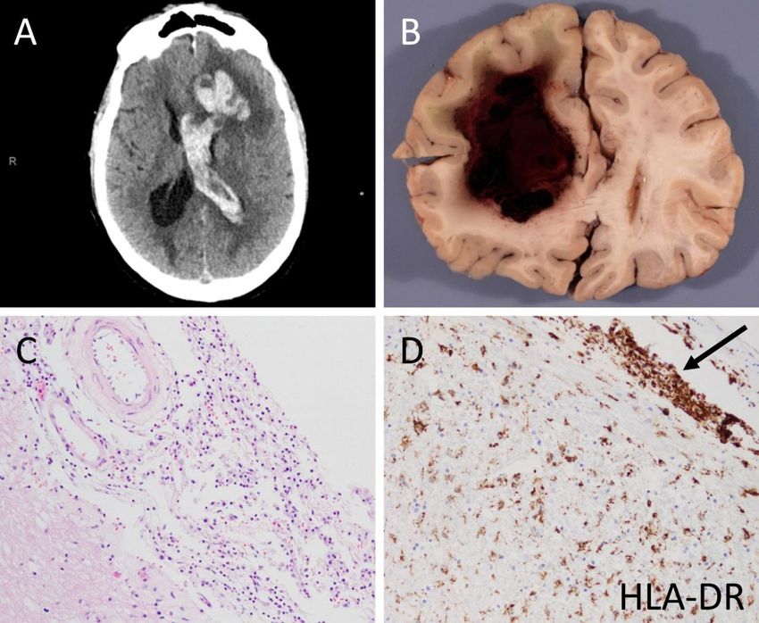
Free Neuropathology 2:7 (2021)                                                                            Levine et al
doi: https://doi.org/10.17879/freeneuropathology-2021-3266                                                page 2 of 6

Introduction                                                 ported in this case were also seen in a younger indi-
                                                             vidual with COVID-19 and fatal ICH for which we
      Intracerebral hemorrhage (ICH) is an uncom-            were unable to acquire consent for publication of
mon complication of infection with severe acute res-         clinical details. The histological similarities and dif-
piratory syndrome coronavirus 2 (SARS-CoV-2) virus,          ferences between these two cases will be high-
affecting 0.5-7.9% of patients hospitalized with             lighted in the text.
COVID-191–3. Therapeutic anticoagulation has been
identified as the most common etiology1–3 and an             Case presentation
important risk factor for ICH in COVID-194. In addi-               This male patient, in his early 70s, presented to
tion, other factors such as micro- and macrovascular         hospital with bilateral pneumonia secondary to
thrombosis, endothelial dysfunction and endo-                COVID-19 infection that was confirmed by nasopha-
theliitis may play important pathogenic roles in this        ryngeal swab testing. Parallel testing for Influenza A,
setting. Despite this plethora of hypothesized fac-          B and Respiratory Syncytial Virus were negative. His
tors, only a few neuropathological reports have in-          prior medical history included hypertension and sig-
cluded detailed histopathological brain evaluations          nificant hip osteoarthritis. He was diagnosed with
of COVID-19 patients with large fatal ICH.                   septic shock and transferred to the Intensive Care
     Post-mortem neuropathological evaluation of             Unit upon admission as he required vasopressors
patients with COVID-19 have shown a range of his-            and mechanical ventilation. His mean arterial pres-
tological changes and have been reviewed else-               sure was targeted to more than 65 mmHg while his
where5,6. The vast majority of cases show relatively         systolic blood pressure (SBP) was kept below a ceil-
mild and non-specific findings including variable and        ing of 180 to 200 mmHg to avoid ischemic brain in-
diffuse micro- and astrogliosis, and mild parenchy-          jury. This was especially relevant in the context of his
mal and leptomeningeal infiltration by chronic in-           shock syndrome and prior history of hypertension.
flammatory infiltrates, which could represent the            Only two SBP episodes over 180 mmHg, both of less
histological features of an encephalopathy associ-           than 30 min in duration, were recorded 4 and 19
ated with severe systemic inflammation rather than           days prior to his terminal ICH.
specific COVID-19-related changes7. In addition,                  During his first week of admission, he devel-
many cases show variable hypoxic-ischemic enceph-            oped renal failure, which was thought clinically sec-
alopathy, infarcts associated with large vessel              ondary to acute tubular necrosis in the context of
thromboembolism and variable hemorrhagic lesions             multisystem organ failure. He was placed on contin-
from microhemorrhages to fatal ICH5,6.                       uous renal replacement therapy (CRRT) one week
                                                             after admission. This required local anticoagulation
      Only three detailed neuropathological reports          with unfractionated heparin titrated to obtain an ac-
of patients with large ICH have been published to            tivated partial thromboplastin time (aPTT) post-
date and include a case of cerebellar hemorrhage             CRRT filter of 60 to 90 seconds. His international nor-
thought to be most likely secondary to hypertensive          malized ratio (INR) hovered around the upper nor-
vasculopathy8, one of hemorrhagic transformation             mal limit of 1.2 during his entire hospitalization.
of a large middle cerebral artery stroke9 and two            Other relevant laboratory findings included an ele-
cases reported by the authors as ICH in the context          vated d-dimer, C reactive protein and procalcitonin.
of lymphocytic panencephalitis, meningitis and dif-          He did not have evidence of pulmonary embolism
fuse petechial hemorrhages10. In the latter, no de-          during his hospitalization.
tails are provided regarding the histological changes
associated with, or the etiopathogenesis of, the ICH              Nearly three weeks into his admission and two
or petechial hemorrhages.                                    weeks after initiation of CRRT, the patient showed
                                                             evidence of decreased level of consciousness. A
     In this report, we expand on the neuropatho-            computerized tomography scan demonstrated a left
logical literature of COVID-19 cases by detailing the        frontal intracerebral hemorrhage with intraventric-
neuropathological findings in an elderly male with           ular extension and subfalcine herniation (Fig. 1A).
COVID-19 and fatal ICH. Some of the findings re-             He died the next day after withdrawal of life-sustain-
Free Neuropathology 2:7 (2021)                                                                                                 Levine et al
doi: https://doi.org/10.17879/freeneuropathology-2021-3266                                                                     page 3 of 6

Figure 1. (A) CT scan showed a large left frontal ICH with intraventricular extension. (B) Coronal sections confirmed this ICH and left-to-
right subfalcine herniation. (C) Leptomeningeal chronic inflammatory infiltrates were most prominent in the brainstem. (D) Immunohisto-
chemistry for HLA-DR, a major histocompatibility class II cell surface receptor, highlighted the leptomeningeal chronic inflammatory infil-
trates (arrow) and the mild-to-moderately increased brainstem microglial activation.

ing therapies. A brain-restricted autopsy was per-                              Microscopic examination revealed mild diffuse
formed 7 days after death to aid in the determina-                        reactive changes (Fig. 1C and D) and focal
tion of the etiology of the brain hemorrhage.                             periventricular lesions at the angles of the ventricles
                                                                          (Fig. 2). The diffuse reactive changes included rather
      Gross examination of the brain revealed a                           mild leptomeningeal chronic inflammatory infil-
weight of 1280 grams, an enlarged left hemisphere,                        trates, parenchymal reactive gliosis and very mildly
subfalcine left-to-right herniation of the left anterior                  increased perivascular CD3 T-lymphocytes. The lep-
frontal lobe and bilateral hippocampal uncal herni-                       tomeningeal inflammatory infiltrates were com-
ations. Coronal sections confirmed a large ICH cen-                       posed predominantly of macrophages and CD3 T-
tered in the left frontal lobe in close proximity to the                  cell lymphocytes found throughout the meninges,
lateral ventricle with extension throughout the ven-                      but were accentuated in the brainstem (Fig. 1C). Re-
tricular system (Fig. 1B). Away from the hemor-                           active gliosis, highlighted by mild-to-moderately in-
rhage, no additional gross pathological changes                           creased immunoreactivity for human leukocyte anti-
were identified.                                                          gen – DR isotype (HLA-DR) and glial fibrillary acidic
Free Neuropathology 2:7 (2021) Levine et al
doi: https://doi.org/10.17879/freeneuropathology-2021-3266 page 4 of 6
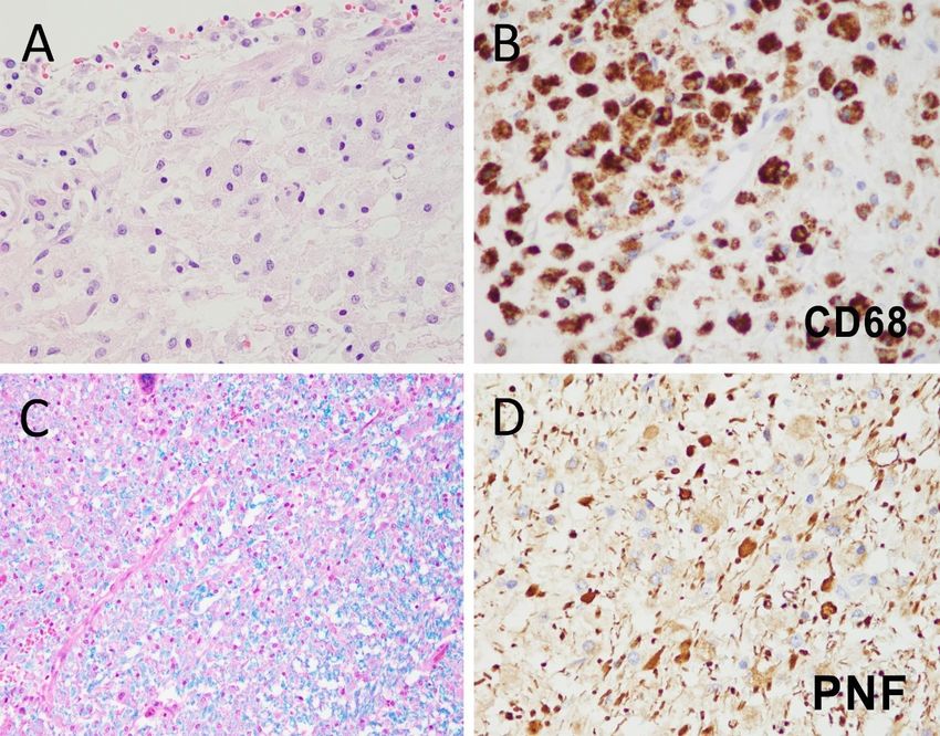
Figure 2. (A) Multifocal periventricular lesions showed loss of ependyma and significant infiltration by macrophages with scant accompa-
nying lymphocytes. (B) CD68 highlighted the significant macrophagic infiltrates. (C) Luxol-fast blue showed mild loss of myelin, while
phosphorylated neurofilament (PNF) IHC (D) showed associated axonal pathology with significant numbers of axonal spheroids.
protein (GFAP), was present predominantly in the axonal spheroids without appreciable demye-
brainstem and olfactory bulb. Similar changes were lination, was also present (Fig. 2C and D). Iron stains
also seen in the younger individual. No microglial performed in several of these lesions were negative.
nodules were identified on hematoxylin and eosin The abundance of phagocytic macrophages and the
(HE) stains. Two possible microglial nodules were absence of hemosiderin suggest that these lesions
identified on HLA-DR immunohistochemistry (IHC) were older than the intracerebral hemorrhage and
in the medulla only. therefore not likely to be a reactive change to intra-
Atypical periventricular lesions (Fig. 2) were ventricular blood. This type of lesion was not seen in
seen at multiple locations including both temporal the younger individual, who instead exhibited multi-
horns, left occipital horn, fourth ventricle and in focal petechial hemorrhages and microscopic is-
close proximity to the anterior left frontal lateral chemic infarcts thought to be most compatible with
ventricle at the edge of acutely hemorrhagic brain embolic lesions. No definite evidence of thrombi or
parenchyma. These lesions consisted of loss of ep- megakaryocytes was identified in either of the
endymal lining, relatively well delimited macro- cases.
phagic and lymphocytic infiltrates (Fig. 2A and B) The neocortex, hippocampi, deep grey nuclei
and reactive vessels with plump endothelial cells. and cerebellum were unremarkable aside from
Axonal pathology, in the form of relatively frequent some mild age-related neurodegenerative pathol-Free Neuropathology 2:7 (2021) Levine et al
doi: https://doi.org/10.17879/freeneuropathology-2021-3266 page 5 of 6
ogy. Pontine white matter tracts only showed sev- periventricular lesion raise the possibility that local
eral microscopic foci of amyloid precursor protein loss of tissue integrity may play a predisposing role
(APP) immunoreactive axonal pathology associated to ICH in COVID-19. A similar situation was observed
with micro- and astrogliosis. CD68 revealed absence in the younger individual, in whom multifocal pete-
of mature phagocytic macrophages in these lesions. chial hemorrhages and microscopic ischemic in-
These were interpreted as foci of axonal damage farcts were identified. These were interpreted as
secondary to traction due to the left frontal and in- most likely due to microemboli in the context of se-
traventricular ICH. No cerebral amyloid angiopathy, vere COVID-19 and therapeutic anticoagulation for
arteriolosclerosis, lacunar infarcts or other vascular extracorporeal membrane oxygenation (ECMO).
changes were identified.
Both patients were locally or systemically anti-
Polymerase chain reaction (PCR) test for the coagulated for therapeutic reasons and the risk of
presence of SARS-CoV-2 RNA in multiple sections ICH in the setting of anticoagulation appears to be
(medulla, hippocampus and olfactory bulbs) of for- increased in patients with COVID-19. A study of 10
malin-fixed paraffin-embedded (FFPE) brain tissue patients with COVID-19 supported on ECMO for
was negative, including representation of the acute respiratory distress syndrome (ARDS) showed
periventricular lesions. Similar results were ob- a markedly increased incidence of hemorrhagic
tained for the younger patient. strokes compared to non-COVID patients on
ECMO12.
Discussion
The diffuse reactive changes identified in both
Our cases showed three main pathological
COVID cases are similar to those reported previously
changes: (1) acute and fatal ICH, (2) diffuse reactive
in the literature and are at least partially explained
changes and (3) additional parenchymal lesions, ei-
as a manifestation of severe illness-related enceph-
ther as multifocal chronic periventricular lesions in
alopathy5–7. Several tissue blocks of both cases were
the elderly individual or as multifocal microhemor-
tested for SARS-CoV-2 by PCR and were negative.
rhages and infarcts in the younger patient. The his-
Neither patient received antiviral therapies that
topathological features of the periventricular lesions
could have decreased SARS-CoV-2 tissue levels. For-
make them most compatible with localized tissue
malin-fixed control lung tissue from an unrelated
necrosis of unclear etiology. They were negative for
COVID-19 autopsy case with a shorter postmortem
SARS-CoV-2 RNA. To our knowledge, this type of le-
interval served as a positive control. Although the
sion has not been studied extensively. A single re-
long post-mortem interval and formalin-fixation
port comparing the neuropathological substrate of
may have interfered with the detection of SARS-
periventricular white matter abnormalities in pa-
CoV-2 in our samples, these results are in line with
tients with major depression and in controls de-
previously published reports that have shown incon-
scribes lesions with similar characteristics to those
sistent and variable detection of SARS-CoV-2 in brain
found in this case. These lesions were interpreted as
parenchyma by PCR and immunohistochemical
corresponding most likely to ischemic insults and
methods7,8. Overall, this would suggest that the
were found both in a patient with major depression
brain is not a site consistently affected by high viral
and a control11. The pathogenesis of these lesions
loads of SARS-CoV-2 and raises the possibility that
awaits further evaluation, but the fact that lesions
direct infection of the CNS tissue may not be the
similar to those seen in our older individual have
main pathogenic mechanism of COVID-19 neuro-
been described in non-COVID cases raises the possi-
logic manifestations.
bility that they are either nonspecific in etiology (e.g.
associated with an episode of severe systemic ill- In summary, the fatal ICH in two cases of
ness) and/or can be brought about by different in- COVID-19 were most likely due to a combination of
jury mechanisms. Furthermore, in our case, it is un- anticoagulation and additional factors affecting the
clear whether these lesions predated the SARS-CoV- integrity of the CNS parenchyma. Additional chronic
2 infection or developed as a consequence of it. inflammatory infiltrates and glial reactive changes
are at least partially explained by severe illness-re-
The close relationship of the acute left frontal
lated encephalopathy.
ICH to changes suggestive of an underlyingFree Neuropathology 2:7 (2021) Levine et al
doi: https://doi.org/10.17879/freeneuropathology-2021-3266 page 6 of 6
References
1. Kvernland, A. et al. Anticoagulation use and Hemorrhagic Stroke in 8. Al-Dalahmah, O. et al. Neuronophagia and microglial nodules in a
SARS-CoV-2 Patients Treated at a New York Healthcare System SARS-CoV-2 patient with cerebellar hemorrhage. Acta Neuropathol
[published online ahead of print, 2020 Aug 24]. Neurocrit Care. (2020) Commun. (2020) 8(1):147. Published 2020 Aug 26.
1-12. https://doi.org/10.1007/s12028-020-01077-0 https://doi.org/10.1186/s40478-020-01024-2
2. Dogra, S. et al. Hemorrhagic stroke and anticoagulation in COVID- 9. Hanley, B. et al. Histopathological findings and viral tropism in UK
19. J Stroke Cerebrovasc Dis. (2020) 29(8):104984. patients with severe fatal COVID-19: a post-mortem study. Lancet
https://doi.org/10.1016/j.jstrokecerebrovasdis.2020.104984 Microbe. (2020) 1(6):e245-e253.
https://doi.org/10.1016/S2666-5247(20)30115-4
3. Benger, M. v Intracerebral haemorrhage and COVID-19: Clinical
characteristics from a case series. Brain Behav Immun. (2020) 88:940- 10. von Weyhern, CH. et al. Early evidence of pronounced brain
944. https://doi.org/10.1016/j.bbi.2020.06.005 involvement in fatal COVID-19 outcomes. Lancet. (2020)
395(10241):e109. https://doi.org/10.1016/S0140-6736(20)31282-4
4. Melmed, KR. et al. Risk factors for intracerebral hemorrhage in
patients with COVID-19 [published online ahead of print, 2020 Sep 24]. 11. Thomas, AJ. et al. A neuropathological study of periventricular
J Thromb Thrombolysis. (2020) 1-8. white matter hyperintensities in major depression. J Affect Disord.
https://doi.org/10.1007/s11239-020-02288-0 (2003) 76(1-3):49-54. https://doi.org/10.1016/s0165-0327(02)00064-2
5. Lou, JJ. et al. Neuropathology of COVID-19 (neuro-COVID): 12. Usman, AA. et al. A Case Series of Devastating Intracranial
clinicopathological update. Free Neuropathol. (2021) 2:2. Hemorrhage During Venovenous Extracorporeal Membrane
https://doi.org/10.17879/freeneuropathology-2021-2993 Oxygenation for COVID-19. J Cardiothorac Vasc Anesth. (2020)
34(11):3006-3012. https://doi.org/10.1053/j.jvca.2020.07.063
6. Mukerji, SS & Solomon, IH. What can we learn from brain autopsies
in COVID-19?. Neurosci Lett. (2021) 742:135528.
https://doi.org/10.1016/j.neulet.2020.135528
7. Deigendesch, N. et al. Correlates of critical illness-related
encephalopathy predominate postmortem COVID-19 neuropathology.
Acta Neuropathol. (2020) 140(4):583-586.
https://doi.org/10.1007/s00401-020-02213-yYou can also read