Bladder-embedded ectopic intrauterine device with calculus
←
→
Page content transcription
If your browser does not render page correctly, please read the page content below
Open Medicine 2020; 15: 501–507
Research Article
Bing-Jian Xiong*, Guang-Jing Tao, Duo Jiang
Bladder-embedded ectopic intrauterine device
with calculus
https://doi.org/10.1515/med-2020-0173 Oceania [4]. Some complications may occur, including
received December 23, 2019; accepted May 8, 2020 expulsion (2–10%) [2], pelvic inflammatory disease (2%)
Abstract: The present study aimed to analyze the data of [5], pelvic actinomycosis [6], and unplanned preg-
embedded intrauterine device (IUD) in the bladder wall with nancy [2].
the additional presence of calculus. This case series study Another rare complication is the IUD shifting and
included 11 female patients with partially or completely embedding itself in the urinary bladder [7]. The
embedded IUD in the bladder wall. Their median age was 34 occurrence of ectopic IUD is not associated with the
(range, 32–39) years. The median duration of IUD placement shape and duration of the use of the IUD [8]. Possible
was 36 (range, 24–60) months. The median duration of causes of ectopic IUD have been suggested [9,10], but no
symptoms was 9 (range, 3–12) months. Six patients under- formal study was performed on the subject because of
went laparoscopy: the operation duration was 129 (range, the rarity of the cases. First, a weak uterine wall after
114–162) min, blood loss was 15 (range, 10–25) mL, the parturition followed by an early and unskillful insertion
hospital stay was 4 (range, 4–4.5) days, the visual analog may lead to the IUD getting embedded in the uterine
scale (VAS) for pain at 6 h after surgery was 3 (range, 2–6), wall and possibly further shifting. Second, abnormal
and the time to removal of the urethral catheter was 7 (range, morphology and the periodic contraction of the uterus
7–8) days. Five patients underwent open surgery: the may lead to the IUD’s ectopic shifting. Third, to reduce
operation duration was 126 (range, 96–192) min, blood loss the side effects of IUD placement, the material and shape
was 30 (range, 20–50) mL, the hospital stay was 7 (range, of IUDs are continuously optimized and improved, but
7–15) days, the VAS was 6 (range, 4–9) at 6 h after surgery, unsuitable material and shape may lead to chronic
and the time to removal of the urethral catheter was 9 (range, incision to the uterine wall during uterine contraction,
8–17) days. The IUD and bladder stones were successfully ultimately leading to the shifting and embedding of the
removed in all 11 (100%) patients. IUD in the posterior wall of the bladder.
So far, this condition has been treated with tradi-
Keywords: cystectomy, intrauterine device, intrauterine tional open cystotomy and IUD removal [11], but it may
device migration, laparoscopy, surgery, urinary bladder, involve a long incision, bleeding, pain, prolonged
urinary bladder calculi immobilization, prolonged hospitalization, and consid-
erable psychological burden. Laparoscopic techniques
generally achieve satisfactory curative results for a wide
variety of benign and malignant conditions of the
1 Introduction bladder [12–14], but only a few studies reported
laparoscopic cystotomy and IUD removal for transloca-
Intrauterine device (IUD) insertion is a routine procedure tion (total or partial) of the IUD in the bladder [15–17].
for female contraception and is considered safe and Shin et al. [15] reported one patient successfully treated
convenient [1–3]. The rate of IUD use in the world using laparoscopic partial cystectomy. Atakan et al. [16]
averages 14.3%, varying from 27% in Asia to 1.8% in reported one case operated by suprapubic cystotomy.
Jin et al. [17] reported one woman operated using a
combination of laparoscopy and air cystoscopy.
* Corresponding author: Bing-Jian Xiong, Department of Urology, Previous studies only report about one or two cases
Ankang City Central Hospital, Ankang, Shaanxi, 725000, China,
of ectopic IUD embedded in the bladder, but 11 such
e-mail: xbj_74_76@163.com, tel: +86-15991056700,
fax: +86-21-57643271
cases were operated at our institution. Therefore,
Guang-Jing Tao, Duo Jiang: Department of Urology, Ankang City considering the rarity of the condition and the lack of
Central Hospital, Ankang, Shaanxi, 725000, China predefined treatment approach, the aim of the present
Open Access. © 2020 Bing-Jian Xiong et al., published by De Gruyter. This work is licensed under the Creative Commons Attribution 4.0
Public License.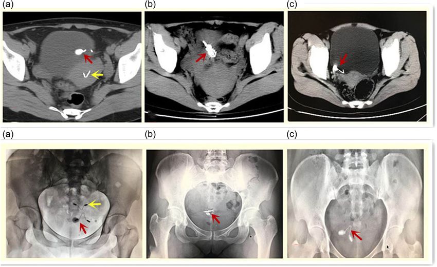
502 Bing-jian Xiong et al.
Wound infection
Wound infection
retrospective study was to analyze the data of all 11
Complications
patients who underwent surgery for an IUD embedded in
the bladder wall with the additional presence of a
calculus and to describe the clinical features and
None
Multiload Cu None
None
None
None
None
None
None
None
surgical outcomes of this rare condition.
Unknown
Unknown
Size (cm) IUD type
Copper t
Gynefix
Gynefix
2 Methods
MYCu
MYCu
MYCu
MYCu
MYCu
(0.9, 1.8)
2.1 Patients
0.9
1.8
1.0
1.6
1.6
1.2
1.2
1.3
1.3
1.7
1.5
1.1
This was a case series of 11 female patients who were
diagnosed with partially or completely embedded IUD in
Bladder foreign body
Bladder foreign body
Bladder foreign body
Bladder foreign body
Imaging diagnosis
the bladder wall with the additional presence of a
Vesical calculus
Vesical calculus
Vesical calculus
Vesical calculus
Vesical calculus
Vesical calculus
Vesical calculus
calculus, and who underwent surgery between January
2008 and December 2017 at the Department of Urology of
Ankang City Central Hospital. This study was approved
by the ethics committee of our Hospital, with a waiver
for individual consent.
symptoms (months)
The inclusion criteria were as follows: (1) diagnosis
of bladder stone caused by ectopic IUD embedded in the
bladder and (2) the ectopic IUD was removed by open
Duration of
surgery or laparoscopic surgery. The exclusion criterion
9 (3, 12)
was an incomplete medical record of the surgery and the
NA
NA
NA
NA
10
12
12
6
9
9
3
patient’s history. The diagnostic criteria for bladder-
embedded IUD were as follows [15–17]: (1) abdominal
pain,
pain
pain
pain
pain, hematuria, and other clinical symptoms consistent
with a vesical lesion and (2) ultrasound, CT, and other
Abdominal
Abdominal
Abdominal
Abdominal
Symptoms
Hematuria
Hematuria
Hematuria
hematuria
imaging examinations showed that the IUD was not in
None
None
None
None
the uterine cavity, but partly or completely embedded in
the bladder wall.
placement (months)
Duration of IUD
G: gravidity, P: parity; IUD: intrauterine device; NA: not applicable.
Ethics statement: This study was approved by the ethics 36 (24, 60)
committee of Ankang City Central Hospital, with a
waiver for individual consent.
60
48
48
28
36
36
36
36
24
39
33
Childbearing
2.2 Surgical techniques
history
G2P2
G2P2
G2P2
G2P2
G2P2
G2P1
G2P1
Table 1: Characteristics of the patients
All interventions were performed by the same senior
G1P1
G1P1
G1P1
G1P1
surgeon, an associate chief physician specialized in laparo-
scopic and endoscopic minimally invasive techniques.
(32, 39)
(years)
For traditional open surgery, the preoperative pre-
Age
Laparoscopy 34
34
39
34
34
32
Laparoscopy 33
33
33
37
paration was routinely performed as per any abdominal
35
35
operation. Spinal anesthesia was used. An 8 cm long
Laparoscopy
Laparoscopy
Laparoscopy
Laparoscopy
incision was made in the middle of the lower abdomen,
Surgery
3 cm above the symphysis pubis. The abdominal wall
Open
Open
Open
Open
Open
was opened layerwise. After the IUD was found at the
most severe site of adhesion, the inflammatory thickened
Total
No
10
adhesion tissues were separated, the posterior wall of
11
8
6
4
9
2
3
7
5
1Bladder-embedded IUD with calculus 503
the bladder was incised, the IUD was removed, the autonomous urination and autonomous activity. Time to
inflammatory tissues adhering to the IUD were excised, discharge was defined as the time between the surgery
and the bladder was sutured. The closure of the and removal of the pelvic drainage tube, normal diet,
peritoneum and the abdominal cavity was performed. and no need for any special treatment.
Finally, an F24 pelvic drainage tube was placed near the All patients were followed routinely over the phone
suture of the pelvic bladder. Catheter drainage of the at 3–6 months after the operation. They were routinely
urine was performed. The urine color was examined, as inquired about whether they had abdominal pain,
well as the pelvic drainage volume. The pelvic drainage incision pain, or abnormal urination. If no complaint
tube was removed 48–72 h after operation. A urethral was made, no additional follow-up was performed by the
catheter was placed and removed 10–12 days after the medical team, and the patient was asked to consult in
operation. case of any symptoms or signs.
For laparoscopy, the supine Trendelenburg position
was adopted after tracheal intubation anesthesia. A
3-lumen urinary catheter was inserted into the bladder. 2.4 Statistical analysis
An incision of about 1.0 cm was made below the
umbilicus. A pneumoperitoneum was established through SPSS 16.0 (SPSS, Chicago, IL, USA) was used for statistical
this incision by needle puncture. A 10 mm trocar was analysis. Only descriptive statistics were used. The
placed at the correct surgical site while making sure that continuous variables were presented as median (range).
there was no iatrogenic injury to the abdominal organs. The categorical variables were expressed as numbers and
The rectus abdominis muscle and anterior superior iliac percentages.
spine were regarded as the reference points to install
three or four trocars. The ectopic IUD was found by using
laparoscopic tools such as separating plier, harmonic 3 Results
scalpel, and needle holder, and it was dissociated from
the tissues. After filling the bladder with about 300 mL of
3.1 Characteristics of the patients
physiological saline, the IUD and tissues in the posterior
wall of the bladder were removed simultaneously.
During the study period, about 8,000 patients were
Continuous suture of the bladder wall was performed
operated for urological diseases at our hospital, including
with 3-0 Vicryl absorbable suture. An extra suture was
11 (0.13%) patients for an IUD that was translocated to the
performed to prevent leakage. A urethral catheter was
bladder and with an accompanying calculus. Five
placed and removed 10–12 days after the operation.
patients underwent open surgery between January 2008
and December 2012, and six patients underwent laparo-
scopy between January 2013 and December 2017 due to
newer surgical equipment and advances in surgical
2.3 Outcomes
techniques. The patients were 34 (range, 32–39) years of
age. The duration of IUD use was 36 (range, 24–60)
The operation duration, bleeding volume, postoperative
months. The duration of symptoms was 9 (range, 3–12)
hospital stay, visual analog scale (VAS) score for pain (at
months (Table 1). The median size of the vesical calculi
6 h after the operation), recovery time, and postoperative
was 1.3 (range, 0.9–1.8) cm, according to imaging (ultra-
complications were extracted from the medical charts.
sound, computed tomography, or plain X-ray; Figure 1).
The operation duration was recorded as the time from
No serious complications such as peritonitis or ileus were
skin incision to suture closure of the skin. The
reported. Surgery was performed successfully in all 11
complications included iatrogenic injury, bleeding,
(100%) patients, and the ectopic IUDs were removed
urinary fistula, wound infection, urinary tract infection,
(Figure 2).
and postoperative pain.
The pain was scored on 10 points using a 10 cm long
VAS marked with “0” and “10” at each end, with 0
representing no pain and 10 points representing the 3.2 Characteristics of the surgeries
most unbearable severe pain imaginable. The recovery
time was defined as the time between surgery and the Six patients underwent laparoscopy: the operation
removal of the urethral catheter after the restoration of duration was 129 (range, 114–162) min, blood loss was504 Bing-jian Xiong et al.
Figure 1: The ectopic IUD was examined by pelvic radiography and computed tomography (CT) and was found to be embedded in the
bladder wall and wrapped with a calculus. (a) A 34-year-old patient, gravida 2, para 2 (case #1). The patient got pregnant after IUD
insertion. After the induced abortion, a new IUD was inserted. The patient suffered from abdominal pain and hematuria for half a year. Two
IUDs can be seen, one is in the uterus (yellow arrow), and the other is embedded in the left posterior wall of the bladder (red arrow). (b) A
33-year-old patient, gravida 2, para 1 (case #2). The patient suffered from abdominal pain and hematuria for 1 year. The ectopic IUD was
completely embedded in the posterior wall of the bladder (red arrow). (c) A 34-year-old patient, gravida 2, para 2 (case #5). The patient
suffered from intermittent hematuria and urodynia for 9 months. The IUD was completely shifted in the bladder (red arrow).
15 (range, 10–25) mL, the hospital stay was 4 (range, caused by an ectopic IUD embedded in the bladder wall
4–4.5) days, the VAS score for pain at 6 h after surgery were retrospectively analyzed. Among them, six patients
was 3 (range, 2–6), and the time to the removal of the underwent laparoscopic surgery and five patients under-
urethral catheter was 7 (range, 7–8) days. Five patients went traditional open surgery. Bladder-embedded ectopic
underwent open surgery: the operation duration was 126 IUD accompanied with calculus is a rare condition, but
(range, 96–192) min, blood loss was 30 (range, the study suggests that it can be treated successfully with
20–50) mL, the hospital stay was 7 (range, 7–15) days, either laparoscopy or open surgery.
the VAS at 6 h was 6 (range, 4–9), and the time to the The clinical symptoms of ectopic IUD may vary
removal of the urethral catheter was 9 (range, 8–17) days among patients but may include colporrhagia, hypogas-
(Table 2). tralgia, abdominal pain, increased urinary frequency,
urinary urgency, urodynia, and hematuria, depending
upon the location of the IUD, and some patients can
have no symptoms after IUD migration [18,19]. The
4 Discussion patients included in the present study showed obvious
lower urinary tract syndrome characterized by urgent
IUD is a common contraceptive method for women of urination, dysuria, and hematuria. Imaging (including
childbearing age. It is safe, effective, and easy to insert. ultrasound, computed tomography, and plain X-ray) and
Nevertheless, some complications may occur and in- cystoscopy provide strong diagnostic support and also
clude migration and embedding into the urinary bladder play a key role in selecting the surgical methods and
wall [7]. In this study, 11 patients with bladder stones approaches [20]. In this study, each IUD was deeplyBladder-embedded IUD with calculus 505
the ectopic IUD without major complications. Laparo-
scopy is usually associated with smaller scars than open
surgery, which is more in line with the aesthetic needs of
younger women, and with smaller blood loss, less pain,
and shorter hospital stay as supported by a previous
study [21]. Under the premise that both laparoscopic and
open surgery can successfully remove the ectopic IUD
and bladder stone, laparoscopy should be preferred,
considering its advantages such as smaller incision, less
blood loss, less pain, and shorter hospital stay. Never-
theless, if the removal of the IUD and bladder stone fails
under laparoscopy, open surgery is needed. For open
surgery of the lower abdomen, spinal anesthesia is
appropriate. General anesthesia can be performed as
well, but it is not necessary and is more expensive.
Therefore, patients underwent open surgery in this study
under spinal anesthesia. Laparoscopy requires the
establishment of a pneumoperitoneum, so all the
patients were operated under general anesthesia.
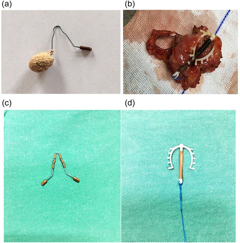
Figure 2: The IUD and associated calculus. (a) The IUD was an This study included six patients who underwent
MYCu, one end of which was wrapped with the calculus. (b) The IUD laparoscopy and suggested that this procedure can be
was embedded in the bladder wall. (c) Normal appearance of the used successfully for the treatment of an IUD embedded
MYCu IUD. (d) Normal appearance of the Multiload Cu IUD.
within the bladder wall with the additional presence of a
calculus. Only some case reports presented laparoscopic
Table 2: Surgical data of the patients surgery for ectopic IUD [15–17,22–26]. Shin et al. [15],
Jin et al. [17], Rahnemai-Azar et al. [23], Santos et al.
No. Operation Bleeding Hospital VAS Recovery [24], Liu et al. [22], Yahsi et al. [25], and Kurdoglu et al.
time (min) volume stay score time [26] each reported one patient successfully operated
(mL) (days) (days) using laparoscopic partial cystectomy or open surgery.
1 114 15 4 6 7 Atakan et al. [16] reported one case operated by
2 120 10 4.5 3 7 suprapubic cystotomy. These reports suggest that both
3 162 25 4 2 8 open and laparoscopic surgeries are appropriate for
4 132 15 4 3 7 ectopic IUD embedded in the bladder wall. Nevertheless,
5 144 17 4 2 7
other methods are also available and could be explored.
6 126 12 4 4 7
Indeed, Sano et al. [27] reported one case operated with
Total 129 15 (10, 25) 4 (4, 4.5) 3 7 (7, 8) endoscopy and laser fragmentation of the calculus
(114, 162) (2, 6) surrounding the IUD. Chai et al. [9] used cystoscopy to
7 126 25 7 4 8 remove the IUD, while Zhang et al. [28] used a
8 108 20 7 8 9
combination of various endoscopes. IUDs are usually
9 96 30 7 9 9
10 174 38 15 6 17
implanted at childbearing age and are recommended
11 192 50 10 5 14 to be removed after menopause. Nevertheless, some
patients do not have their IUD removed after meno-
Total 126 30 7 (7, 15) 6 9 (8, 17) pause, for various reasons, and IUD displacement can
(96, 192) (20, 50) (4, 9) happen at an old age. If there is no symptom, we
VAS: visual analog scale. recommend conservative treatments. If the patients have
hematuria, abdominal pain, or recurrent urinary tract
embedded in the bladder wall and could not be removed infections, they should choose surgical treatment. Since
by hysteroscopy or cystoscopy. The largest vesical surgeries are always associated with some risks such as
calculus was 1.8 cm. Hence, the patients had to undergo adhesion or iatrogenic injury, it has been suggested that
proper surgery. In the present study, both open surgery patients without symptoms or patients with high surgical
and laparoscopic surgery were successful in removing risk factors, especially elderly patients, might be more506 Bing-jian Xiong et al.
suitable for conservative treatment [18]. The traditional [2] Stoddard A, McNicholas C, Peipert JF. Efficacy and safety of
method of suturing the bladder is to suture the whole long-acting reversible contraception. Drugs. 2011;71(8):
layer of the incision first and then suture the sarco- 969–80. doi. 10.2165/11591290-000000000-00000.
[3] Curtis KM, Tepper NK, Jatlaoui TC, Berry-Bibee E, Horton LG,
plasmic layer to prevent leakage. Laparoscopic suturing
Zapata LB, et al. U.S. medical eligibility criteria for contra-
of the bladder is more difficult. At our center, we use a ceptive use. MMWR Recomm Rep. 2016;65(3):1–103.
continuous suture from the left side of the incision, with doi. 10.15585/mmwr.rr6503a1.
the assistant holding the end of the suture to maintain [4] Buhling KJ, Zite NB, Lotke P, Black K, Group IW. Worldwide use
the tension before knotting. Then, we reverse to suture of intrauterine contraception: a review. Contraception.
2014;89(3):162–73. doi. 10.1016/j.contraception.2013.11.011.
from the right side of the incision using the same
[5] Aoun J, Dines VA, Stovall DW, Mete M, Nelson CB, Gomez-
method. Using this method, one suture is enough, as per Lobo V. Effects of age, parity, and device type on complica-
our observation. Nevertheless, this method has not been tions and discontinuation of intrauterine devices. Obstet
reported before and should be examined more closely. In Gynecol. 2014;123(3):585–92. doi. 10.1097/
addition, new products such as barbed sutures, which AOG.0000000000000144.
[6] Westhoff C. IUDs and colonization or infection with
were not available at our center during the study period,
actinomyces. Contraception. 2007;75(Suppl 6):S48–50.
might provide satisfactory results [29].
doi. 10.1016/j.contraception.2007.01.006.
This study has limitations. This was a retrospective [7] Markovitch O, Klein Z, Gidoni Y, Holzinger M, Beyth Y.
study of all patients IUD getting embedded in the Extrauterine mislocated IUD: is surgical removal mandatory?
bladder wall with the additional presence of a calculus Contraception. 2002;66(2):105–8.
treated at one hospital. The number of patients was too [8] Andersson K, Ryde-Blomqvist E, Lindell K, Odlind V, Milsom I.
Perforations with intrauterine devices. Report from a Swedish
small to be able to compare open versus laparoscopic
survey. Contraception. 1998;57(4):251–5.
surgeries. Due to the rarity of the condition, the wide [9] Chai W, Zhang W, Jia G, Cui M, Cui L. Vesical transmigration of
range of IUD available on the market, and the conditions an intrauterine contraceptive device: a rare case report and
specific to each patient, the selection of laparoscopy literature review. Med (Baltim). 2017;96(40):e8236. doi.
versus open surgery should be tailored to each specific 10.1097/MD.0000000000008236.
case. Finally, based on our observation, a calculus forms [10] Nouira Y, Rakrouki S, Gargouri M, Fitouri Z, Horchani A.
Intravesical migration of an intrauterine contraceptive device
when the IUD comes in contact with urine. We did
complicated by bladder stone: a report of six cases. Int
observe even rarer cases of ectopic IUD without Urogynecol J Pelvic Floor Dysfunct. 2007;18(5):575–8. doi.
contemporary calculus, but they were not included, as 10.1007/s00192-006-0157-z.
per the inclusion criteria, and no data were collected. [11] Mechanism of action, safety and efficacy of intrauterine
Long-term studies with larger sample sizes are needed to devices. Report of a WHO Scientific Group. WHO Technical
Report Series. Geneva: World Health Organization;
confirm the results.
1987.
An ectopic IUD getting embedded in the bladder wall [12] Arnold MR, Lu CD, Thomas BW, Sachdev G, Cunningham KW,
with the additional presence of a calculus is rare. Both Vaio R, et al. Advancing the use of laparoscopy in trauma:
open surgery and laparoscopy can be used to remove the repair of intraperitoneal bladder injuries. Am Surg.
ectopic IUD and bladder stone successfully. 2019;85(12):1402–4.
[13] Morii Y, Osawa T, Suzuki T, Shinohara N, Harabayashi T,
Ishikawa T, et al. Cost comparison between open radical
Acknowledgments: The authors acknowledge the help of
cystectomy, laparoscopic radical cystectomy, and robot-
all the study participants. assisted radical cystectomy for patients with bladder cancer:
a systematic review of segmental costs. BMC Urol.
Disclosure statement: None. 2019;19(1):110. doi. 10.1186/s12894-019-0533-x.
[14] Shi H, Li J, Li K, Yang X, Zhu Z, Tian D. Minimally invasive
versus open radical cystectomy for bladder cancer:
Funding: None.
a systematic review and meta-analysis. J Int Med Res.
2019;47(10):4604–18. doi. 10.1177/0300060519864806.
[15] Shin DG, Kim TN, Lee W. Intrauterine device embedded into
the bladder wall with stone formation: laparoscopic removal is
References a minimally invasive alternative to open surgery. Int
Urogynecol J. 2012;23(8):1129–31. doi. 10.1007/s00192-011-
[1] Espey E, Ogburn T. Long-acting reversible contraceptives: 1632-8.
intrauterine devices and the contraceptive implant. Obstet [16] Atakan RH, Kaplan M, Ertrk E. Intravesical migration of
Gynecol. 2011;117(3):705–19. doi. 10.1097/ intrauterine device resulting in stone formation. Urology.
AOG.0b013e31820ce2f0. 2002;60(5):911.Bladder-embedded IUD with calculus 507
[17] Jin C, Fan Y, Zhang Q, Wang Y, Wu S, Jin J. Removal of foreign device embedded in intestine. JSLS. 2014;18(3):e2014.00122.
bodies embedded in the urinary bladder wall by a combination doi. 10.4293/JSLS.2014.00122.
of laparoscopy and carbon dioxide cystoscopic assistance: [24] Santos AP, Wetzel C, Siddiqui Z, Harper DS. Laparoscopic
case report and literature review. Investig Clin Urol. removal of migrated intrauterine device. BMJ Case
2016;57(6):449–52. doi. 10.4111/icu.2016.57.6.449. Rep. 2017;2017:bcr2017221342. doi. 10.1136/bcr-2017-
[18] Ucar MG, Sanlikan F, Ilhan TT, Gocmen A, Celik C. Management 221342.
of intra-abdominally translocated contraceptive devices, is [25] Yahsi S, Aktas BK, Erbay G, Salar R, Gokkaya CS. Intravesical
surgery the only way to treat this problem? J Obstet Gynaecol. migration of intrauterine device mimicking bladder stone on
2017;37(4):480–6. doi. 10.1080/01443615.2016.1268577. radiologic imaging: a case report. Indian J Surg.
[19] Aydogdu O, Pulat H. Asymptomatic far-migration of an 2015;77(Suppl 1):97–99. doi. 10.1007/s12262-014-1176-5.
intrauterine device into the abdominal cavity: a rare entity. [26] Kurdoglu Z, Ceylan K, Kurdoglu M, Guler A, Sahin HG. Ectopic
Can Urol Assoc J. 2012;6(3):E134–6. doi. 10.5489/cuaj.11100. intrauterine device in the bladder of a pregnant woman. Case
[20] Boortz HE, Margolis DJ, Ragavendra N, Patel MK, Kadell BM. Rep Med. 2010;2010:181032. doi. 10.1155/2010/181032.
Migration of intrauterine devices: radiologic findings and [27] Sano M, Nemoto K, Miura T, Suzuki Y. Endoscopic treatment of
implications for patient care. Radiographics. intrauterine device migration into the bladder with stone
2012;32(2):335–52. doi. 10.1148/rg.322115068. formation. J Endourol Case Rep. 2017;3(1):105–7.
[21] Zi D, Duan K, Fu KA, Mengyue Y, Hanlin Y, Guan X. Single- doi. 10.1089/cren.2017.0038.
incision laparoscopic surgery for removal of ectopic iud with [28] Zhang NN, Zuo N, Sun TS, Yang Q. An effective method
bladder repair. J Minim Invasive Gynecol. 2019;26(7):S57. combining various endoscopes in the treatment of intravesical
[22] Liu L, Liu H, Zhang X. Intravesical migration of a Chinese migrated intrauterine device. J Minim Invasive Gynecol.
intrauterine device and secondary stone formation: diagnostic 2020;27(3):582. doi. 10.1016/j.jmig.2019.07.024.
investigation and laparoscopic management. Int Urogynecol J. [29] Agrusa A, Di Buono G, Buscemi S, Cucinella G, Romano G.
2015;26(11):1715–6. doi. 10.1007/s00192-015-2735-4. Gulotta G,3D laparoscopic surgery: a prospective clinical trial.
[23] Rahnemai-Azar AA, Apfel T, Naghshizadian R, Cosgrove JM, Oncotarget. 2018;9(25):17325–33. doi. 10.18632/
Farkas DT. Laparoscopic removal of migrated intrauterine oncotarget.24669.You can also read