Acrylamide-induced changes of granulopoiesis in porcine bone marrow - Sciendo
←
→
Page content transcription
If your browser does not render page correctly, please read the page content below
J Vet Res 65, 2021
                                                                                             DOI:10.2478/jvetres-2021-0040

             Acrylamide-induced changes of granulopoiesis
                       in porcine bone marrow

                                       Dominika Grzybowska, Anna Snarska
                                         Department and Clinic of Internal Diseases
                                                Faculty of Veterinary Medicine
                                  University of Warmia and Mazury, 10-719 Olsztyn, Poland
                                                  anna.snarska@uwm.edu.pl

                                   Received: February 1, 2021        Accepted: June 30, 2021

Abstract

      Introduction: Due to the widely documented and diverse toxic effects of acrylamide, the authors decided to evaluate the
impact of high and low doses of this compound on the process of granulopoiesis in porcine bone marrow. Material and Methods:
The experiment was conducted on 15 Danish Landrace pigs at the age of 8 weeks. The animals were randomly assigned into three
equal groups (n = 5). Control animals received empty gelatine capsules as placebo. Animals in the first experimental group
(the LD group) received a low dose of acrylamide of 0.5 μg/kg b.w./day, and animals in the second experimental group (the HD
group) received a tenfold higher dose of acrylamide of 5 μg/kg b.w./day. Placebo and acrylamide capsules were administered with
feed every morning for 28 days. Bone marrow was collected into tubes without an anticoagulant twice – before the first capsule
administration (day 0) and on the 28th day of the study. After drying and staining, bone marrow smears were subjected to detailed
cytological evaluation under a light microscope. Results: Changes in cell morphology, i.e. degenerative changes in the cellular
nuclei, were observed in both experimental groups. Both low and high doses of acrylamide decreased the number of segmented
eosinophils, neutrophilic and segmented metamyelocytes, neutrophils, as well as basophils and basophilic metamyelocytes.
Conclusion: Acrylamide at doses of 0.5 μg/kg b.w./day and 5 μg/kg b.w./day clearly influences porcine granulopoiesis.

Keywords: acrylamide, pig, bone marrow, granulopoiesis, granulocytes.

Introduction                                                       rats. Long-term exposure to AA can damage the central
                                                                   nervous system and give signs of neurological disorders
     Acrylamide (AA) is a vinyl monomer, from                      (11). Jones et al. (9) also confirmed its neurotoxicity in
which polyacrylamides are synthesised. It is a colourless          research on workers who had frequent contact with
and odourless compound widely distributed in the                   acrylamide. They showed a clear relationship between
environment, which forms naturally in high-                        the level of acrylamide haemoglobin adducts and
carbohydrate products, mainly potatoes and cereals,                symptoms suggesting damage to the central nervous
when these are subjected to thermal processing at                  system. The toxic effect of AA can also manifest in
temperatures higher than 120°C (13, 18). It has a fairly           reduced fertility, increased risk of heart diseases and
well-known biological activity, as indicated by the                increased incidence of atherosclerosis (24). Acrylamide
harmonised classification of this compound (CAS                    also affects erythropoiesis in bone marrow (21).
Registry Number 79-06-1). Acrylamide is absorbed into                    Since the discovery of acrylamide’s presence in
the body through the digestive tract, respiratory system           food, many studies have indicated that this compound
and skin (26).                                                     participates in the development of neoplasms (14).
     The first studies on the toxicity of this compound            Despite the association that many studies show between
began in the late 1970s and 1980s (3, 10). Since then,             the occurrence of cancer in animals and AA in the diet,
numerous studies have confirmed its diverse harmful                none of the studies conducted in humans have clearly
activities. Yener and Dıkmenlı (22) demonstrated that              demonstrated the direct influence of this compound on
AA can cause genotoxicity in rats and mice. Research by            the formation of specific types of cancer. However, they
Manière et al. (12) indicated that it causes severe DNA            undoubtedly indicate that AA is associated with
changes in blood, brain, bone marrow and liver tissue in           increased cancer frequency.

© 2021 D. Grzybowska et al. This is an open access article distributed under the Creative Commons Attribution-
NonCommercial-NoDerivs license (http://creativecommons.org/licenses/by-nc-nd/3.0/)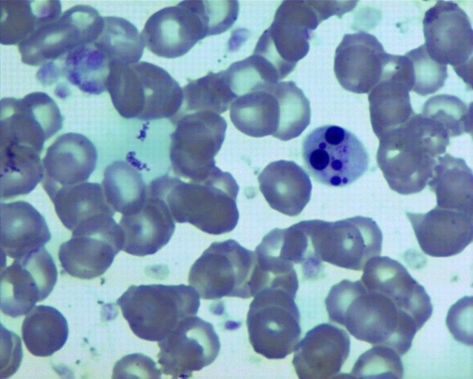
D. Grzybowska et al./J Vet Res/65 (2021)

      Due to the lifelong constant exposure of humans                   orally with feed in the morning for 28 days. After this
and animals to the effects of low doses of acrylamide and               time, animals of all groups were administered azaperone
despite the availability of extensive toxicological data on             at 4 mg/kg b.w. I.M. (Stresnil, Jansen Pharmaceutica
the compound, it is reasonable to conduct more research                 N.V., Geel, Belgium) and euthanised after 15 min using
on how strong and diverse the impacts of AA on human                    a lethal dose of 0.6 mL/kg b.w. I.V. of sodium pentobarbital
and animal organisms really are, and whether exposure                   (Morbital, Biowet Puławy, Puławy, Poland).
to it could be a more serious threat to human and animal                      Sample collection. Two bone marrow samples
health than we currently believe. The research model choice             were taken from all animals: on day 0 (a day before
was based on the general recognition that the domestic                  beginning AA administration), and on the 28th day of the
pig is a scientific model adapted to humans (25).                       experiment. Bone marrow was sampled from the lateral
      It is necessary to expand knowledge on the impact                 condyle of the femur under local anaesthesia with
of repeated exposure to acrylamide, especially in the                   1.5 mg/kg b.w. I.M. of xylazine hydrochloride (Rompun,
aspect of assessing interspecies differences in sensitivity             Bayer, Leverkusen, Germany), and 2.2 mg/kg b.w. I.M.
to this xenobiotic, and therefore we decided to evaluate                of zolazepam and tiletamine (Zoletil, Virbac, Carros,
the impact of high and low doses of this compound on                    France), using Jamshidi bone marrow needles (Synthes,
the process of granulopoiesis in porcine bone marrow.                   Salzburg, Austria). Bone marrow samples were
                                                                        collected into two tubes without anticoagulant and used
                                                                        to prepare bone marrow smears.
Material and Methods                                                          Cytological evaluation. The smears were stained
                                                                        with the May–Grunwald–Giemsa method and evaluated
      Animals and design of the experiment. The study                   under an Eclipse 80i light microscope (Nikon, Tokyo,
was conducted on 15 eight-week old Danish Landrace                      Japan) using a SH-96/24D haematological counter
pigs weighing approximately 20 kg. All pigs were kept                   (Alchem, Toruń, Poland). The number of particular cells
under standard laboratory conditions, had free access to                from the granulocytic cell line were defined per 1,000 bone
water, and were fed a commercial grain mixture. After                   marrow cells, which is a standard method of evaluation
seven days of acclimatisation, animals were randomly                    of bone marrow smears.
assigned to one of three groups: the control (C) group                        Statistical analysis. Statistical analysis was
(n = 5) of animals receiving empty gelatine capsules,                   performed using ANOVA and post-hoc Bonferroni tests
a low dose (LD) group (n = 5) of animals receiving                      with Statistica 10 software (StatSoft Inc, Tulsa, OK,
capsules with the tolerable daily intake (TDI) dose of AA               USA). The differences were considered statistically
(0.5 μg/kg b.w./day) used at > 99% purity (Sigma-Aldrich,               significant at P ≤ 0.05.
St. Louis, MO, USA), and a high dose (HD) group
(n = 5) of animals receiving capsules with a tenfold
higher dose of AA (5 μg/kg b.w./day). The lower dose                    Results
used in the study is a dose recognised in many countries
as the TDI or reference dose for acrylamide, and is                          Cytological evaluation of bone marrow smears
considered to be safe for humans and animals. To ensure                 before AA administration (day 0) did not show any
that pigs received the appropriate dose of AA, they were                significant differences in the number and morphology
weighed once a week. Capsules were administrated                        of all types of cells between all three groups (Table 1).

Table 1. The average number of cells from the granulocytic cell line per 1,000 porcine bone marrow cells (mean ± SD)
before acrylamide administration
 Cell type                           Control group                Low dose group                High dose group
 Myeloblasts                         2.720 ± 0.504                2.980 ± 0.336                 2.820 ± 0.344
 Promyelocytes                       2.080 ± 0.656                2.160 ± 0.672                 2.240 ± 0.192
 Myelocyte                           3.600 ± 0.120                3.520 ± 0.144                 3.200 ± 0.240
 Metamyelocyte                       6.500 ± 1.000                6.360 ± 0.568                 6.360 ± 0.888
 Band neutrophils                    15.180 ± 2.584               13.620 ± 1.064                14.520 ± 1.576
 Neutrophilic granulocytes           12.400 ± 2.200               13.380 ± 2.224                12.820 ± 2.296
 Eosinophilic myelocytes             1.740 ± 0.416                1.320 ± 0.384                 1.620 ± 0.264
 Eosinophilic metamyelocytes         1.960 ± 0.552                2.260 ± 0.568                 2.200 ± 0.280
 Band eosinophils                    2.640 ± 1.728                2.820 ± 1.624                 2.780 ± 1.176
 Eosinophilic granulocytes           1.300 ± 0.720                1.560 ± 1.272                 1.960 ± 1.632
 Basophilic myelocytes               0.000                        0.000                         0.000
 Basophilic metamyelocytes           0.060 ± 0.048                0.020 ± 0.032                 0.080 ± 0.064
 Band basophiles                     0.280 ± 0.136                0.320 ± 0.184                 0.375 ± 0.225
 Basophilic granulocytes             0.360 ± 0.192                0.280 ± 0.136                 0.300 ± 0.160
 Hypersegmented granulocytes         0.000                        0.000                         0.000
 Total granulocytes                  50.820 ± 1.184               50.600 ± 2.76                 51.220 ± 0.896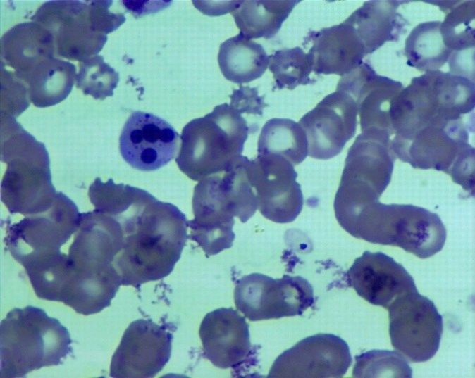
D. Grzybowska et al./J Vet Res/65 (2021)
Table 2. The average number of cells from the granulocytic cell line per 1,000 porcine bone marrow cells (mean ±SD)
on the 28th day of the experiment
Cell type Control group Low dose group High dose group
Myeloblasts 2.680 ± 0.576a,b 1.160 ± 0.512a 1.080 ± 0.104b
Promyelocytes 2.100 ± 0.68a,b 1.460 ± 0.728a 1.320 ± 0.504b
Myelocyte 3.540 ± 0.088a 2.180 ± 0.896a,c 3.300 ± 1.600c
Metamyelocyte 6.540 ± 1.208a,b 3.840 ± 1.568a,c 3.120 ± 1.824b,c
Band neutrophils 15.940 ± 2.872a,b 8.620 ± 1.304a,c 6.520 ± 1.944b,c
Neutrophilic granulocytes 12.460 ± 2.192b 10.920 ± 1.104c 18.680 ± 9.016b,c
Eosinophilic myelocytes 1.740 ± 0.456 1.180 ± 0.776 0.820 ± 0.384
Eosinophilic metamyelocytes 2.220 ± 0.464 2.320 ± 1.464 0.840 ± 0.568
Band eosinophils 2.960 ± 1.952b 3.300 ± 2.200c 1.420 ± 1.112b,c
Eosinophilic granulocytes 2.000 ± 1.480a 2.460 ± 1.632a,c 2.120 ± 1.096a,c
Basophilic myelocytes 0.020 ± 0.032 0.000 0.000
Basophilic metamyelocytes 0.080 ± 0.064 0.000 0.040 ± 0.048
Band basophiles 0.260 ± 0.192a,b 0.060 ± 0.072a,c 1.420 ± 1.112b,c
Basophilic granulocytes 0.320 ± 0.184 a,b 0.040 ± 0.064a,c 0.080 ± 0.064b,c
Hypersegmented granulocytes 0.000b 0.000c 0.500 ± 0.640b,c
Total granulocytes 52.860 ± 2.528a,b 37.520 ± 8.784a 44.680 ± 3.144b
a
– statistically significant difference between control and low dose group (P ≤ 0.05)
b
– statistically significant difference between control and high dose group (P ≤ 0.05)
c
– statistically significant difference between low dose and high dose group (P ≤ 0.05)
However, there was a significant decrease in the total
number of granulocytes in experimental pigs at the end
of the experiment (Table 2). The number of myeloblasts,
promyelocytes, myelocytes, and basophilic granulocytes
declined significantly (P ≤ 0.05) after 28 days of
receiving AA (Table 2), the high doses of which affected
some cells differently to the low doses. Acrylamide used
in low doses decreased the total of neutrophilic
granulocytes and band eosinophils. However, used in
high doses it increased the numbers of those cells.
Basophilic myelocytes appeared only in the control
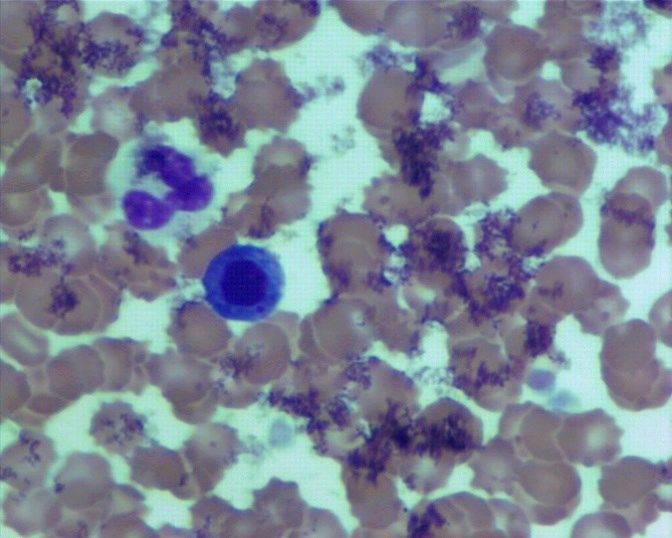
group at the end of the experiment (Fig. 3). Our research
also showed very clear changes in the morphology of
granulocytes consisting in strong condensation and
fragmentation of chromatin in cell nuclei in the HD
group (Figs. 1 and 2). Fig. 2. Representative cytological image of a bone marrow smear
stained using the May–Grunwald–Giemsa method showing
condensation and fragmentation of the nucleus of a neutrophil in the
HD group. Magnification 1,000×
Fig. 1. Representative cytological image of a bone marrow smear
stained using the May–Grunwald–Giemsa method showing
condensation and fragmentation of the nucleus of a neutrophil in the Fig. 3. Representative cytological image of a bone marrow smear
HD group. Magnification 1,000× stained using the May–Grunwald–Giemsa method showing a basophilic
myelocyte in the C group. Magnification 1,000×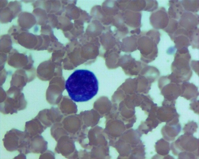
D. Grzybowska et al./J Vet Res/65 (2021)

                                                                        administration of acrylamide. A study by Shler et al. (16)
                                                                        showed the effect of AA on the development of
                                                                        inflammation, manifested by an increase in the number
                                                                        of leukocytes and the development of leucocytosis.
                                                                        An increase in the number of leukocytes and precursor
                                                                        cells of granulocytes may indicate the activation of the
                                                                        immune system and consequent inflammation.
                                                                              Results regarding acrylamide administration
                                                                        published so far indicate bone marrow hyperplasia in
                                                                        rats (6). Developing anaemia and thrombopaenia as well
                                                                        as an increase in the number of leukocytes were
                                                                        observed in mice in research by Raju et al. (15).
                                                                        However, the results described in this publication
                                                                        indicate decreased activity of the porcine granulocytic
                                                                        cell line under the influence of AA. Only high doses of
Fig. 4. Representative cytological image of a bone marrow smear         acrylamide caused an increase in the number of
stained using the May–Grunwald–Giemsa method showing a myeloblast
in the LD group. Magnification 1,000×                                   neutrophilic granulocytes and band basophils and the
                                                                        appearance of hypersegmented granulocytes in pigs. The
                                                                        observed changes in the morphology of cells (e.g. chromatin
Discussion                                                              condensation) may be a sign of cell degeneration and
                                                                        activation of an apoptotic pathway. Low doses of AA led
      Expansion of knowledge on the impact of repeated                  to a statistically significant increase in the number of
exposure of human and animal organisms to acrylamide                    eosinophils; however, despite those increases the total
is paramount, especially in the aspect of interspecies                  granulocytic count significantly decreased in both
differences in sensitivity to its toxic activity. The                   experimental groups to resemble bone marrow
sparseness of research on its influence on haematopoiesis               hypoplasia in the granulocytic cell line. The presented
occurring in bone marrow prompted the authors of this                   results undoubtedly indicate that regardless of the
publication to address this topic and model it in the                   animal species (pigs, rats, or mice), AA influences the
domestic pig. Due to this species’ phylogenetic                         processes of haematopoiesis and it can have either
similarity to humans, it is often used as an animal model               a stimulatory or inhibitory effect on certain cell lines
(25). However, most experiments on the effects of AA                    depending on the studied species.
on mammals were conducted on rodents (4, 5, 19).                              A study by Hammad et al. (8) in rats showed
      Since acrylamide is a compound widely distributed                 an increase in the number of white blood cells in all
in the environment, most humans are exposed to it in                    groups receiving acrylamide. The authors of the present
varying amounts in food and other sources such as                       publication observed a decrease in the total number of
tobacco smoke (7). The WHO estimated that the total                     granulocytes in pigs. Our research also showed that there
daily intake of AA from food ranges between 0.3 and                     were very clear changes in the morphology of granulocytes,
0.8 μg/kg b.w. (20). Livestock and pets are also at risk                consisting in strong condensation and fragmentation of
of its negative effects as inhabitants of the same                      cell nuclei in the group receiving high doses of AA. Such
environment as humans, and it could be assumed that                     changes are often observed in animals (especially dogs
daily exposure to AA in animals is similar to that in                   and cats) in the course of inflammation and cancer,
humans. However, considering humans’ consumption of                     particularly leukaemia (1, 17). Changes in the morphology
a wide range of highly processed food, human exposure                   of neutrophils may indicate a significant influence of AA
to acrylamide could be even higher, and its effect on                   on the processes of haematopoiesis, however, we cannot
bone marrow even more detrimental. Literature data                      state whether these changes will generate long-term
indicate that besides damaging bone marrow,                             disturbances of this process in the granulocytic cell line.
acrylamide toxicity can manifest in skeletal muscle                     Therefore, whether AA can cause such strong changes
atrophy, distended urinary bladders, increased                          in the morphology of granulocytes as to qualify it to the
prevalence of duct ectasia in preputial glands,                         group of agents that can cause severe disturbances of
haematopoietic cell proliferation in the spleen,                        haematopoiesis is a question to stimulate interest in
hepatocyte degeneration and liver necrosis, mesenteric                  continued research. However, it is certain that the usage
lymph node cellular infiltration and pituitary gland                    of this compound may adversely affect the human and
hyperplasia (23).                                                       animal body.
      Research conducted by Dobrzyńska (4) on mice                            The results obtained during the present investigation
shows toxic AA activity by dose-dependent increase in                   clearly show that acrylamide suppresses granulopoiesis,
DNA damage of somatic and germ cells. The results of                    which manifests in a decreased number of most types of
the study by Benziane et al. (2) indicate that in Wistar                cells from the granulocytic cell line and changes in cell
rats, an increase in white blood cell system components,                morphology. It was seen that different doses can have
in particular leukocytes, was noted after oral                          different effects on certain cell types. Moreover, the
D. Grzybowska et al./J Vet Res/65 (2021)
results of this research may be a valuable source of 11. LoPachin R.M.: Acrylamide neurotoxicity: neurological,
information about the harmfulness of acrylamide to the morphological and molecular endpoints in animal models.
In: Chemistry and Safety of Acrylamide in Food. Advances in
process of granulopoiesis in humans and animals, Experimental Medicine and Biology, vol 561, edited by M.
especially in view of the high utility of the domestic pig Friedman, D. Mottram, Springer, Boston, MA, 2005, pp. 21–37,
as a scientific model adopted for research applicable to doi: 10.1007/0-387-24980-X_2.
humans. 12. Manière I., Godard T., Doerge D.R., Churchwell M.I., Guffroy M.,
Laurentie M., Poul J.M.: DNA damage and DNA adduct
formation in rat tissues following oral administration of
Conflict of Interests Statement: The authors declare acrylamide. Mutat Res 2005, 580, 119–129, doi: 10.1016/
that there is no conflict of interests regarding the j.mrgentox.2004.10.012.
publication of this article. 13. Mottram D.S., Wedzicha B.L., Dodson A.T.: Acrylamide is
formed in the Maillard reaction. Nature 2002, 419, 448–449, doi:
Financial Disclosure Statement: This study was 10.1038/419448a.
14. Mucci L.A., Wilson K.M.: Acrylamide intake through diet and
financed by the KNOW (Leading National Research human cancer risk. J Agric Food Chem 2008, 56, 6013–6019, doi:
Centre) Scientific Consortium “Healthy Animal – Safe 10.1021/jf703747b.
Food”. 15. Raju J., Roberts J., Taylor M., Patry D., Chomyshyn E., Caldwell D.,
Cooke G., Mehta R.: Toxicological effects of short-term dietary
Animal Rights Statement: The research was carried acrylamide exposure in male F344 rats. Environ Toxicol
Pharmacol 2015, 39, 85–92, doi: 10.1016/j.etap.2014.11.009.
out in accordance with EU Directive 2010/63/EU for 16. Shler A.F.M., Kawa A., Heshu S.R., Hemn H.O.: The
animal experiments and with the approval of the Local pathophysiological effects of acrylamide in Albino Wister Rats.
Ethical Committee for Experiments on Animals in Int J Med Res Health Sci 2016, 5, 42–48.
Olsztyn (Approval no. 11/2017). 17. Tan E., Abrams-Ogg A.C.G., Defarges A., Bienzle D.: Automated
analysis of bone marrow aspirates from dogs with haematological
disorders. J Comp Pathol 2014, 151, 67–79, doi:
10.1016/j.jcpa.2014.02.005.
References 18. Tareke E., Rydberg P., Karlsson P.: Analysis of acrylamide,
a carcinogen formed in heated foodstuffs. J Agric Food Chem
1. Azakami D., Saito A., Ochiai K., Ishiwata T., Takahashi K., 2002, 50, 4998–5006, doi: 10.1021/jf020302f.
Kaji N., Kaji D., Kaji N., Michishita M.: Chronic basophilic 19. Twaddle N.C., Churchwell M.I., McDaniel L.P., Doerge D.R.:
leukaemia in a dog. J Comp Pathol 2019, 166, 5–8, doi: 10.1016/ Autoclave sterilization produces acrylamide in rodent diets:
j.jcpa.2018.10.170. implications for toxicity testing. J Agric Food Chem 2004, 52,
2. Benziane A.B., Bouras A.D., Mezaini A., Belhadri A., Benali M:. 4344–4349, doi: 10.1021/jf0497657.
Effect of oral exposure to acrylamide on biochemical and 20. World Health Organisation: FAO/WHO Consultations on the
hematologic parameters in Wistar rats. Drug Chem Toxicol 2019, health implications of acrylamide in food. Summary report of
42, 157–166, doi: 10.1080/01480545.2018.1450882. a meeting held in Geneva, 25–27 June, 2002. https://apps.who.
3. Bull R.J., Robinson M., Laurie R.D., Stoner G.D., Greisiger E., int/iris/bitstream/handle/10665/67372/a76870.pdf;sequence=1.
Meier J.R., Stober J.: Carcinogenic effects of acrylamide in Sencar 21. Yener Y.: Effects of long term low dose acrylamide exposure on
and A/J mice. Cancer Res 1984, 44, 107–111. rat bone marrow polychromatic erythrocytes. Biotech Histochem
4. Dobrzyńska M.: Assessment of DNA damage in multiple organs 2013, 88, 356–360, doi: 10.3109/10520295.2013.790561.
from mice exposed to X-rays or acrylamide or a combination of 22. Yener Y., Dıkmenlı M.: Increased micronucleus frequency in rat
both using the comet assay. In vivo 2007, 21, 657–662. bone marrow after acrylamide treatment. Food Chem Toxicol
5. El-Tohamy A.A., Bayomy A.A.: Effects of long term low dose 2009, 47, 2120–2123, doi: 10.1016/j.fct.2009.05.037
acrylamide exposure on rat bone marrow polychromatic 23. Zamani E., Shaki F., Abedian Kenari S., Shokrzadeh M.:
erythrocytes. Arab J Biotech 2008, 11, 29–38, doi: Acrylamide induces immunotoxicity through reactive oxygen
10.3109/10520295.2013.790561. species production and caspase-dependent apoptosis in mice
6. European Food Safety Agency Panel on Contaminants in the Food splenocytes via the mitochondria-dependent signaling pathways.
Chain (CONTAM): EFSA Scientific Opinion on acrylamide in Biomed Pharmacother 2017, 94, 523–530, doi: 10.1016/j.biopha.
food. EFSA J 2015, 13, 4104, doi: 10.2903/j.efsa.2015.4104. 2017.07.033.
7. Gargas M.L., Kirman C.R., Sweeney L.M., Tardiff R.G.: 24. Zenick H., Hope E., Smith M.K.: Reproductive toxicity associated
Acrylamide: consideration of species differences and nonlinear with acrylamide treatment in male and female rats. J Toxicol
processes in estimating risk and safety for human ingestion. Food Environ Health 1986, 17, 457–472, doi: 10.1080/
Chem Toxicol 2009, 47, 760–768, doi: 10.1016/j.fct.2008.12.032. 15287398609530840.
8. Hammad A.Y., Osman M.E., Abdelgadir W.S.: Histopathological 25. Zhong C., Wu J., Izpisua Belmonte J.C.: Pig Chimeric Model with
assessment and hematotoxicity of dietary acrylamide on Wistar Human Pluripotent Stem Cells. Methods Mol Biol 2019, 2005,
rats. Int J Life Sci 2013, 7, 21–25, doi: 10.3126/ijls.v7i1.8018. 101–124, doi: 10.1007/978-1-4939-9524-0_8.
9. Jones K., Garfitt S., Emms V., Warren N., Cocker J., Farmer P.: 26. Zödl B., Schmid D., Wassler G., Gundacker C., Leibetseder V.,
Correlation of haemoglobin-acrylamide adducts with airborne Thalhammer T., Ekmekcioğlu C.: Intestinal transport and
exposure: an occupational survey. Toxicol Lett 2006, 162, metabolism of acrylamide. Toxicol 2007, 232, 99–108, doi:
174–180, doi: 10.1016/j.toxlet.2005.09.016. 10.1016/j.tox.2006.12.014.
10. Kesson C.M., Baird A.W., Lawson D.H.: Acrylamide poisoning.
Postgrad Med J 1997, 53, 16–17, doi: 10.1136/pgmj.53.615.16.You can also read