Araloside C Prevent Myocardial Cell Apoptosis Through Regulating PI3K/AKt to Relieve Heart Failure
←
→
Page content transcription
If your browser does not render page correctly, please read the page content below
Araloside C Prevent Myocardial Cell Apoptosis
Through Regulating PI3K/AKt to Relieve Heart
Failure
Xingsheng Zhao ( xingsheng_z@163.com )
Inner Mongolia People’s Hospital
Yu Ren
Inner Mongolia People’s Hospital
Hongkun Ren
Inner Mongolia People’s Hospital
Yun Wu
Inner Mongolia People’s Hospital
Xi Liu
Inner Mongolia People’s Hospital
Hua Chen
Inner Mongolia People’s Hospital
Chun Ying
Inner Mongolia People’s Hospital
Research Article
Keywords: Araloside C, heart failure, PI3K/Akt
Posted Date: June 23rd, 2021
DOI: https://doi.org/10.21203/rs.3.rs-620532/v1
License: This work is licensed under a Creative Commons Attribution 4.0 International License.
Read Full License
Page 1/11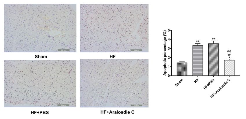
Abstract
Background Araloside C (AsC), a natural saponin isolated from Aralia elata, has a wide range of anti-
inflammatory properties and has been found in recent years to have heart-protective effects. Present
study aimed to determine the effects of AsC on myocardial cell apoptosis through regulating PI3K/AKt.

Methods and Results Statistical analyses were performed using GraphPadPrism7.0 software. The
differences between two groups and multiple groups were analyzed using t-test and one-way ANOVA,
respectively. In vivo results showed that AsC administration could improve cardiac functions and
apoptotic rate in HF model through PI3K/AKt signaling pathway, including increasing left ventricular
ejection fraction (LVEF) and left ventricular fraction shortening (LVFS), and decreasing left ventricular end
systolic diameter (LVESD) and left ventricular end diastolic diameter (LVEDD) in detection of myocardial
function, inhibiting LDH, CK, CK-MB, CK and HBDH in biochemical index level assessment, inhibiting BNP,
ANG II, IL-1b, IL-4, IL-6 and TNF-a in immunological index level. ASC regulates the expression of key
apoptotic molecules, including increasing the expression of Bcl-2 and Bax. ASC also regulates
phosphorylation of p-PI3K and p-Akt.

Conclusion This study suggested for the first time that AsC could partially regulate the PI3K/AKt
signaling pathway to prevent myocardial cell apoptosis. This study provided a basis for further research
on effective substances in the treatment of HF.

Introduction
In recent years, the research on the treatment of heart failure (HF) with traditional Chinese medicine has
attracted wide attention1. In particular, salvia miltiorrhiza, berberine, ginseng, astragalus and other
traditional Chinese medicines, including salvianolic acid A, salvianolic acid B, tanshinone IIA, ginsenoside
Re, berberine, flavonoids, have been proved to improve cardiac hypertrophy, reduce cell apoptosis and
inflammatory response, and protect heart function, has an important role2–5. Araloside C (AsC), a natural
saponin isolated from Aralia elata, has a wide range of anti-inflammatory properties and has been found
in recent years to have heart-protective effects. Luo et al studied on atherosclerotic mice, AsC can
regulate macrophage polarization through sirt1-mediated autophagy, thereby reducing the formation of
plaque area in atherosclerotic mice6. Moreover, inhibiting oxidative stress and Ca2+ overload by regulating
HSP90 can effectively reduce hypoxia/reoxygenation of H9c2 cardiomyocyte ischemia/reperfusion (I/R)
injury7–9.

However, the therapeutic effect of AsC in rats with HF has not been reported so far, and its internal
mechanism has not been reported. Therefore, we will focus on AsC to study its effect on cardiac function
in rats with HF. Sun et al. found that the HSP90/Akt pathway induces cardiac hypertrophy and cell death,
affecting the course of HF10. Therefore, we will study whether AsC can inhibit cell death, improve cardiac
function and delay the occurrence and development of HF through the PI3K/AKt signaling pathway
regulated apoptosis.

                                                   Page 2/11
Results
AsC repairs cardiac function in HF rats
In detection of myocardial function, we found that compared with Sham group, the heart function of rats
in the HF group was significantly decreased, with LVEF (Fig. 1C) and LVFS (Fig. 1D) decreased, and
LVEDD (Fig. 1A) and LVESD (Fig. 1B) increased (P < 0.01), indicating the success of the model
establishment. Compared with the HF group, the HF + PBS group showed no significant change in the
ultrasonic indexes of cardiac function, while the HF + AsC group showed increased LVEF (Fig. 1C) and
LVFS (Fig. 1D), and decreased LVESD (P < 0.01), indicating significant improvement in cardiac function
after AsC administration.
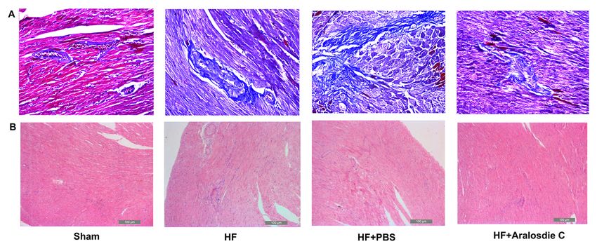
Pathological changes of cardiac muscle in HF rats inhibited
by ASC
Description of Masson staining results: myocardial cell fibers in sham group were evenly and neatly
arranged, with clear transverse lines, and a small amount of blue-dyed fibrous tissue was observed
between the cells (Fig. 2A). In the HF and HF + PBS groups, myocardial fibers were fractured, the cells
showed granular degeneration to varying degrees, and a large number of blue-stained fibrous tissues
were observed in the tissues. The Model + AsC group showed a significant reduction in the blue-stained
fibrous tissue of myocardial tissue.
HE staining results (Fig. 2B): compared with Sham group, HF and HF + PBS rat had disordered
arrangement of cardiomyocytes, hypertrophy of cardiomyocytes, and enlarged nuclei. Compared with the
HF group, the Model + AsC group significantly reduced the degree of myocardial cell hypertrophy and
nuclear enlargement, while the Model + PBS group showed no significant changes.
AsC reduced plasma biochemical index level
Myocardial enzyme (CK, CK-MB, LDH, AST and HBDH) had become biomarkers for the diagnosis of
myocardial injury10,11. Myocardial injury caused the release of LDH, AST, CK, CK-MB, and HBDH into
bloodstream. The expression of LDH (Fig. 3A), CK (Fig. 3C), CK-MB (Fig. 3D), and HBDH (Fig. 3E)
decreased significantly after AsC intervention compared to HF and HF + PBS groups. However, the
expression of AST (Fig. 3B) had no significantly change after AsC intervention in HF rats (Fig. 3B). Above
all monitoring of biochemical indexes showed that the drug intervention had obvious improvement on HF
in rats.
AsC reduced plasma Immunological index level
ELISA were used to detect the expression of inflammatory factors (IL-1β, IL-6, IL-4 and TNF-α) in the
experiments. The results showed that sham group had significant difference in expression of BNP
(Fig. 4A), Ang II (Fig. 4B), IL-1β (Fig. 4C), IL-4 (Fig. 4D), IL-6 (Fig. 4E), and TNF-α (Fig. 4F) compared to
Page 3/11other groups (P < 0.05). There was no significant change between HF group and HF + PBS group. The
expression of indicators decreased significantly after AsC intervention compared to HF and HF + PBS
groups. Above all these results suggested that AsC treatment might decrease the expression of
inflammatory factors to improve heart function in HF rats.
ASC inhibits cardiomyocyte apoptosis in HF rats
Apoptosis of HF rat cardiomyocytes was detected by TUNEL assay to evaluate the antiapoptotic effect of
AsC on cardiomyocytes (Fig. 5). The results showed that compared with sham rats, the percentage of
cardiomyocyte apoptosis in HF and HF + PBS rats was significantly increased. However, percentage of
apoptotic myocardial cells was significantly lower after the intervention of the AsC, which indicated that
the AsC had the effect of preventing the apoptosis of heart cells.
AsC promotes PI3K/Akt signaling pathway activation
Total protein was extracted from cardiac tissue and WB was used to detect the changes in the levels of
myocardial protein p/t-PI3K, p/t-Akt, Bcl-2, Bax, Caspase-3 and cytochrome C (Fig. 6A). WB analysis
results (Fig. 6B) showed that compared with sham group, the expressions of P-PI3K, P-Akt and Bcl-2 were
significantly reduced in the HF and HF + PBS groups and significantly increased after drug intervention.
However, the expression of Bax increased compared with sham group, and decreased after AsC
intervention. There were no statistically significant differences in the expression of other proteins between
these groups.
Discussion
There are more than 4 million patients with HF in China, and the fatality rate is much higher than that in
developed countries13. The research on the mechanism of HF has been in progress, and apoptosis has
been proved to be helpful to the development of HF. Cardiomyocyte apoptosis occurs in a variety of
cardiovascular diseases, such as myocardial infarction and I/R14,15. ASC is one of the most abundant
triterpenoids isolated from A. elata, which has been clearly shown to stimulate cardiac activity16,17.
However, the cardioprotective effect of AsC and its mechanism remain unclear. In this study, we
demonstrated that ASC could protect HF cardiomyocytes from apoptosis and inhibit the activation of Akt
in HF myocardium. The results showed that AsC had a protective effect on myocardial cell apoptosis in
rats with HF by inhibiting the overexpression of PI3K/Akt signaling pathway.
It has been reported that HF is not only due to the decrease of myocardial contractility, but also to the
increase of apoptotic cells19. Apoptosis plays an important role in ventricular remodeling and HF19,20. Bcl-
2 protein family determines the commitment of cells to apoptosis, and the activation of Caspase-3
triggers the execution of apoptosis. There are both pro apoptotic and anti-apoptotic proteins in Bcl-2
protein family. Bax promotes the formation of membrane pores in the form of oligomers, and releases
pro-apoptotic substances into the cytoplasm to play a pro-apoptotic role, while Bcl-2 inhibits apoptosis by
Page 4/11blocking the oligomerization of pro-apoptotic proteins21. The expression of Bcl-2, Bax, caspase-3 and
cytochrome C were detected by Western blot. The balance between Bcl-2 and Bax was disrupted, while
the expression of Caspase-3 and cytochrom C was not changed, suggesting that the apoptotic pathway
was activated in the HF model. AsC treatment can increase the level of Bax, inhibit the expression of Bcl-
2, but not affect the expression of Caspase-3.
In this study, the rat model of HF was established to explore the effects of PI3K/Akt signaling pathway on
cardiac apoptosis. PI3K/Akt, as an important signal transduction pathway, plays an important role in cell
survival, apoptosis and proliferation21.
The expression of p-PI3K and p-Akt increased in ASC treatment group, but decreased in HF rats. It
suggests that AsC may play an anti-apoptotic role by regulating P-PI3K and P-Akt, rather than directly
interacting with PI3K and Akt. Some ASC components have been confirmed to have anti apoptotic effect.
For example, quercetin, luteolin and tanshinone IIA have been shown to have anti-apoptotic effects on
myocytes22–24. In future studies, we will verify the role of these potential active ingredients through in
vitro and in vivo experiments. In conclusion, this study explored the protective effect of ASC in HF animal
model and apoptosis model. The results showed that AsC could partially regulate the PI3K/Akt signaling
pathway to inhibit myocardial cell apoptosis. This study provides a basis for further study of effective
substances for the treatment of HF.
Materials And Methods
Establishment of the HF Model in Rats and Grouping
The HF model was established by abdominal aortic constriction. After feeding adult Wister male rats for
1 week, pentobarbital sodium anesthetized rats, and then the abdominal aorta was separated 1cm above
the left renal artery through a median incision. Abdominal aorta was sutured with no. 22 needle 4-0 silk
thread to form abdominal aortic stenosis (about 50% ~ 60%). Abdominal dissection was performed. At
the same time, 1×10⁵ U penicillin was intraperitoneally injected to prevent infection. The rats were
monitored daily after surgery, and after 10 weeks, AsC medication interfered with HF (2.5 mg/kg/day) for
4 weeks.
The rats were divided into four groups (sham group, HF model, HF+PBS and HF+ AsC group) and five of
each group. Sham group rats received DMEM 70μl for control.
All rats were used for subsequent experiments in accordance with the Laboratory Animal Management
Regulations and Animal Ethical Requirements before modeling. Animal experimental protocols were
approved by the Ethics Committee of the Inner Mongolia People’s Hospital and the study was carried out
in compliance with the ARRIVE guidelines. Rats were sacrificed and anesthetized by intraperitoneal
injection of pentobarbital sodium solution (1%) at a dose of 50mg/kg. We tried our best to reduce the
number of animals that are used and reduce their suffering.
Page 5/11Detection of myocardial function
Ten weeks after the last administration, the rats in each group were anesthetized. After removing the
chest hair and applying the coupling agent, the VisualsonicVevo 2100 imaging system was used to
evaluate the preoperative and postoperative cardiac function through echocardiography. Mice were
anesthetized with 2.5l /min isoflurane before evaluation. Left ventricular end diastolic diameter (LVEDD),
left ventricular end systolic diameter (LVESD), left ventricular fraction shortening (LVFS) and left
ventricular ejection fraction (LVEF) were calculated with Vevo analysis software25.
Histological examination in rats
The rat heart was dissected, fixed with 4% formalin for 24 hours, embedded in paraffin, and cut into 5μm-
thick slices. Then the slices were stained with Masson's trichrome staining (Solarbio, Beijing, China) and
hematoxylin-eosin (HE) staining to observe heart tissue morphology. Image analysis software (Image-Pro
Plus v4.0, Media Cybernetics, USA) was used to calculate the area occupied by collagen
Analysis of serum biochemical indexes
The contents of creatine kinase (CK), creatine kinase isoenzyme (CK-MB), aspartate aminotransferase
(AST),lactate dehydrogenase (LDH) and hydroxybutyrate dehydrogenase (HBDH) in plasma were
determined by automatic chemical analyzer (Labospect-008, Hitachi High-Tech Diagnostics (Shanghai)
Ltd., Japan)
ELISA Analysis
The levels of BNP, TNF-α, AngII, IL-6, IL-4, IL-1b in plasma were quantified using Glucagon Quantikine
ELISA Kit (Elabscience Biotechnology Co., Ltd, Wuhan, China). Analyze according to manufacturer's
instructions.
Apoptosis Assay
According to the manufacturer's instructions, cardiomyocyte apoptosis was detected by TUNEL (EMD
Millipore, Billerica, MA). The apoptosis of cardiomyocytes was brown. The average percentage of
apoptotic cells in 5 randomly selected fields (under magnification ×40) was calculated by Olympus
microscope.
Western blotting (WB)
Total proteins were extracted from cardiac tissue and the levels of cardiac proteins P/t-PI3K, P/t-AKt, Bcl-
2, Bax, Caspase-3 and cytochrome C were detected by WB.
Statistical analysis
Page 6/11All statistical analyses were performed using GraphPadPrism7.0 software. The differences between two
groups and multiple groups were analyzed using t-test and one-way ANOVA, respectively. P < 0.05 was
considered a statistically significant difference.
Declarations
Competing interests
The authors declare that they have no competing interests.
Acknowledgements
1. Inner Mongolia Autonomous Region Science and Technology Innovation Guidance Project
(KCBJ2018039).
2. Inner Mongolia Autonomous Region Science and Technology Project (201702118)
References
1. Zhang F, Zhang Y, Li X, et al. Research on Q-markers of Qiliqiangxin capsule for chronic heart failure
treatment based on pharmacokinetics and pharmacodynamics association. Phytomedicine :
international journal of phytotherapy and phytopharmacology. 2018;44:220-230.
2. Zhou R, Gao J, Xiang C, et al. Salvianolic acid A attenuated myocardial infarction-induced apoptosis
and inflammation by activating Trx. Naunyn-Schmiedeberg's archives of pharmacology. 2019.
3. Liu H, Liu W, Qiu H, et al. Salvianolic acid B protects against myocardial ischaemia-reperfusion injury
in rats via inhibiting high mobility group box 1 protein expression through the PI3K/Akt signalling
pathway. Naunyn-Schmiedeberg's archives of pharmacology. 2019.
4. Gao S, Li L, Li L, et al. Effects of the combination of tanshinone IIA and puerarin on cardiac function
and inflammatory response in myocardial ischemia mice. Journal of molecular and cellular
cardiology. 2019;137:59-70.
5. Zeng Z, Pan Y, Wu W, et al. Myocardial hypertrophy is improved with berberine treatment via long
non-coding RNA MIAT-mediated autophagy. The Journal of pharmacy and pharmacology.
2019;71(12):1822-1831.
6. Luo Y, Lu S, Gao Y, et al. Araloside C attenuates atherosclerosis by modulating macrophage
polarization via Sirt1-mediated autophagy. Aging. 2020;12(2):1704-1724.
7. Wang M, Tian Y, Du YY, et al. Protective effects of Araloside C against myocardial
ischaemia/reperfusion injury: potential involvement of heat shock protein 90. Journal of cellular and
molecular medicine. 2017;21(9):1870-1880.
8. Du Y, Wang M, Liu X, et al. Araloside C Prevents Hypoxia/Reoxygenation-Induced Endoplasmic
Reticulum Stress via Increasing Heat Shock Protein 90 in H9c2 Cardiomyocytes. Frontiers in
pharmacology. 2018;9:180.
Page 7/119. Wang M, Wang R, Xie X, Sun G, Sun X. Araloside C protects H9c2 cardiomyoblasts against oxidative
stress via the modulation of mitochondrial function. Biomedicine & pharmacotherapy = Biomedecine
& pharmacotherapie. 2019;117:109143.
10. Sun X, Sun Y, Jiang P, Qi G, Chen X. Crosstalk between endothelial cell-specific calpain inhibition and
the endothelial-mesenchymal transition via the HSP90/Akt signaling pathway. Biomedicine &
pharmacotherapy = Biomedecine & pharmacotherapie. 2020;124:109822.
11. Yu B, Wang W. Cardioprotective Effects of Morroniside in Rats Following Acute Myocardial
Infarction. Inflammation. 2018;41(2):432-436.
12. Liu ZF, Zhang X, Qiao YX, et al. Neuroglobin protects cardiomyocytes against apoptosis and cardiac
hypertrophy induced by isoproterenol in rats. International journal of clinical and experimental
medicine. 2015;8(4):5351-5360.
13. Go AS, Mozaffarian D, Roger VL, et al. Executive summary: heart disease and stroke statistics--2013
update: a report from the American Heart Association. Circulation. 2013;127(1):143-152.
14. Foglio E, Puddighinu G, Germani A, Russo MA, Limana F. HMGB1 Inhibits Apoptosis Following MI
and Induces Autophagy via mTORC1 Inhibition. Journal of cellular physiology. 2017;232(5):1135-
1143.
15. Wu Z, Qi Y, Guo Z, Li P, Zhou D. miR-613 suppresses ischemia-reperfusion-induced cardiomyocyte
apoptosis by targeting the programmed cell death 10 gene. Bioscience trends. 2016;10(4):251-257.
16. Zhang JX, Tian Y, Sun GB, Sun XB, Xu XD. Research progress in chemcial constituents of saponins
from Aralia elata and their pharmacological activities. Chinese Traditional & Herbal Drugs.
2013;44(6):770-779.
17. Sokolov S. [The influence of saponins of Manchurian aralia on the electric activity of the brain].
Biulleten' eksperimental'noi biologii i meditsiny. 1965;60(8):73-77.
18. Emanuel K, Mackiewicz U, Pytkowski B, Lewartowski B. Properties of ventricular myocytes isolated
from the hypertrophied and failing hearts of spontaneously hypertensive rats. Journal of physiology
and pharmacology : an official journal of the Polish Physiological Society. 1999;50(2):243-258.
19. Hojo Y, Saito T, Kondo H. Role of apoptosis in left ventricular remodeling after acute myocardial
infarction. Journal of cardiology. 2012;60(2):91-92.
20. P.M. vEV, T.A. BA, Leo H, Crijns HJ, Doevendans PA, J. DWL. Myocyte apoptosis in heart failure.
Cardiovascular Research. 2005(1):1.
21. Li C, Wang T, Zhang C, Xuan J, Su C, Wang Y. Quercetin attenuates cardiomyocyte apoptosis via
inhibition of JNK and p38 mitogen-activated protein kinase signaling pathways. Gene.
2016;577(2):275-280.
22. Chang H, Li C, Huo K, et al. Luteolin Prevents H2O2-Induced Apoptosis in H9C2 Cells through
Modulating Akt-P53/Mdm2 Signaling Pathway. BioMed research international. 2016;2016:5125836.
23. Zhang Z, Li Y, Sheng C, Yang C, Chen L, Sun J. Tanshinone IIA inhibits apoptosis in the myocardium
by inducing microRNA-152-3p expression and thereby downregulating PTEN. American journal of
translational research. 2016;8(7):3124-3132.
Page 8/1124. Filippone SM, Samidurai A, Roh SK, et al. Reperfusion Therapy with Rapamycin Attenuates
Myocardial Infarction through Activation of AKT and ERK. Oxidative medicine and cellular longevity.
2017;2017:4619720.
Figures
Figure 1
Detection of myocardial function.
Figure 2
Histological examination in rats.
Page 9/11Figure 3
AsC reduced plasma biochemical index level
Page 10/11Figure 4
AsC reduced plasma Immunological index level.
Figure 5
AsC reduced plasma immunological index level.
Figure 6
Total protein was extracted from cardiac tissue and WB was used to detect the changes in the levels of
myocardial protein.
Page 11/11You can also read