Clinical Study Effects of Vonoprazan Compared with Esomeprazole on the Healing of Artificial Postendoscopic Submucosal Dissection Ulcers: A ...
←
→
Page content transcription
If your browser does not render page correctly, please read the page content below
Hindawi
Gastroenterology Research and Practice
Volume 2018, Article ID 1615092, 6 pages
https://doi.org/10.1155/2018/1615092

Clinical Study
Effects of Vonoprazan Compared with Esomeprazole on the
Healing of Artificial Postendoscopic Submucosal Dissection
Ulcers: A Prospective, Multicenter, Two-Arm, Randomized
Controlled Trial

          Yasuaki Ishii ,1 Hiroaki Yamada,1 Takeshi Sato,1 Soichiro Sue,1 Hiroaki Kaneko,1
          Kuniyasu Irie,1 Tomohiko Sasaki,1 Toshihide Tamura,1 Ryosuke Ikeda,2 Takehide Fukuchi,2
          Ryosuke Kobayashi,2 Makomo Makazu,2 Chiko Sato,2 Kingo Hirasawa,2 Masaaki Kondo ,1
          Wataru Shibata,1,3 and Shin Maeda 1
          1
            Department of Gastroenterology, Yokohama City University Graduate School of Medicine, 3-9 Fukuura, Kanazawa-ku,
            Yokohama 236-0004, Japan
          2
            Division of Endoscopy, Yokohama City University Medical Center, 4-57 Urafune-cho, Minami-ku, Yokohama 232-0024, Japan
          3
            Advanced Medical Research Center, Yokohama City University, 3-9 Fukuura, Kanazawa-ku, Yokohama 236-0004, Japan

          Correspondence should be addressed to Shin Maeda; smaeda@yokohama-cu.ac.jp

          Received 3 October 2017; Accepted 6 December 2017; Published 18 February 2018

          Academic Editor: Tatsuya Toyokawa

          Copyright © 2018 Yasuaki Ishii et al. This is an open access article distributed under the Creative Commons Attribution
          License, which permits unrestricted use, distribution, and reproduction in any medium, provided the original work is
          properly cited.

          Background. Vonoprazan affords more clinical benefits than proton pump inhibitors (PPIs) during the healing of
          gastroduodenal ulcers. However, it remains controversial whether vonoprazan is more effective than PPIs when used to
          heal artificial ulcers arising after endoscopic submucosal dissection (ESD). Aim. This study investigated the effects of
          vonoprazan compared with esomeprazole on the healing of post-ESD artificial ulcers. Methods. Sixty patients who
          underwent gastric ESD between May 2015 and May 2017 were randomized to treatment with vonoprazan (V group) or
          esomeprazole (E group) for 8 weeks. Upper endoscopy was performed at 4 and 8 weeks after ESD, and drug effects were
          estimated based on the ulcer healing rates and shrinkage rates. Results. Fifty-three patients were analyzed. The respective
          4- and 8-week ulcer healing rates did not differ significantly between V and E groups (8.0 versus 11.5%, P = 0 669; 88.9
          versus 84.6%, P = 0 420). Similarly, the respective 4- and 8-week ulcer shrinkage rates did not differ significantly between
          V and E groups (96.8 versus 97.5%, P = 0 656; 100 versus 100%, P = 0 257). Conclusion. The healing of artificial ulcers
          after ESD did not differ using vonoprazan or esomeprazole. Both vonoprazan and esomeprazole were effective when used
          to promote artificial ulcer healing after ESD.

1. Introduction                                                      prevents post-ESD bleeding, proton pump inhibitors (PPIs)
                                                                     are widely prescribed as the first-line therapy for artificial
Endoscopic submucosal dissection (ESD), which was devel-             ulcers developing after ESD [3–5].
oped in Japan in the late 1990s, has been performed in many              Recently, a novel potassium-competitive acid blocker
countries in recent years; with ESD, the 5-year survival rate of     (P-CAB) termed vonoprazan (TAKECAB; Takeda Pharma-
patients with early gastric cancer (EGC) exceeds 90% [1].            ceutical Co. Ltd., Tokyo, Japan) was developed. P-CAB
    Several complications of ESD are known, the most                 exhibits a more powerful and longer antisecretory effect on
important of which is post-ESD bleeding [2]. As ulcer healing        H+/K+-ATPase than do PPIs. P-CAB was reported to be
2                                                                                              Gastroenterology Research and Practice

                                                              Patients who informed
                                                           consent for this study (n = 60)

                                                                    Randomized

                       0 wk                                             ESD

                                                               20 mg omeprazole twice daily
                      1 day
                                                                 Upper endoscopy

                                          V group (n = 30)                                   E group (n = 30)
                                       vonoprazan 20 mg/day                              esomeprazple 20 mg/day
                                                 +                                                  +
                                       rebamipide 300 mg/day                              rebamipide 300 mg/day

                       4 wk                                        Upper endoscopy

                       8 wk                                        Upper endoscopy

Figure 1: Patient flow chart. Sixty patients who gave written informed consent were randomly divided into two groups. All patients were
given injections of 20 mg of omeprazole on the day of ESD and the next day. Oral administration of the test medications commenced on
day 2 after ESD. Ulcer areas were evaluated via upper endoscopy on the next day and 4 weeks and 8 weeks after ESD, respectively.

more effective than PPIs in the healing of gastroduodenal                    Patients diagnosed with EGC or gastric adenoma and
ulcers [6, 7]. Thus, P-CAB would be expected to afford better            treated via ESD at either hospital were recruited. We
healing of artificial ulcers developing after ESD.                       included patients who were ≥ 20 years of age and pro-
    We began to investigate the effect of P-CAB on the                   vided written informed consent. Conversely, our exclusion
healing of post-ESD artificial ulcers in March 2015 (trial               criteria were (i) continuous prescription of any medicine
UMIN000016835). To date, four comparative studies have                  that could interact with vonoprazan or esomeprazole
examined the extent of artificial ulcer healing afforded by               (e.g., another PPI or an H2 receptor blocker); (ii)
P-CAB compared with PPIs [8–11]. However, the results                   prescription of NSAIDs, steroids, anticoagulants, and/or
were controversial, and further work was required. Here,                antithrombotic agents; (iii) pregnancy; (iv) any serious
we examined the effects of vonoprazan compared with those                disease rendering ESD difficult; (v) a past history of
of esomeprazole on the healing of post-ESD artificial ulcers             resection of the upper gastrointestinal tract; or (vi) con-
in a prospective, multicenter, two-arm, randomized con-                 sidered incompetent by a doctor.
trolled trial (RCT) and found that the extent of healing of                 A total of 60 patients were randomly (and equally)
artificial ulcers after ESD was identical when either P-CAB              divided into a vonoprazan group (V group) and an esome-
or PPI was prescribed.                                                  prazole group (E group) using QMinim Online Minimiza-
                                                                        tion (http://qminim.sourceforge.net) prior to ESD. We
                                                                        divided age, sex, Helicobacter pylori infection, and diabetes
2. Methods                                                              into stratification. Neither the physicians nor the patients
                                                                        were blinded to group status. All patients were given injec-
2.1. Study Design and Patients. We conducted a prospective              tions of 20 mg of omeprazole twice daily on the day of ESD
study between May 2015 and May 2017 at two university                   and on the next day. Two days after ESD, 20 mg of vonopra-
hospitals (Yokohama City University Hospital, Yokohama,                 zan and 300 mg of rebamipide (V group) or 20 mg of esome-
Japan, and Yokohama City University Medical Center,                     prazole and 300 mg of rebamipide (E group) were prescribed
Yokohama, Japan). The study protocol was approved by                    orally (daily) for 8 weeks. E group is the regular follow-up of
the ethics review boards of both hospitals, and the trial               each hospital. To evaluate the sizes and conditions of all
was registered with the University Hospital Medical Infor-              artificial ulcers, the patients underwent upper endoscopy on
mation Network (number MIN000016835) and performed                      the day after ESD and at 4 and 8 weeks later (Figure 1). The
in accordance with the Declaration of Helsinki.                         major and minor axes of the ulcers were measured
Gastroenterology Research and Practice 3
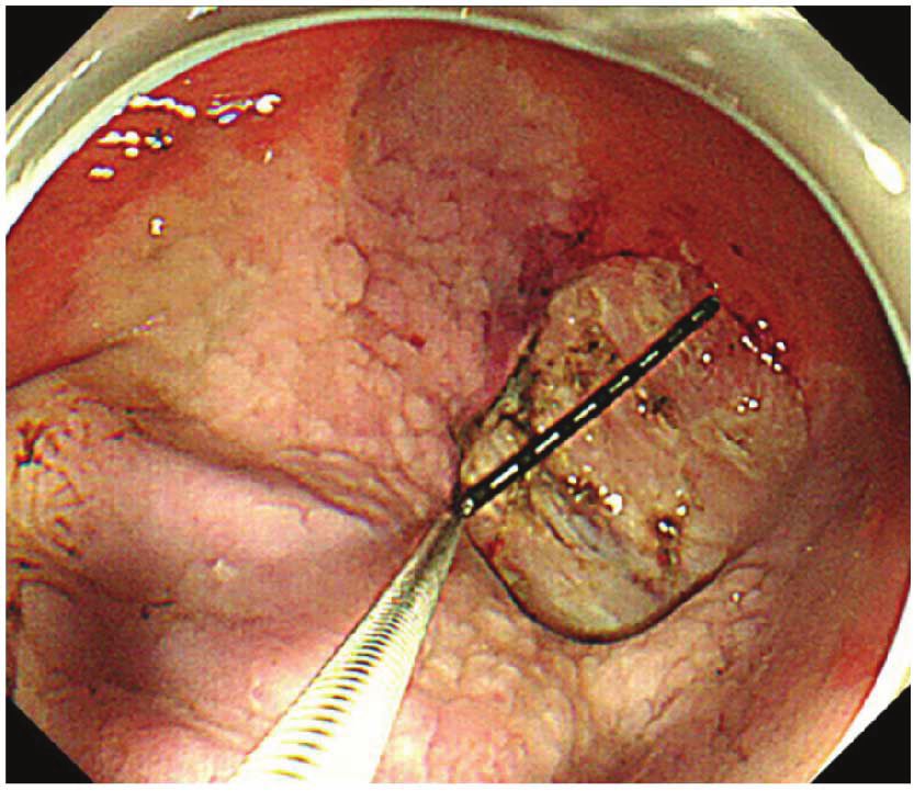
endoscopically (M2-4K; Olympus Corp., Tokyo, Japan)
(Figure 2). Assuming that each ulcer was an ellipse, the ulcer
area (in mm2) was calculated using the following formula
[12]: major axis/2 × minor axis/2 × π.
2.2. ESD. We performed ESD using a single-channel endo-
scope (GIF-Q260J; Olympus Corp.) or via multiangle
two-channel endoscopy (GIF-2TQ260M; Olympus Corp.).
The injection solution contained glycerol, hyaluronic acid
sodium (0.4% [w/v]), and 0.001% (w/v) epinephrine and
was locally injected into the submucosal layer using a dispos-
able 23-gauge needle (Top Corp., Tokyo, Japan). The
ITknife2 (KD-611L; Olympus Corp.) was the primary cutting
device used, but we occasionally employed a DualKnife
(KD-650U; Olympus Corp.). An electrosurgical current
was applied with the aid of an electrosurgical generator
(VIO300D or ICC200; ERBE Elektromedizin GmbH, Figure 2: The major and minor axes of the artificial ulcers were
Tubingen, Germany). Ulcers that developed after ESD were measured endoscopically.
carefully examined endoscopically and any visible vessels
clipped (EZ Clip; Olympus Corp.) and/or heat-coagulated
Table 1: Participating institutions and their contributions to the
using hemostatic forceps (Coagrasper, FD410LR; Olympus two patient groups.
Corp.) in all patients in both groups.
V group E group
2.3. Endpoint. Our primary endpoint was the shrinkage Participating institutions
(exclude) (exclude)
rate of the artificial ulcers 4 and 8 weeks after ESD. The
Yokohama City University
shrinkage rate was calculated using the following formula: 7 (1) 11 (4)
Hospital
ESD specimen area − ulcerated area at 4 or 8 weeks af ter E
Yokohama City University
SD / ESD specimen area × 100 % . The ulcer healing rate 23 (2) 19 (0)
Medical Center
(scarring at stage S1 or S2) was also noted. In addition,
we compared the post-ESD bleeding rates. Post-ESD Total 30 (3) 30 (4)
bleeding was defined as a clinical episode of hematemesis
and/or melena and/or a decline in the hemoglobin level Their distributions between the two participating institutions
to below 2 g/dL. are shown in Table 1. We randomly divided the enrolled
patients into two groups of 30 patients each. We excluded 3
2.4. Statistical Analysis. We used Fisher’s exact test to com- patients of the V group and 4 patients of the E group, finally
pare quantitative variables (sex, Helicobacter pylori infec- analyzing 27 and 26 patients, respectively. The reasons for
tion status, diabetes, and tumor location). The Mann– exclusion in the V group (n = 3) were surgical treatment
Whitney U test was employed to compare age, ulcer area, (n = 1), a suspected allergic reaction (n = 1), and a perfora-
and ulcer cure rate. P values < 0.05 were considered to tion (n = 1); in the E group (n = 4), the reasons for exclu-
reflect statistical significance. Patient ages and ESD speci- sion were surgical treatment (n = 3) and loss to follow-up as
men areas are presented as medians with interquartile defined in the protocol (n = 1). Drug compliance exceeded
ranges (IQRs). The IQR is a measure of statistical disper- 90% in all patients. Several clinical characteristics that delay
sion, being the difference between the 75th and 25th per- ulcer healing have been reported [5, 14–16]. Therefore, we
centiles, or between the upper and lower quartiles. analyzed the data with respect to age, sex, H. pylori infection
All statistical analyses were performed with the aid of status, the presence of diabetes mellitus, and tumor location
EZR software (Saitama Medical Center, Jichi Medical Uni- but found no significant difference between the two groups
versity, Saitama, Japan), which is a graphical user interface in terms of any factor (Table 2). We also analyzed tumor size,
for R (The R Foundation for Statistical Computing, Vienna, specimen size, and procedure duration, which are known as
Austria). More precisely, EZR is a modified version of the R risk factors of post-ESD bleeding [2], but found no significant
commander with additional statistical functions used fre- difference between the two groups in terms of any factor
quently by biostatisticians [13]. (Table 2). H. pylori infection rate in this study was lower than
that of Japanese general population, because H. pylori has
3. Results been eradicated before ESD in some patients.
3.1. Characteristics of the Patients and Lesions. In consider-
ation of the cure healing rate of vonoprazan and esomeprazole 3.2. Ulcer Healing and Shrinkage Rate. The median areas of
in the previous report, it was judged that there was a significant the ESD artificial ulcers were 961.8 (IQR: 707.7–1380.0)
difference in 72 patients, and this study was conducted. Inter- mm2 and 880.8 (IQR: 588.8–1638.8) mm2 in the V group
mediate analysis was carried out when 60 patients finished, and E group, respectively. We performed endoscopic
because there was no difference, this study was terminated. follow-up at 4 and 8 weeks after ESD. The proportions of
4                                                                                                              Gastroenterology Research and Practice

                                                       Table 2: Characteristics of all parameters analyzed.

Characteristic                                                                             Total (n = 53)     V group (n = 27) E group (n = 26) P value
Age (years) median (IQR)                                                                    70.2 (66.76)         70 (65.3–75)            70 (66–75.3)     0.957∗∗
Sex (male), n (%)                                                                             55 (87.3)            23 (85.2)              22 (84.6)        0.728∗
Helicobacter pylori infection (positive), n (%)                                               15 (28.3)             7 (26.0)               8 (30.8)        0.766∗
Diabetes, n (%)                                                                               8 (12.7)              3 (11.1)               5 (19.2)        0.467∗
Location (upper, middle, lower)                                                               28, 20, 7            12, 10, 5              14, 10, 2        0.596∗
Location (greater curvature, lesser curvature, anterior wall, posterior wall)               13, 7, 11, 10          7, 13, 3, 4            6, 6, 8, 6       0.157∗
Tumor size (mm), range, n (>20 mm)                                                        14.1 (4–38), 12       14.9 (4–38), 7        13.2 (4–35), 5       0.745∗
Specimen size (mm), range, n (>30 mm)                                                    40.2 (26–80), 40 40.6 (30–54), 22 39.8 (26–80), 18 0.352∗
Procedure duration (min), range, n (>60 min)                                             43.3 (8–103), 14       45.5 (9–103), 8      41.1 (8–100), 6       0.757∗
IQR: interquartile range (a measure of variability, based on dividing a dataset into quartiles). A P value < 0.05 was considered to reflect significance. ∗ Significant
by Fisher’s exact test. ∗∗ Significant by the Mann–Whitney U test.

                                          Table 3: Ulcer evaluations after ESD, and the absence of post-ESD bleeding.

                                                                 Total (n = 53)             V group (n = 27)                E group (n = 26)              P value
Ulcer stage after 4 weeks (scar, n)                                     5                            2                               3                    0.669∗∗
Ulcer stage after 8 weeks (scar, n)                                    46                           24                              22                     0.42∗∗
Shrinkage rate after 4 weeks (%), range                         95.4 (72.0–100)             96.8 (72.0–100)                  97.5 (75.8–100)              0.656∗∗
Shrinkage rate after 8 weeks (%)                                       100                         100                              100                   0.257∗∗
ESD specimen area (mm2) median (IQR)                         942.3 (588.8–1452.3)        961.8 (707.8–1380.0)             880.8 (588.8–1638.8)            0.612∗∗
Post-ESD bleeding                                                      0                             0                               0
IQR: interquartile range (a measure of variability, based on dividing a dataset into quartiles). A P value < 0.05 was considered significant. ∗∗ Significant by the
Mann–Whitney U test.

                                                NS
                                                                                    the V and E groups, respectively (Table 3). The 4-week
                                                                                    shrinkage rates of artificial ulcers were 96.8% (range: 72.0–
                         100
                                                                                    100%) in the V group and 97.5% (range: 75.8–100%) in the
                                                                                    E group, thus not significantly different between the V and
                          95                                                        E groups (P = 0 656) (Figure 3). The 8-week shrinkage rates
                                                                                    of artificial ulcers were 100% in both groups, thus lacking
                                                                                    any significant difference (P = 0 257) (Table 3). In terms of
                          90                                                        delayed bleeding, no obvious bleeding was observed in either
    Shrinkage rate (%)

                                                                                    group. As the healing of artificial ulcers after ESD did not dif-
                                                                                    fer when vonoprazan or esomeprazole was prescribed, both
                          85
                                                                                    drugs effectively aided artificial ulcer healing after ESD.

                          80                                                        4. Discussion
                                                                                    We sought to clarify the effects of vonoprazan and esomepra-
                          75                                                        zole on the healing of post-ESD artificial ulcers by designing a
                                                                                    prospective, multicenter, two-arm RCT. We found no signif-
                                                                                    icant difference between vonoprazan and esomeprazole in
                               V group                       E group                terms of artificial ulcer healing after ESD. Statistically, no fac-
                                         4 weeks after ESD                          tor associated with delayed ulcer healing was evident. As the
Figure 3: The shrinkage rates of ulcers of the V group and E group 4
                                                                                    study was performed at two centers, nine physicians (ranging
weeks after ESD.                                                                    from beginners to experts) performed ESD, but their various
                                                                                    skill levels did not affect the results.
                                                                                         Of the four previous reports comparing artificial ulcer
subjects with ulcer scarring of stage S1 or S2 after ESD were                       healing using P-CAB and PPIs, three found that P-CAB
8.0% (2/27) and 11.5% (3/26) at 4 weeks (P = 0 669) and                             was superior to PPIs in terms of post-ESD ulcer shrinkage
88.9% (24/27) and 84.6% (22/26) at 8 weeks (P = 0 420) in                           rates [10, 11] or late bleeding rates [8]. However, no obvious
Gastroenterology Research and Practice                                                                                               5

differences in ulcer shrinkage rate were found in either our       images can be captured from flat surfaces only, the accuracies
present study or another report [9]. Differences among the         of our area measurements may have been compromised. The
results of the five studies (including this study) are probably    reason that we used our previously reported method [12] is
due to differences in protocols, patient selection, and other      that the region just after the ESD lies closest to the ESD
variables. We excluded patients who were taking NSAIDs,           sample area (data not shown).
steroids, anticoagulants, and/or antithrombotic agents to             In conclusion, we found no difference in terms of
minimize situations where other medications might influ-           artificial ulcer healing after ESD using rebamipide in
ence the effects of P-CAB or PPIs. Therefore, it may be that       combination with either vonoprazan or esomeprazole in
we found no significant difference because we selectively           low-risk patients with late bleeding. Vonoprazan strongly
excluded patients with comorbidities (which the other stud-       suppresses gastric acid secretion, but there was no great
ies did not). In particular, excluding patients on antithrom-     need for the use of this drug during the healing of artifi-
botic therapy may explain the absence of bleeding in our          cial ulcers after ESD; esomeprazole exerted a sufficient
study [17]. Although we cannot mention about post-ESD             therapeutic effect. No post-ESD bleeding was observed.
bleeding assertively in our study, we analyzed several factors    We consider that both vonoprazan and esomeprazole
(tumor size, specimen size, and procedure duration), which        effectively aided artificial ulcer healing and esomeprazole
are known as risk factors of post-ESD bleeding [2]. As a          was superior to vonoprazan in view of cost benefit in
result, there was no significant difference in any of them          low-risk patients.
(Table 2). Another difference is that our study did not feature
a monotherapy protocol; we added oral rebamipide because a        Conflicts of Interest
previous meta-analysis showed that treatment with PPIs plus
rebamipide was superior to PPIs monotherapy in terms of           No author has any conflict of interest to declare concerning
the healing of ESD-induced ulcers over 4 weeks, particularly      the content of this manuscript.
large ulcers [18]. As patients should not be disadvantaged
during clinical trials, we considered that monotherapy would      Acknowledgments
not be ethical. Also, the median ESD specimen area in the
present study was only 942 mm2, thus small compared to            The authors would like to express their gratitude to the staff
those of previous reports (1256 mm2 [10] and 1114.7 mm2           of the Endoscopy Department at Yokohama City University
[11]). The low rate of H. pylori infection may also be a factor   Hospital and the staff of the Endoscopy Department at Yoko-
that did not differ significantly in ulcer shrinkage rate.          hama City University Medical Center for their assistance.
    Vonoprazan action is barely affected by the CYP2C19
genotype, and the drug affords more potent and longer anti-        References
secretory effects on H+/K+-ATPase than do conventional
                                                                   [1] T. Akasaka, T. Nishida, S. Tsutsui et al., “Short-term outcomes
PPIs [19, 20]. Therefore, vonoprazan has been widely
                                                                       of endoscopic submucosal dissection (ESD) for early gastric
prescribed in Japan for the treatment of gastric ulcers,               neoplasm: multicenter survey by Osaka University ESD study
for H. pylori eradication, and for the treatment of gastro-            group,” Digestive Endoscopy, vol. 23, no. 1, pp. 73–77, 2011.
esophageal reflux disease in the time since its release in          [2] T. Toyokawa, T. Inaba, S. Omote et al., “Risk factors for perfo-
2015. Although vonoprazan inhibits acid production irre-               ration and delayed bleeding associated with endoscopic sub-
spective of CYP2C19 status more potently than does                     mucosal dissection for early gastric neoplasms: analysis of
esomeprazole [20], CYP2C19 status is associated with                   1123 lesions,” Journal of Gastroenterology and Hepatology,
approximately a threefold lower intrinsic clearance of the             vol. 27, no. 5, pp. 907–912, 2012.
S-isomer of esomeprazole compared with the R-isomer                [3] T. Kato, H. Araki, F. Onogi et al., “Clinical trial: rebamipide
and omeprazole. Esomeprazole is less affected by metabo-                promotes gastric ulcer healing by proton pump inhibitor after
lism than are the other PPIs [21]. In fact, as we found no             endoscopic submucosal dissection—a randomized controlled
significant difference between the drugs in the present                  study,” Journal of Gastroenterology, vol. 45, no. 3, pp. 285–
study, it may be unnecessary to strongly inhibit gastric               290, 2010.
acid secretion during artificial ulcer healing.                     [4] S. Fujiwara, Y. Morita, T. Toyonaga et al., “A randomized con-
    The limitations of our study include the mode of admin-            trolled trial of rebamipide plus rabeprazole for the healing of
istration of drugs and the method that we used to measure              artificial ulcers after endoscopic submucosal dissection,” Jour-
ulcer area. We did not simply compare the effects of vono-              nal of Gastroenterology, vol. 46, no. 5, pp. 595–602, 2011.
prazan and esomeprazole because we gave rebamipide and             [5] M. Kobayashi, M. Takeuchi, S. Hashimoto et al., “Contributing
                                                                       factors to gastric ulcer healing after endoscopic submucosal
omeprazole intravenously. However, we considered that we
                                                                       dissection including the promoting effect of rebamipide,”
should not disadvantage the patients. Also, monotherapy is             Digestive Diseases and Sciences, vol. 57, no. 1, pp. 119–126,
not prescribed in actual clinical practice. As various treat-          2012.
ments are available, we concluded that our protocol was the        [6] Y. Hori, A. Imanishi, J. Matsukawa et al., “1-[5-(2-Fluor-
most appropriate. We earlier thoroughly evaluated the                  ophenyl)-1-(pyridin-3-ylsulfonyl)-1H-pyrrol-3-yl]-N-methyl-
method that we used to measure ulcers. The stomach can                 methanamine monofumarate (TAK-438), a novel and potent
be easily expanded and contracted by supplying, and then               potassium-competitive acid blocker for the treatment of acid-
removing, gas via an endoscope. However, the stomach is                related diseases,” The Journal of Pharmacology and Experi-
of course a three-dimensional structure. As endoscopic                 mental Therapeutics, vol. 335, no. 1, pp. 231–238, 2010.
6                                                                        Gastroenterology Research and Practice

 [7] K. Otake, Y. Sakurai, H. Nishida et al., “Characteristics of the
     novel potassium-competitive acid blocker vonoprazan fuma-
     rate (TAK-438),” Advances in Therapy, vol. 33, no. 7,
     pp. 1140–1157, 2016.
 [8] T. Kagawa, M. Iwamuro, S. Ishikawa et al., “Vonoprazan
     prevents bleeding from endoscopic submucosal dissection-
     induced gastric ulcers,” Alimentary Pharmacology and Ther-
     apeutics, vol. 44, no. 6, pp. 583–591, 2016.
 [9] K. Takahashi, Y. Sato, J. Kohisa et al., “Vonoprazan 20 mg vs
     lansoprazole 30 mg for endoscopic submucosal dissection-
     induced gastric ulcers,” World Journal of Gastrointestinal
     Endoscopy, vol. 8, no. 19, pp. 716–722, 2016.
[10] D. Maruoka, M. Arai, S. Kasamatsu et al., “Vonoprazan is
     superior to proton pump inhibitors in healing artificial ulcers
     of the stomach post-endoscopic submucosal dissection: a pro-
     pensity score-matching analysis,” Digestive Endoscopy, vol. 29,
     no. 1, pp. 57–64, 2017.
[11] I. Tsuchiya, Y. Kato, E. Tanida et al., “Effect of vonoprazan on
     the treatment of artificial gastric ulcers after endoscopic sub-
     mucosal dissection: prospective randomized controlled trial,”
     Digestive Endoscopy, vol. 29, no. 5, pp. 576–583, 2017.
[12] S.-Y. Lee, J. J. Kim, J. H. Lee et al., “Healing rate of EMR-
     induced ulcer in relation to the duration of treatment with
     omeprazole,” Gastrointestinal Endoscopy, vol. 60, no. 2,
     pp. 213–217, 2004.
[13] Y. Kanda, “Investigation of the freely available easy-to-use
     software ‘EZR’ for medical statistics,” Bone Marrow Trans-
     plantation, vol. 48, no. 3, pp. 452–458, 2013.
[14] K. Niimi, M. Fujishiro, O. Goto et al., “Prospective single-arm
     trial of two-week rabeprazole treatment for ulcer healing after
     gastric endoscopic submucosal dissection,” Digestive Endos-
     copy, vol. 24, no. 2, pp. 110–116, 2012.
[15] J. H. Cheon, J. H. Kim, S. K. Lee, T. I. Kim, W. H. Kim, and
     Y. C. Lee, “Helicobacter pylori eradication therapy may facili-
     tate gastric ulcer healing after endoscopic mucosal resection:
     a prospective randomized study,” Helicobacter, vol. 13, no. 6,
     pp. 564–571, 2008.
[16] H. Isomoto, S. Shikuwa, N. Yamaguchi et al., “Endoscopic
     submucosal dissection for early gastric cancer: a large-scale
     feasibility study,” Gut, vol. 58, no. 3, pp. 331–336, 2009.
[17] J. Dong, K. Wei, J. Deng et al., “Effects of antithrombotic
     therapy on bleeding after endoscopic submucosal dissection,”
     Gastrointestinal Endoscopy, vol. 86, no. 5, pp. 807–816, 2017.
[18] J. Wang, X. Guo, C. Ye et al., “Efficacy and safety of proton
     pump inhibitors (PPIs) plus rebamipide for endoscopic sub-
     mucosal dissection-induced ulcers: a meta-analysis,” Internal
     Medicine, vol. 53, no. 12, pp. 1243–1248, 2014.
[19] H. Yamasaki, N. Kawaguchi, M. Nonaka et al., “In vitro metab-
     olism of TAK-438, vonoprazan fumarate, a novel potassium-
     competitive acid blocker,” Xenobiotica, vol. 47, no. 12,
     pp. 1027–1034, 2016.
[20] T. Kagami, S. Sahara, H. Ichikawa et al., “Potent acid inhibition
     by vonoprazan in comparison with esomeprazole, with refer-
     ence to CYP2C19 genotype,” Alimentary Pharmacology and
     Therapeutics, vol. 43, no. 10, pp. 1048–1059, 2016.
[21] P. Lindberg, D. Keeling, J. Fryklund, T. Andersson,
     P. Lundborg, and E. Carlsson, “Esomeprazole — enhanced
     bio-availability, specificity for the proton pump and inhibition
     of acid secretion,” Alimentary Pharmacology and Therapeutics,
     vol. 17, no. 4, pp. 481–488, 2003.
MEDIATORS         of

                                                                                   INFLAMMATION

The Scientific                                 Gastroenterology                                                              Journal of
World Journal
Hindawi Publishing Corporation
                                               Research and Practice
                                               Hindawi
                                                                                   Hindawi
                                                                                                                             Diabetes Research
                                                                                                                             Hindawi
                                                                                                                                                              Disease Markers
                                                                                                                                                              Hindawi
                                                                                   www.hindawi.com        Volume 2018
http://www.hindawi.com
www.hindawi.com                  Volume 2018
                                        2013   www.hindawi.com   Volume 2018                                                 www.hindawi.com    Volume 2018   www.hindawi.com            Volume 2018

Journal of                                                                                                                                                    International Journal of
Immunology Research
Hindawi
                                                                                                                                                              Endocrinology
                                                                                                                                                              Hindawi
www.hindawi.com                  Volume 2018                                                                                                                  www.hindawi.com            Volume 2018

                                                                 Submit your manuscripts at
                                                                    www.hindawi.com

                                                                                                                                                              BioMed
PPAR Research
Hindawi
                                                                                                                                                              Research International
                                                                                                                                                              Hindawi
www.hindawi.com                  Volume 2018                                                                                                                  www.hindawi.com            Volume 2018

                                                                                                                             Journal of
                                                                                                                             Obesity

                                                                               Evidence-Based
Journal of                                     Stem Cells                      Complementary and                                                              Journal of
Ophthalmology
Hindawi
                                               International
                                               Hindawi
                                                                               Alternative Medicine
                                                                               Hindawi                                       Hindawi
                                                                                                                                                              Oncology
                                                                                                                                                              Hindawi
www.hindawi.com                  Volume 2018   www.hindawi.com   Volume 2018   www.hindawi.com                 Volume 2018   www.hindawi.com    Volume 2018   www.hindawi.com            Volume 2013

                                                                               Parkinson’s
                                                                               Disease

Computational and
Mathematical Methods
in Medicine
                                               Behavioural
                                               Neurology
                                                                                                                             AIDS
                                                                                                                             Research and Treatment
                                                                                                                                                              Oxidative Medicine and
                                                                                                                                                              Cellular Longevity
Hindawi                                        Hindawi                         Hindawi                                       Hindawi                          Hindawi
www.hindawi.com                  Volume 2018   www.hindawi.com   Volume 2018   www.hindawi.com                 Volume 2018   www.hindawi.com    Volume 2018   www.hindawi.com            Volume 2018

You can also read