A step-by-step guide to placement of the LAP-BAND adjustable gastric banding system
←
→
Page content transcription
If your browser does not render page correctly, please read the page content below
The American Journal of Surgery 184 (2002) 26S–30S

       A step-by-step guide to placement of the LAP-BAND adjustable
                           gastric banding system
                                     George A. Fielding, M.D.a*, Jeff W. Allen, M.D.b
            a
              Wesley Hospital and Royal Brisbane Hospital, Brisbane, Australia, Suite 93, 30 Chasely Street, Auchenflower 4066, QLD, Australia
        b
            Department of Surgery, University of Louisville School of Medicine, Center for Advanced Surgical Technologies, and Norton Hospital,
                                                                 Louisville, Kentucky, USA

Abstract
    The early promise of laparoscopic adjustable gastric banding was tempered by reports of high rates of gastric herniation or prolapse.
These complications are a function of the operative technique used early on. At the time, in the early 1990s, the LAP-BAND device
(INAMED Health, Santa Barbara, CA) was placed lower on the stomach, near the first short gastric vessel. The required perigastric
dissection was difficult and variable in its extent, depending on the width of the stomach and where the surgeon began the dissection. To
combat these problems, a new surgical method for placement of the band has evolved. Called the pars flaccida technique, it emphasizes
minimal dissection and placement of the LAP-BAND out of the lesser sac. This leads to a higher position of the band, away from the body
of the stomach. The technique serves to make band placement simple, safe, reproducible, and easily teachable, as well as to decrease the
rate of gastric herniation or prolapse. Keeping the band out of the lesser sac, away from the peristalsing stomach, minimizing dissection of
the attachments to the stomach, paying strict attention to gastric-to-gastric suturing, and leaving all fluid out of the band until at least 6 weeks
after surgery appear to be the most important factors in reducing the incidence of this complication. © 2002 Excerpta Medica Inc. All rights
reserved.

The technique for placement of the LAP-BAND (INAMED                             ened at completion of the procedure by injecting 2 mL of
Health, Santa Barbara, CA) has evolved considerably since                       sterile saline. This constellation of factors— big pouch, en-
its debut in September 1993 [1,2]. The early promise of a                       try into the lesser sac, variable dissection length, and a tight
minimally invasive procedure that actively restricts intake                     band at onset—led to an unacceptably high rate of gastric
and controls hunger was moderated by reports of high rates                      herniation of 10% to 15% [3–10].
of gastric herniation† through the band as well as the occa-                       To combat these problems, a new surgical method for
sional erosion of the band through the stomach [3–10].                          placement of the band has evolved. Called the pars flaccida
Furthermore, there has been a dichotomy of results for                          technique, it emphasizes minimal dissection and placement
weight loss between Europe, Australia, and Mexico on the                        of the LAP-BAND out of the lesser sac. This leads to a
one hand [9,11–17] and the United States on the other                           higher position of the band, away from the body of the
[18 –20].                                                                       stomach. The operation has been modified and developed in
    We feel that the complication of gastric herniation                         an attempt to make band placement simple, safe, reproduc-
through the band was a function of the operative technique                      ible, and easily teachable, as well as to decrease the rate of
used early in the experience. At the time, in the early 1990s,                  gastric herniation.
the device was placed lower on the stomach, near the first
short gastric vessel. The required perigastric dissection was
difficult and variable in its extent. It was necessary to use a                 Operative technique
pressure gauge, the gastrotenometer, to ensure the band was
not placed too tightly. Usually the band was partially tight-                      A multidisciplinary team sees patients preoperatively,
                                                                                over the 6 to 8 weeks before surgery. Patients are admitted
                                                                                on the day of surgery. In the operating room, the patient is
   * Corresponding author: Tel.: 07 3371 9521; fax: 07 3870 5466.
   E-mail address: Gfielding@wesley.com.au                                      placed supine in reverse Trendelenburg position, with the
   †
     The phenomenon of gastric herniation or prolapse is known as “slip”        legs together. No stirrups are used. The surgeon stands on
and represents a complication that usually requires surgical repair.            the patient’s right side, with the monitors at the head of the

0002-9610/02/$ – see front matter © 2002 Excerpta Medica Inc. All rights reserved.
doi:10.1016/S0002-9610(02)01176-5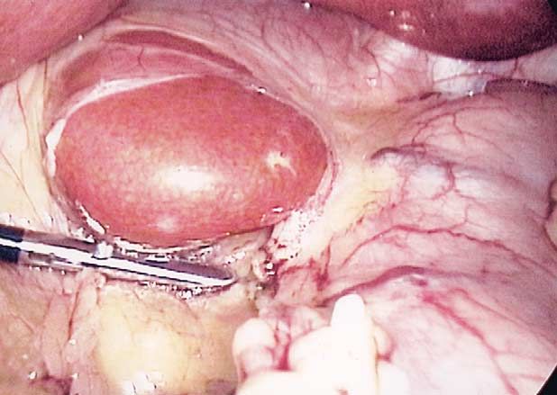
G.A. Fielding, J.W. Allen / The American Journal of Surgery 184 (2002) 26S–30S 27S
bed on either side. Some surgeons prefer a lithotomy posi- may be encountered in this location as well and should be
tion; this is also acceptable. preserved, if possible.
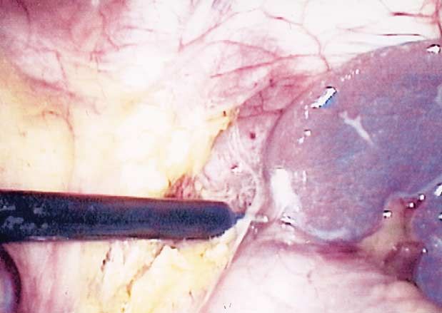
Visual access is gained with a 10-mm Optiview trocar After incising the pars flaccida, the right crus is seen
(Ethicon Endo-Surgery, Cincinnati, OH), using a 0° lapa- inferomedial to the caudate lobe of the liver, curving to the
roscope. The Optiview is placed laterally just below the left right to disappear in the retroperitoneal fat (Fig. 5). We
costal margin. The abdomen is insufflated to 15 mm Hg, and believe it is important to also identify the vena cava, which
additional ports are placed under direct vision, including a lies just to the right and parallel to the right crus. With large
5-mm right subcostal port, a 15-mm right-upper-quadrant amounts of intra-abdominal adipose tissue, the crus and
paramedian port (an 18-mm port may be used), and a 5-mm cava can be mistaken for one another.
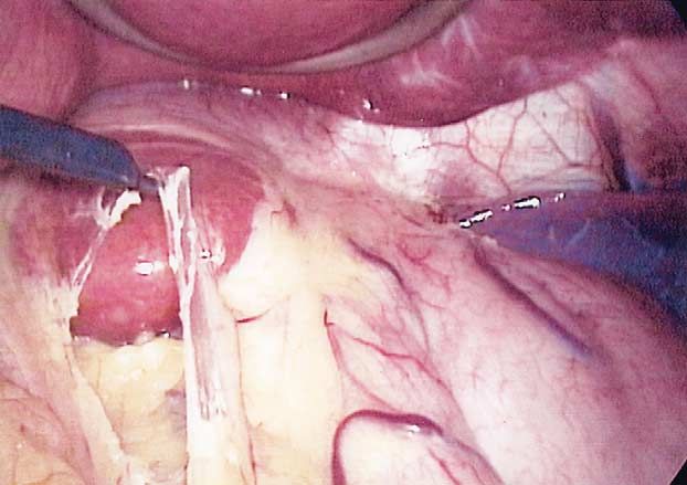
left-upper-quadrant midaxillary line. Although patient posi- After identification of the right crus, the peritoneum just
tioning, port location, and surgeon location will vary by medial to the crus is incised (Fig. 6). (This is usually at the
surgeon preference, we recommend port placement similar level of the constant vein at the start of the lesser curve of
to that used by the surgeon when performing laparoscopic the stomach.) This is the second key step. The inferolateral
Nissen fundoplication. placement of the 5-mm port gives a flat approach of the
A Nathanson liver retractor (Cook Medical, Queensland, instrument to this dissected area, free of torque or any force.
Australia) is inserted through a 5-mm skin incision in the The grasper is very gently inserted through the scored peri-
subxiphoid location and curved up to retract the left hepatic toneum into the space medial to the crus, behind the esoph-
lobe. This retractor has variably sized arms to cope with agus (Fig. 7). Using virtually no force, it is passed to the left
even huge hepatic lobes and is attached to a fixed arm on the and emerges at the angle of His that was previously dis-
table. It is inserted directly into the abdomen rather than sected. The course of the grasper is usually a short distance,
through a port and generally needs no further attention once only 3 to 4 cm (Fig. 8).
in place (Fig. 1). The end-tag of the band is brought up to meet the now
The LAP-BAND is primed with sterile saline and in- retrogastric grasper and is pulled through, encircling the
stomach (Fig. 9). Given the minimal dissection performed
serted into the abdomen via the 15-mm port. It is pushed
and the shape of the LAP-BAND, it is often necessary to
inferiorly on the left side, where it remains ready for later
tease some of the peritoneal attachments medial to the crus
retrieval.
to allow the widened portion of the band to be pulled
A long (45-cm) atraumatic grasper is placed in the
completely around the stomach. The band is locked in place,
groove between the greater curvature of the stomach and the
and a 5-mm instrument is passed between the band and the
spleen. Using this instrument, the omentum covering the
stomach. It should pass freely. If it does not, the band is
angle of His is swept inferiorly. This maneuver places the
likely too constricting, and any fatty tissue between the band
fundus of the stomach on a stretch. The camera position in
and the stomach should be removed.
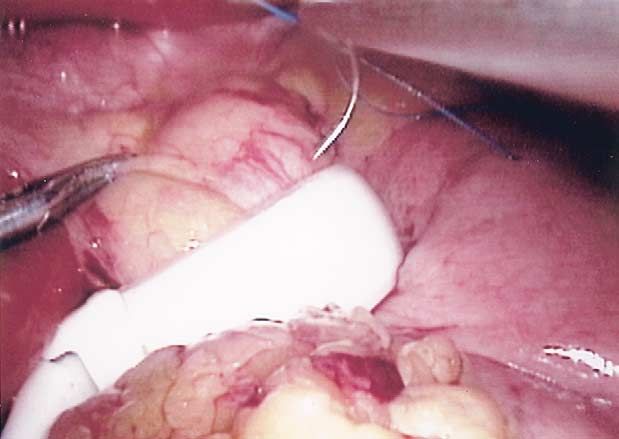
the left upper quadrant, looking cephalad, provides an un- The camera is placed in the 15-mm port. Suturing is done
paralleled view of the angle of His. The assistant then holds through the ports that are now on either side of the laparo-
the grasper retracting the omentum. When correctly posi- scope. A permanent suture, such as 2/0 Novafil (Tyco
tioned, the handle of the grasper lies cephalocaudal, tilted Healthcare, Mansfield, MA) or 2/0 Ethibond威 (Ethicon,
forward in a straight line (Fig. 2). The key is to sweep the Somerville, NJ), is employed to secure the band in place.
omentum, not to pull it. This sweeping retraction is the first Gastric-to-gastric sutures are used (Fig. 10). These should
essential step in the procedure. be placed so that the stomach above and below the band are
The surgeon places another long, atraumatic grasper approximated, but without undue tension. The suturing
through the right lateral port and a diathermy hook through causes the band to be covered, but the true function is to
the 15-mm port. The grasper pulls the fundus inferiorly and prevent herniation of the stomach upward through the band.
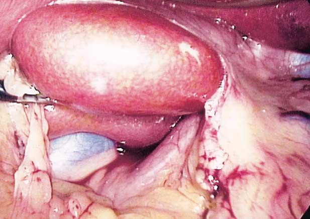
to the right to further expose the angle of His. A large fat Even though the device is placed high up on the stomach,
pad precluding safe placement of the band should be ex- the mobile fundus can still migrate through the band without
cised. The peritoneum lateral to the gastroesophageal angle this proper fixation. Suturing should be carried as far pos-
is incised and swept posteriorly, freeing the fundus of the terolateral as possible because it has been our experience
stomach off the diaphragm (Fig. 3). This is well above that this is the most likely location of a slipped band. Either
(cephalad) the first short gastric vessel in most cases. a running or interrupted suture is used, based on surgeon
The 30° laparoscope is then rotated to the right to view preference. With either technique, 3 to 5 bites will usually
the lesser omentum, also known as the pars flaccida. This is complete the job. It is important not to evert the stomach
usually quite easily seen with the caudate lobe shining over the buckle of the LAP-BAND, for it is here that
through. The surgeon retracts the stomach to the left and erosions are more likely to occur (Fig. 11).
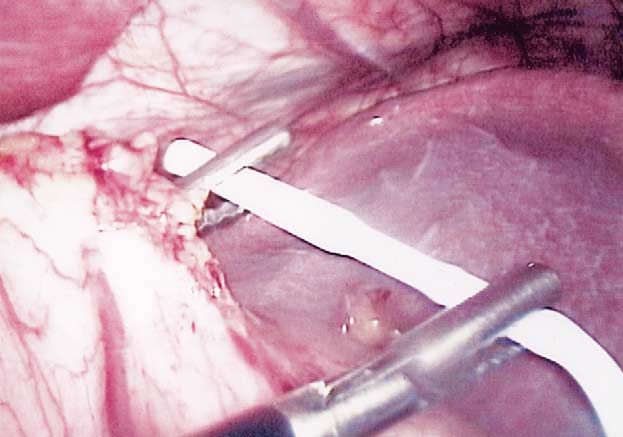
incises the nearly transparent pars flaccida (Fig. 4). The After suturing is complete, the liver retractor is removed,
hepatic branch of the vagus nerve is present at the superior and the camera is again placed in the Optiview. The tubing
aspect and should be spared except in patients who have had is pulled out through the 15-mm port, and the laparoscopic
antecedent cholecystectomy. An aberrant left hepatic artery part of the procedure is complete.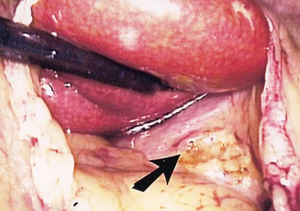
28S                                    G.A. Fielding, J.W. Allen / The American Journal of Surgery 184 (2002) 26S–30S

Fig. 1. A Nathanson liver retractor is inserted through a 5-mm skin incision       Fig. 4. The surgeon retracts the stomach to the left and incises the nearly
in the subxiphoid location and curved up to retract the left hepatic lobe.         transparent pars flaccida.

Fig. 2. A long (45-cm) atraumatic grasper is placed in the groove between          Fig. 5. After incising the pars flaccida, the right crus is seen inferomedial
the greater curvature of the stomach and the spleen. Using this instrument,        to the caudate lobe of the liver, curving to the right to disappear in the
the omentum covering the angle of His is swept inferiorly.                         retroperitoneal fat.

Fig. 3. The surgeon places another long, atraumatic grasper through the
right lateral port and a diathermy hook through the 15-mm port. The grasper        Fig. 6. After identification of the right crus, the peritoneum just medial to
pulls the fundus inferiorly and to the right to further expose the angle of His.   the crus is incised.
The peritoneum lateral to the gastroesophageal angle is incised and swept
posteriorly, freeing the fundus of the stomach off the diaphragm.
G.A. Fielding, J.W. Allen / The American Journal of Surgery 184 (2002) 26S–30S 29S
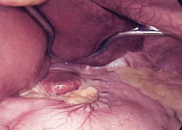
Fig. 7. The grasper is gently inserted through the scored peritoneum into Fig. 9. The end-tag of the band is brought up to meet the now retrogastric
the space medial to crus, behind the esophagus. grasper and is pulled through, encircling the stomach.
Fig. 8. Using virtually no force, the grasper is passed to the left and Fig. 10. Gastric-to-gastric sutures are used. These should be placed so that
emerges at the angle of His that was previously dissected. The course of the the stomach above and below the band are approximated, but without
grasper is usually a short distance, only 3 to 4 cm. undue tension.
Fig. 11. Properly positioned gastric band.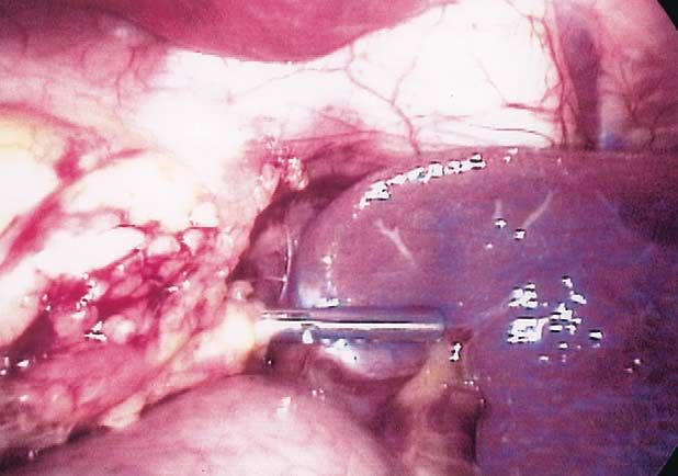
30S                           G.A. Fielding, J.W. Allen / The American Journal of Surgery 184 (2002) 26S–30S

    The 15-mm port incision is extended lateral and deep                band until at least 6 weeks after placement appear to be the
down to the rectus sheath. Narrow Deaver retractors facil-              most important factors in reducing the prolapse rate.
itate this, as the sheath is often farther down than antici-
pated. The anterior fascia is incised approximately 2 cm
lateral to the fascial defect. The tubing is connected to the           References
access port, and, in turn, the access port is affixed to the
fascia. This is accomplished by placing an Ethibond suture               [1] Belachew M, Legrand MJ, Vincent V. History of LAP-BAND: from
in each corner of the incised fascia and placing these into                  dream to reality. Obes Surg 2001;11:297–302.
the 4 holes on the access port. The access port is then                  [2] Belachew M, Legrand MJ, Defechereux TH, et al. Laparoscopic
                                                                             adjustable silicone gastric banding in the treatment of morbid obesity:
parachuted down onto the rectus sheath. The sutures are                      a preliminary report. Surg Endosc 1994;8:1354 –1356.
tied. The tubing is simply slid back into the abdomen, and               [3] Wiesner W, Schlumpf R, Schob O, et al. Gastric pouch dilatation:
the wounds are closed.                                                       complications after laparoscopic implantation of a silicone gastric
    The band is left empty. An upper gastrointestinal radio-                 band in pathologic obesity. Rofo Fortschr Geb Rontgenstr Neuen
graph using water-soluble contrast is performed the next                     Bildgeb Verfahr 1998;169:479 – 483.
                                                                         [4] Carbajo Caballero MA, Martin del Olmo JC, Blanco Alvarez JI, et al.
morning to exclude gastric perforation, malposition, and                     Intragastric migration of laparoscopic adjustable gastric band (LAP-
obstruction. Once this is reviewed, the patient can be dis-                  BAND) for morbid obesity. J Laparoendosc Adv Surg Tech A 1998;8:
charged. A diet plan of liquids for 2 weeks, slushy food for                 241–244.
2 weeks, and normal food for 2 weeks is initiated.                       [5] Silechia G, Restuccia A, Elmore U, et al. Laparoscopic adjustable
                                                                             silicone gastric banding: prospective evaluation of intragastric migra-
                                                                             tion of the LAP-BAND. Surg Laparosc Endosc Percutan Tech 2001;
                                                                             11:229 –234.
Discussion                                                               [6] Capizzi FD, Boschi S, Brulatti M, et al. Laparoscopic adjustable esopha-
                                                                             gogastric banding: preliminary results. Obes Surg 2002;12:391–394.
   Working at the Wesley and Royal Brisbane Hospitals (in                [7] Niville E, Dams A, Vlasselaers J. LAP-BAND erosion: incidence and
Australia), the senior author (GAF) has placed 1,041 LAP-                    treatment. Obes Surg 2001;11:744 –747.
BAND devices. Of these, 79 (7.6%) have been associated                   [8] Abu-Abeid S, Szold A. Laparoscopic management of LAP-BAND
                                                                             erosion. Obes Surg 2001;11:87– 89.
with gastric prolapse. With the exception of 2 patients who              [9] Weiner R, Wagner D, Bockhorn H. Laparoscopic gastric banding for
presented with severe pain, these have had an indolent                       morbid obesity. J Laparoendosc Adv Surg Tech A 1999;9:23–30.
presentation typified by reflux, increasing food intolerance,           [10] Favretti F, Cadiere GB, Segato G, et al. Laparoscopic adjustable
and dysphagia to solids. Management includes band defla-                     silicone gastric banding (LAP-BAND): how to avoid complications.
                                                                             Obes Surg 1997;7:352–358.
tion, barium swallow to assess pouch size and position,
                                                                        [11] Angrisani L, Alkilani M, Basso N, et al. Laparoscopic Italian expe-
admission for intravenous rehydration, and surgical reposi-                  rience with the LAP-BAND. Obes Surg 2001;11:307–310.
tion or replacement.                                                    [12] Dargent J. Laparoscopic adjustable gastric banding: lessons from the
   Between February 1996 and December 1998, 480 bands                        first 500 patients in a single institution. Obes Surg 1999;9:446 – 452.
were inserted using the older, perigastric method. To date,             [13] O’Brien PE, Brown WA, Smith A, et al. Prospective study of a
                                                                             laparoscopically placed, adjustable gastric band in the treatment of
there have been 64 (15%) cases of prolapse, occurring at an
                                                                             morbid obesity. Br J Surg 1999;86:113–118.
average of 11 months (range: 4 to 52 months) after surgery.             [14] Fielding GA, Rhodes M, Nathanson LK. Laparoscopic gastric band-
   The change to the pars flaccida technique occurred in                     ing for morbid obesity: surgical outcome in 335 cases. Surg Endosc
December 1998. Using this technique, 561 bands have been                     1999;13:550 –554.
inserted in Australia and 107 in the United States at Norton            [15] de Wit LT, Mathus-Vliegen L, Hey C, et al. Open versus laparoscopic
                                                                             adjustable silicone gastric banding: a prospective randomized trial for
Hospital in Louisville, Kentucky (JWA). In this group, there
                                                                             treatment of morbid obesity. Ann Surg 1999;230:800 – 805.
have been 12 (1.8%) cases of prolapse.                                  [16] Belachew M, Belva PH, Desaive C. Long-term results of laparoscopic
   The LAP-BAND offers all the well-known advantages of                      adjustable gastric banding for the treatment of morbid obesity. Obes
laparoscopy: same-day admission, early discharge, reduced                    Surg 2002;12:564 –568.
pain, and early return to normal activities. The band is easily         [17] Vertruyen M. Experience with LAP-BAND system up to 7 years.
                                                                             Obes Surg 2002;12:569 –572.
adjusted and, if necessary, easily removed.
                                                                        [18] Rubenstein RB. Laparoscopic adjustable gastric banding at a US
   The high rate of gastric herniation seen early in the                     center with up to 3-year follow-up. Obes Surg 2002;12:380 –384.
history of the LAP-BAND is not without supporting evi-                  [19] Greenstein RJ, Martin Ll, MacDonald KJ, et al. The USA LAP-
dence, especially from US experience [20]. The evolving                      BAND Study Group. The LAP-BAND system as surgical therapy for
technical modifications described here have markedly de-                     morbid obesity: intermediate results of the USA, multicenter, pro-
                                                                             spective study [abstract]. Surg Endosc 1999;13(suppl):S1.
creased the prolapse rate. Keeping the band out of the lesser
                                                                        [20] LAP-BAND Adjustable Gastric Banding (LAGB) System—
sac, away from peristalsing stomach; minimizing dissection                   P000008. Available at: http://www. fda.gov/cdrh/pdf/p000008.htm
of the attachments to the stomach; paying strict attention to                2002: Center for Devices and Radiological Health. Accessed August
gastric-to-gastric suturing; and leaving all fluid out of the                23, 2002.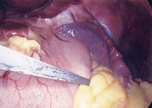
You can also read