Combined Descemet-stripping automated endothelial keratoplasty and phacoemulsification with toric intraocular lens implantation for treatment of ...
←
→
Page content transcription
If your browser does not render page correctly, please read the page content below
CASE REPORT

    Combined Descemet-stripping automated
endothelial keratoplasty and phacoemulsification
      with toric intraocular lens implantation
 for treatment of failed penetrating keratoplasty
          with high regular astigmatism
   Vincenzo Scorcia, MD, Andrea Lucisano, MD, Jacqueline Beltz, FRANZCO, Massimo Busin, MD

                  We present the case of a 57-year-old woman who had combined Descemet-stripping automated
                  endothelial keratoplasty (DSAEK) and phacoemulsification with implantation of a toric intraocular
                  lens (IOL). Surgery was intended to treat a cataract developing in a post-penetrating keratoplasty
                  (PKP) eye with high astigmatism and endothelial decompensation. Six months after uneventful
                  surgery, the cornea was clear and the corrected distance visual acuity was 20/20 with a refraction
                  of C0.25 1.00  10 (from 3.00 8.50  12 preoperatively). The internal topography map
                  (OPD-Scan) showed an IOL rotation of 4 degrees. The endothelial cell loss was 15% of the
                  eye-bank value. Descemet-stripping automated endothelial keratoplasty combined with phaco-
                  emulsification and toric IOL implantation is a relatively simple and very effective procedure for
                  eyes with endothelial failure and high post-PKP astigmatism. The speed of visual rehabilitation
                  and final visual acuity achieved with this approach was superior to that obtained with other
                  surgical procedures.
                 Financial Disclosure: Dr. Busin received royalties from Moria in 2008, 2009, and 2010, and his
                 travel expenses were partly reimbursed by Moria from 2005 to 2011. No other author has a financial
                 or proprietary interest in any material or method mentioned.
                 J Cataract Refract Surg 2012; 38:716–719 Q 2012 ASCRS and ESCRS

Currently, Descemet-stripping automated endothelial                   corrected by implantation of a toric intraocular lens
keratoplasty (DSAEK) is the surgical procedure of                     (IOL) alone (phakic IOL) or in combination with phaco-
choice for the treatment of endothelial failure after                 emulsification.7–11 We report the outcome of a combined
penetrating keratoplasty (PKP).1–5 However, if high                   procedure of DSAEK and phacoemulsification with
astigmatism is present as a result of the PKP, it remains             implantation of a hydrophilic acrylic bitoric IOL to treat
substantially unaffected by DSAEK and visual acuity is                high regular corneal astigmatism in a patient with
unlikely to improve postoperatively despite the recov-                cataract in a failed PKP.
ery of corneal clarity.5,6 Recently, moderate or high
astigmatism resulting from PKP has been successfully                  CASE REPORT
                                                                      A 57-year-old woman presented to our institution complain-
                                                                      ing of a progressive decrease in visual acuity in her left eye.
                                                                      Penetrating keratoplasty for keratoconus had been per-
Submitted: August 5, 2011.                                            formed in that eye in 1995. Since then, the acuity had been
Final revision submitted: September 22, 2011.                         affected by anisometropia and high regular astigmatism,
Accepted: September 22, 2011.                                         which could be only partly corrected by spectacles. The
                                                                      patient was intolerant to hard contact lenses.
From the Department of Ophthalmology (Scorcia, Lucisano, Busin),         On presentation, the uncorrected distance visual acuity
Magna Graecia University, Catanzaro and the Department of             (UDVA) and corrected distance visual acuity (CDVA) were
Ophthalmology (Beltz, Busin), Villa Igea Hospital, Forlı, Italy.     20/400 and 20/200, respectively, with a manifest refraction
                                                                      of 3.00 8.50  12. Slitlamp examination showed a full-
Corresponding author: Vincenzo Scorcia, MD, Via dei Crociati 40,      thickness graft (8.5 mm in diameter) with mild edema and
88100 Catanzaro, Italy. E-mail: vscorcia@libero.it.                   a dense nuclear cataract (Figure 1, A). Endothelial cell

716    Q 2012 ASCRS and ESCRS                                                                                0886-3350/$ - see front matter
       Published by Elsevier Inc.                                                                             doi:10.1016/j.jcrs.2012.01.014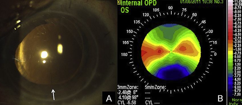
CASE REPORT: DSAEK AND PHACO WITH TORIC IOL IMPLANTATION IN FAILED PKP WITH HIGH ASTIGMATISM 717
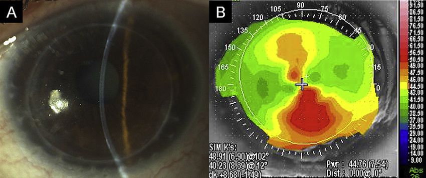
Figure 1. A: Preoperative digital slitlamp im-
age of the anterior segment showing corneal
graft edema and dense nuclear lens opacity.
B: Topographic anterior axial curvature map
showing the high degree of astigmatism re-
sulting from the previous PKP.
density could not be evaluated as the noncontact specular placed on a Barron punch (Katena Products, Inc.), endothe-
microscope (Topcon SP2000P, Topcon Corp.) could not lial side up, and trephined to 9.0 mm in diameter. An ante-
obtain a useful picture of the endothelial surface. Corneal rior chamber maintainer was placed at the 12 o’clock
topography (OPD-Scan, Nidek Co. Ltd.) showed high astig- position for continuous irrigation, and the donor tissue
matism with a regular asymmetrical bowtie pattern was inserted into the anterior chamber with the pull-
(Figure 1, B); keratometry values (K1/K2) were 48.91/ through technique using the Busin glide (Moria). Both the
40.23 @ 102. clear corneal tunnel and the side entries were sutured
Before surgery, the patient was asked to sit at the slitlamp. tightly with interrupted 10-0 nylon sutures, and the ante-
After the beam was set horizontally, a sterile ink pencil was rior chamber was filled with air injected through the tem-
used to mark the 0- and 180-degree positions, thus compen- poral side entry. Triamcinolone acetonide and gentamicin
sating for cyclorotation. Local anesthesia was administered were injected subconjunctivally at the end of the procedure.
with a peribulbar injection of a mixture of lidocaine The patient was pressure-patched overnight and instructed
hydrochloride 2.0% and bupivacaine hydrochloride 0.5%. to lie supine for 6 to 8 hours. Postoperatively, dexametha-
Standard phacoemulsification was performed through sone phosphate 0.1% and tobramycin antibiotic eyedrops
a 2.2 mm clear corneal tunnel centered on the steepest were administered initially every 2 hours and then tapered
corneal axis previously marked using a Mendez ring. over 3 to 4 months. All sutures were removed 3 weeks after
The customized hydrophilic acrylic bitoric IOL (AT TOR- surgery. No intraoperative or postoperative complications
BI 709M-Acri.Confort 646TCL, Carl Zeiss Meditec AG) was occurred.
manufactured based on the biometric data obtained by By 1 month postoperatively, the UDVA was 20/30 and
partial coherence interferometry (PCI) (IOLMaster, Carl the CDVA was 20/25 with a refraction of C0.75 1.50
Zeiss Meditec AG). The IOL power was C12.5 C8.0, which 10. At the last follow-up, 6 months postoperatively, the cor-
included an intended overcorrection of 0.75 D to compensate neal graft was transparent and the central endothelial cell
for the mild hyperopic shift usually seen after DSAEK density, measured with a noncontact specular microscope,
surgery. The IOL was implanted in the capsular bag and was 2210 cells/mm2, representing a 15% cell loss from
then rotated until proper alignment with the planned axis the initial eye-bank count (2600 cells/mm2). The CDVA
was achieved. The pupil was constricted with acetylcholine had improved to 20/20 with a minimal change in refraction
chloride (Miochol-E), and the clear corneal tunnel was wid- (C0.25 1.00 10) compared with the 1-month measure-
ened to 4.0 mm. The anterior chamber was filled with air, ment. The keratometry readings had not changed substan-
and the Descemet membrane and endothelium were peeled tially from the preoperative values. The alignment of the
off in a single piece. At this point, a small peripheral iridec- toric IOL was evaluated by slitlamp examination and fur-
tomy was performed at the 6 o’clock position with a vitrec- ther confirmed by the wavefront analysis and internal re-
tome to prevent the possible onset of pupillary block. fractive map obtained with the topography system that
The donor lenticule, prepared using the automated la- showed a postoperative IOL rotation of 4 degrees from
mellar therapeutic keratoplasty system (Moria), was then the intended axis (Figure 2).
Figure 2. A: One week postoperatively, the dig-
ital slitlamp image of the anterior segment
shows a transparent corneal graft. The IOL is
aligned with the steepest corneal meridian
(white arrow). B: Six months after surgery, the
internal refractive map obtained with the to-
pography system shows the power and axis
of the implanted toric IOL.
J CATARACT REFRACT SURG - VOL 38, APRIL 2012718 CASE REPORT: DSAEK AND PHACO WITH TORIC IOL IMPLANTATION IN FAILED PKP WITH HIGH ASTIGMATISM
DISCUSSION curvature in this post-PKP eye compared with the ef-
Several authors have recently reported that a triple fect in an unoperated eye. Therefore, we decided not
procedure combining DSAEK, phacoemulsification, to consider the theoretical calculations of surgically in-
and IOL implantation allows rapid visual rehabilita- duced astigmatism during the IOL calculation, as to
tion with minimal surgically induced astigmatism date these have not been validated in the setting of
and/or ametropia.5,12 However, performing this triple PKP. However, in cases such as this, the aim of surgery
procedure in an eye with a full-thickness edematous is to obtain a debulking effect, that is, to achieve a post-
graft has the potential limitation of not correcting the operative refractive error that can be corrected with
high astigmatism that may be present in up to 20% spectacles, and the above-mentioned variables have
of cases.13 In these cases, after the cornea has cleared had a negligible effect in this respect. Minor residual
following DSAEK, additional surgery is required to ametropia, including astigmatism, could then be ad-
allow spectacle correction. Incisional surgery and/or dressed, if required, with more accurate procedures
excimer laser procedures (both photorefractive kera- involving an excimer laser treatment.
tectomy and laser in situ keratomileusis) have been In conclusion, this case describing implantation of
used in the past but have had variable refractive re- a toric IOL at the time of combined cataract surgery
sults, ie, undercorrection, overcorrection, and/or re- and DSAEK in a post-PKP eye with high astigmatism
gression of effect over time, as well as relatively high shows that a 1-stage approach is at least as effective as
rates of other complications, including perforation, other 2-stage or even 3-stage approaches. This proce-
wound gaping, infection, and loss of CDVA.14–19 dure reduced postoperative recovery to a period of
Another option would be to combine cataract sur- several weeks and allowed excellent final CDVA.
gery with a repeat PKP, but this approach would
expose the patient to a renewed higher immunologic REFERENCES
risk as well as other PKP-related possible complica- 1. Price MO, Price FW Jr. Descemet’s stripping with endothelial
tions. In addition, at least 1 year would be required keratoplasty; comparative outcomes with microkeratome-
before all sutures could be removed and final refrac- dissected and manually dissected donor tissue. Ophthalmology
2006; 113:1936–1942
tion achieved. 2. Terry MA, Shamie N, Chen ES, Hoar KL, Friend DJ. Endothelial
Recently, phacoemulsification and implantation of keratoplasty; a simplified technique to minimize graft dislocation,
a customized toric IOL have been suggested as a safe iatrogenic graft failure, and pupillary block. Ophthalmology
and effective procedure for the correction of high 2008; 115:1179–1186
astigmatism resulting from PKP, pellucid marginal de- 3. Busin M, Bhatt PR, Scorcia V. A modified technique for Desce-
met membrane stripping automated endothelial keratoplasty to
generation, and keratoconus.10,20,21 Our case demon- minimize endothelial cell loss. Arch Ophthalmol 2008;
strates that toric IOLs have an additional indication 126:1133–1137. Available at: http://archopht.ama-assn.org/cgi/
in eyes with full-thickness corneal grafts that are visu- reprint/126/8/1133.pdf. Accessed September 28, 2011
ally impaired due to the development of cataract as 4. Price FW Jr, Price MO. Endothelial keratoplasty to restore clarity
well as the presence of endothelial failure and high to a failed penetrating graft. Cornea 2006; 25:895–899
5. Covert DJ, Koenig SB. Descemet stripping and automated en-
post-PKP astigmatism. In these eyes, a toric IOL can dothelial keratoplasty (DSAEK) in eyes with failed penetrating
be implanted at the time of combined DSAEK and keratoplasty. Cornea 2007; 26:692–696
phacoemulsification, thus effectively treating all 6. Straiko MD, Terry MA, Shamie N. Descemet stripping auto-
conditions negatively affecting vision with a single mated endothelial keratoplasty under failed penetrating
procedure. As with the procedures reported in the keratoplasty: a surgical strategy to minimize complications.
Am J Ophthalmol 2011; 151:233–237
past,11,22 the toric IOL in our case exhibited minimal 7. Sun X-Y, Vicary D, Montgomery P, Griffiths M. Toric intraoc-
rotation from the intended position and good stability ular lenses for correcting astigmatism in 130 eyes. Ophthal-
despite the additional maneuvers required during mology 2000; 107:1776–1781; discussion by RM Kershner,
DSAEK, particularly the complete air fill of the ante- 1781–1782
rior chamber and its consequent pronounced 8. Tehrani M, Stoffelns B, Dick HB. Implantation of a custom
intraocular lens with a 30-diopter torus for the correction of
deepening. high astigmatism after penetrating keratoplasty. J Cataract
A potential limitation of our approach concerns Refract Surg 2003; 29:2444–2447
the calculation of the toric IOL. The accuracy of this 9. Tahzib NG, Cheng YYY, Nuijts RMMA. Three-year follow-up
calculation may have been negatively affected by analysis of Artisan toric lens implantation for correction of
2 factors. First, the corneal edema may have prevented postkeratoplasty ametropia in phakic and pseudophakic eyes.
Ophthalmology 2006; 113:976–984
the PCI from measuring as precisely as in the presence 10. de Sanctis U, Eandi C, Grignolo F. Phacoemulsification and cus-
of a clear cornea. Second, the 4.0 mm wide clear cor- tomized toric intraocular lens implantation in eyes with cataract
neal tunnel that we routinely use for DSAEK may and high astigmatism after penetrating keratoplasty.
have caused an unpredictable change in corneal J Cataract Refract Surg 2011; 37:781–785
J CATARACT REFRACT SURG - VOL 38, APRIL 2012CASE REPORT: DSAEK AND PHACO WITH TORIC IOL IMPLANTATION IN FAILED PKP WITH HIGH ASTIGMATISM 719
11. Holland E, Lane S, Horn JD, Ernest P, Arleo R, Miller KM. 18. Busin M, Arffa RC, Zambianchi L, Lamberti G, Sebastiani A.
The AcrySof toric intraocular lens in subjects with cataracts Effect of hinged lamellar keratotomy on postkeratoplasty eyes.
and corneal astigmatism; a randomized, subject-masked, Ophthalmology 2001; 108:1845–1851; discussion by ED
parallel-group, 1-year study. Ophthalmology 2010; 117: Donnenfeld, 1851–1852
2104–2111 19. Kwitko S, Marinho DR, Rymer S, Ramos Filho S. Laser in situ
12. Terry MA, Shamie N, Chen ES, Phillips PM, Shah AK, Hoar KL, keratomileusis after penetrating keratoplasty. J Cataract
Friend DJ. Endothelial keratoplasty for Fuchs’ dystrophy with Refract Surg 2001; 27:374–379
cataract; complications and clinical results with the new triple 20. Visser N, Gast STJM, Bauer NJC, Nuijts RMMA. Cataract sur-
procedure. Ophthalmology 2009; 116:631–639 gery with toric intraocular lens implantation in keratoconus:
13. Vail A, Gore SM, Bradley BA, Easty DL, Rogers CA, a case report. Cornea 2011; 30:720–723
Armitage WJ, on behalf of collaborating surgeons. Conclusions 21. Luck J. Customized ultra-high-power toric intraocular lens
of the corneal transplant follow-up study. Br J Ophthalmol 1997; implantation for pellucid marginal degeneration and cataract.
81:631–636. Available at: http://www.ncbi.nlm.nih.gov/pmc/ J Cataract Refract Surg 2010; 36:1235–1238
articles/PMC1722292/pdf/v081p00631.pdf. Accessed Septem- 22. Chang DF. Comparative rotational stability of single-piece
ber 28, 2011 open-loop acrylic and plate-haptic silicone toric intraocular
14. Solomon A, Siganos CS, Frucht-Pery J. Relaxing incision lenses. J Cataract Refract Surg 2008; 34:1842–1847
guided by videokeratography for astigmatism after keratoplasty
for keratoconus. J Refract Surg 1999; 15:343–348
15. Koay PYP, McGhee CNH, Crawford GJ. Effect of a standard
paired arcuate incision and augmentation sutures on postkera- First author:
toplasty astigmatism. J Cataract Refract Surg 2000; 26: Vincenzo Scorcia, MD
553–561
16. Amm M, Duncker GIW, Schro € der E. Excimer laser correction of Department of Ophthalmology,
high astigmatism after keratoplasty. J Cataract Refract Surg Villa Igea Hospital, Forlì, Italy
1996; 22:313–317
17. Chang DH, Hardten DR. Refractive surgery after corneal
transplantation. Curr Opin Ophthalmol 2005; 16:251–255
J CATARACT REFRACT SURG - VOL 38, APRIL 2012You can also read