Some of the ophthalmic surgeries performed at the DMV Center.
←
→
Page content transcription
If your browser does not render page correctly, please read the page content below
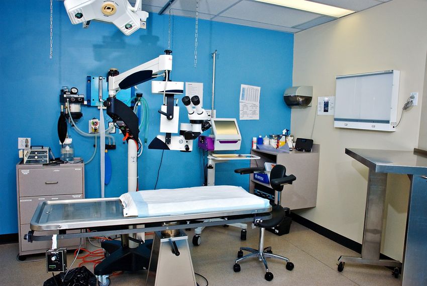
Some of the ophthalmic surgeries performed at the DMV Center.
This document presents some types of the surgeries performed by the ophthalmology service at the
DMV veterinary center as well as the equipment used during these surgeries. The equipment used is
similar to the one used in human ophthalmology.

View of the ophthalmic surgical room

Surgeries of the cornea:

For these surgeries, the animal needs to be anesthetized and an operating microscope and tiny surgical
instruments are necessary.
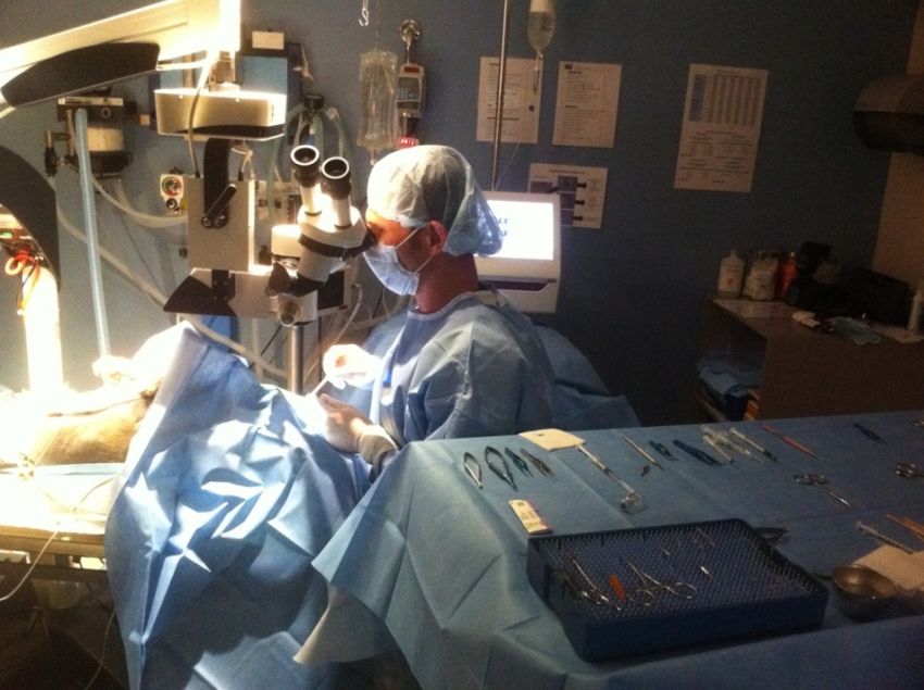
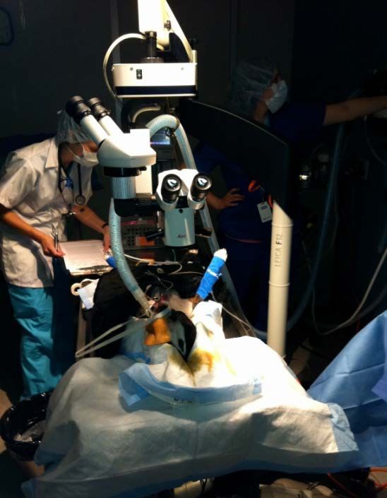
View of a dog anesthetized and placed on its back for a
surgery of the cornea where an operating microscope is
needed (left picture) and Dr Ollivier performing a corneal
surgery through the operating microscope (right picture).
Conjunctival graft to treat corneal ulcers in animals.
A corneal ulcer is a break, a wound in one or more layers of the cornea. Normally, when there is a defect
or ulcer on the surface of the eye (of the epithelium), the cells at the outer edges grow into the center
and healing is often complete within 2-4 days. When the ulcer is deeper, it takes more time to heal.
An infected corneal ulceration (infected wound of the clear, front part of the eye) poses a special problem
because bacteria that are present are actually delaying the healing process and even digesting the corneal
tissues (this is called a melting ulcer). This can lead to corneal perforation (full thickness whole) and
cause blindness if not treated promptly. Depending on a variety of factors, your pet may need a surgically
placed biodegradable graft, conjunctival graft, corneal graft or corneal glue to provide protection and
support for the cornea as it heals.
A conjunctival graft represents a technique commonly performed to treat deep corneal ulcers or corneal
perforations. It allows an immediate coverage/filling of the corneal defect with the pink tissue around the
eye (conjunctiva) used as a bandage over a wound. This tissue is attached to the cornea with very thin
stitches. The graft covering the ulcer will remain in place and will form a scar but the base of the graft
(the pedicle) can be trimmed 2 months after surgery to stop the vascularisation of the graft in order to
reduce the corneal scar and increase the visual field.
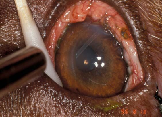
Corneal ulcer almost Same eye: the
perforating in a dog conjunctival graft
that needs surgical is sutured over
repair.. the corneal ulcer.
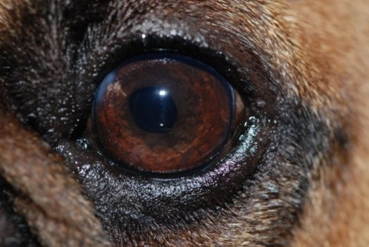
Same eye 3 weeks
Same eye 8 weeks
after the surgery
after the surgery.
after trimming of
the pedicule.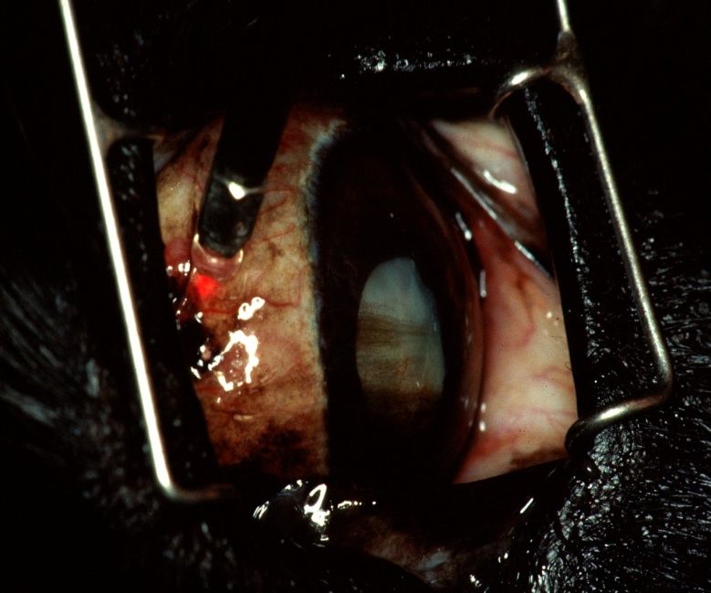
Glaucoma surgeries
Glaucoma is defined as increased pressure within the eye that causes degenerative changes in the optic
nerve and retina with subsequent blindness. The goals of glaucoma treatment are to save as much vision
as possible for as long as possible and to keep the patient comfortable. The treatment program is
determined by the type of glaucoma, the severity and duration; the pet's other medical problems, and
the possibility of saving vision. Treatment ranges from using eye drops at home, to hospitalization for
intense therapy, to possible surgery. A treatment plan for your pet will be discussed with you as the pet
is evaluated and as the response to treatment is noted. Usually, the treatment will start with topical and
maybe systemic medication. If the medial treatment is not enough to control the pressure, a type of
surgery might be recommended for your animal. This document will present the various surgical
alternatives possible in case of glaucoma.

Transscleral Diode Laser Cyclophotocoagulation (CPC)
A laser is used to destroy cells of the ciliary body, which is the gland located inside of the eye that
produces fluid. This procedure is estimated to control eye pressure in 60-70% of patients for a period of
6 months. Complications include post-laser pressure spikes, bleeding, and corneal ulcers. The laser
surgery is non-invasive (laser energy is delivered by a probe placed on the surface of the eye; however a
short episode of general anesthesia is required to perform the procedure. Most pets must stay in the
hospital for pressure monitoring for 1 to 3 days after laser surgery.

                                                              Glaucomatous eye treated by diode laser
                                                              (left picture),the diode laser (right picture).

Aqueous shunt implant (gonioimplant)
A small tube is implanted inside the eye. This tube, or shunt, sits partially inside the eye and partially
outside the eye under the mucous membrane (conjunctiva) around the eye to allow fluid to leave the
eye when pressure increases above a specific level. The success rate for this procedure to control
pressure is similar to laser surgery. When laser treatment is combined with placement of a
gonioimplant, the estimated success rate per eye is maximized. Complications include scarring around
the shunt causing back-up of the fluid in the eye and dislocation of the shunt. General anesthesia is
required along with overnight monitoring.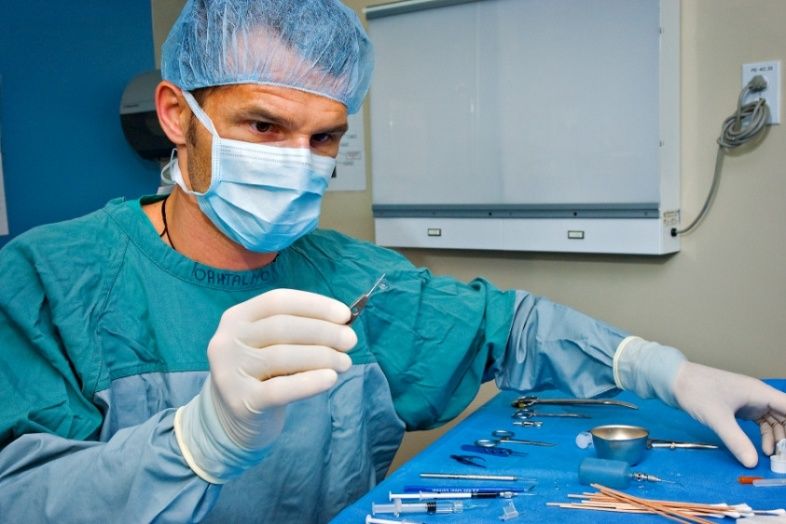
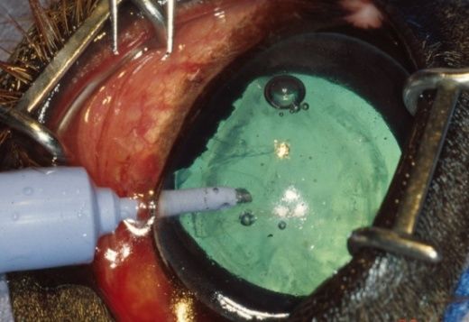
A glaucomatous eye treated by a gonioimplant (left picture and picture in the middle) and the
   gonioimplant (right picture).

Cataract surgery by phacoemulsification and artificial lens implantation
No medication will keep cataracts from becoming worse or cause a cataract to “clear-up”. Medical
treatment (usually eye drops) is often used to control the inflammation caused by the cataract or to open
the pupil to increase vision.

Removal of the cataract (surgery) is still the only method of improving vision in a patient with cataracts.
Whether or not cataract surgery would be helpful for any individual patient depends upon many factors.
Several differences between a human eye and a dog's eye should be considered in determining if
cataract surgery is advisable for your pet.

Before cataract surgery the eyes need to be carefully evaluated. Active problems within the eye must be
controlled before surgery is considered. Surgery is performed under general anesthesia (gas with
oxygen). Certain laboratory tests are done to learn of any other internal medical problems which may
require treatment.

An artificial lens can be inserted in the bag of the natural lens to improve the visual acuity of the dog.

The cataract surgery: the same technique as in humans is performed in animals. The opaques masses are removed
from the bag of the lens by phacoemulsification and an artificial lens can be then inserted in the emptied bag.

 For this surgery , the animal is anesthetized , placed on its back and an operating microscope, a
 phacoemulsification machine and fine surgical instruments are needed, like in human medicine.
View during a cataract surgery: the animal is anesthetized and placed on its back, a operating microscope
and a phaco machine (digital screen in the back) are used during this surgery (left picture) . The phaco
machine (right picture).
Dr Ollivier holding an artificial lens during cataract surgery (left picture) and an artificial lens (right eye)
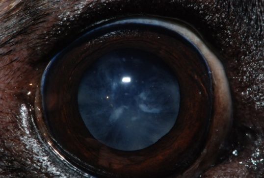
An eye with an immature cataract The same eye 24 hours after the surgery
By Dr Franck OLLIVIER, Dipl. ACVO & ECVO. The same eye 3 weeks after the surgery
Centre vétérinaire DMV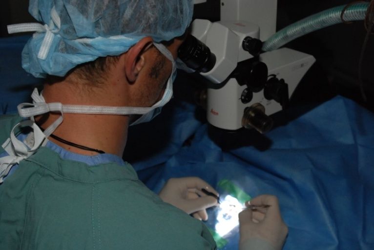
You can also read