Thermo Scientific Tundra Cryo-TEM: 100kV Cryo-TEM dedicated for Single Particle Analysis
←
→
Page content transcription
If your browser does not render page correctly, please read the page content below
1330                                                                                                                        Microsc. Microanal. 27 (Suppl 1), 2021
            doi:10.1017/S1431927621004967                                                                                               © Microscopy Society of America 2021

            Thermo Scientific™ Tundra Cryo-TEM: 100kV Cryo-TEM dedicated for Single
            Particle Analysis
            Zuzana Hlavenková1, Dimple Karia2, Miloš Malínský3, Daniel Němeček4, Fanis Grollios5, Vojtěch
            Doležal1, Ondřej Sháněl1, Abhay Kotecha6, Markéta Červinková7, Lingbo Yu1 and Anke Mulder8
            1
              Thermo Fisher Scientific, United States, 2Thermo Fisher Scientific, Eindhoven, Noord-Brabant,
            Netherlands, 3Thermo Fisher Scientific, Brno, Jihomoravsky kraj, Czech Republic, 4Thermo Fisher
            Scientific, Czech Republic, 5Thermo Fisher Scientific, Eindhoven, Netherlands, 6Thermo Fisher
            Scientific, Noord-Brabant, Netherlands, 7Thermo Fisher Scientific, Brno-Cernovice, Czech Republic,
            8
              Thermo Fisher Scientific, Oregon, United States

            Single Particle Analysis (SPA) application of cryo-electron microscopy (cryo-EM) has become a well-
            established method for determination of the 3D structure of wide variety of proteins and their complexes,
            revealing the mechanism of their function and showing their interactions with known and novel drugs[1].
            However, as the popularity of this technique increases, so does the need for greater efficiency and
            accessibility from not only microscopy experts but also from scientists of different scientific backgrounds
            and with little to no cryo-EM experience.

            For the most part, the operation of an electron microscope for high-resolution cryo-EM data collection
            still requires an experienced person. Training of a skilled EM operator can take months and is perceived
            as a significant bottleneck in broader adoption of cryo-EM.

            The Thermo Scientific Tundra Cryo-TEM is a new transmission electron microscope operating at 100kV
            high tension dedicated to SPA[2] which has been specifically developed for new users from biochemistry
            and biology labs. The Tundra Cryo-TEM brings a new level of automation and user guidance for
            microscope operation and SPA data collection; moreover, the Tundra Cryo-TEM fits into a standard
            laboratory room, thereby reducing costs associated with room renovation.

            Benchmark cryo-EM measurements using apo-ferritin demonstrates that Tundra Cryo-TEM can achieve
            2.6Å resolution of a reconstructed 3D map (Figure 1). At this resolution, de novo protein structures can
            be determined, and important biological questions answered. Data was collected with pixel size of
            0.75Å, for about 17 hours and processed using Relion 3.1[3]. The data was collected using Thermo
            Scientific™ EPUTM software with pre-defined settings. We used a new functionality in EPU that
            automatically checks and refines optical alignments and provides system status for high-quality data
            acquisition. Furthermore, data quality was monitored on-the-fly using EPU quality monitor.

            Data was collected on a new scintillator-based camera CETA-F with speed enhancement dedicated for
            operation at 100kV high tension. It has 4 times better sensitivity with respect to CETATM 16M. CETA-F
            also brings the possibility of dose fractionation mode as implemented on the Falcon camera. Dose
            fractionation allows storage of image frames for correction of beam induced motion. in a post processing
            pipeline.

            Additionally, a new objective lens was developed for Tundra Cryo-EM to decrease the spherical and
            chromatic aberrations at 100-kV acceleration voltage and boost signal at high resolution frequencies.

Downloaded from https://www.cambridge.org/core. IP address: 46.4.80.155, on 07 Aug 2021 at 11:41:21, subject to the Cambridge Core terms of use, available at
https://www.cambridge.org/core/terms. https://doi.org/10.1017/S1431927621004967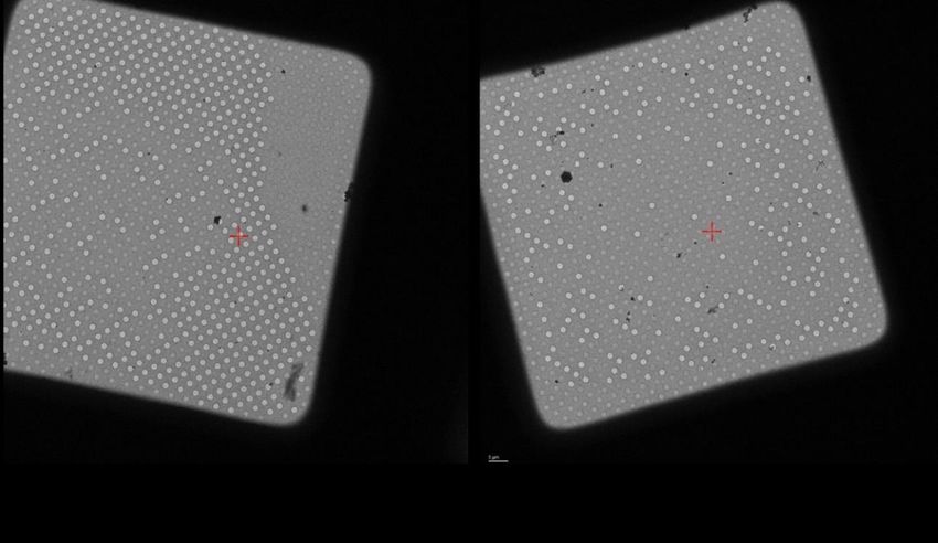
Microsc. Microanal. 27 (Suppl 1), 2021                                                                                                                                1331

            To load the sample into the microscope, we have introduced a novel semi-automated loading technology
            (SAL) on Tundra. Samples can be loaded into the microscope within minutes with minimum ice
            contamination on the grid (Figure 2). SAL allows a new, iterative way of working to optimize of sample
            concentration and vitrification conditions that can quickly qualify samples for high resolution data
            collection. SAL also has a fully guided workflow on the on-screen display (OSD), to guide users with
            different levels of expertise.

            All these new features that are introduced within the Tundra Cryo-TEM allows novice as well as expert
            users to achieve relevant resolution of their biological samples while keeping an accessible price point.
            This would make cryo-TEM accessible to many scientists across all life science branches.

            Figure 1. Fig.1 Structure of Apoferritin protein determined at 100 keV. a) 3D reconstruction of apoferritin
            at 2.6Å resolution, b) Gold-standard FSC plot corresponding to the calculated map, showing the
            correlation between the phase-randomized (red), unmasked (green) and masked (blue) half-maps, c)
            Electron density of the 2.6 Å resolution map showing the apoferritin α-helix

Downloaded from https://www.cambridge.org/core. IP address: 46.4.80.155, on 07 Aug 2021 at 11:41:21, subject to the Cambridge Core terms of use, available at https://www.cambridge.org/core/terms.
https://doi.org/10.1017/S1431927621004967
1332                                                                                                                       Microsc. Microanal. 27 (Suppl 1), 2021

            Figure 2. Fig. 2 Grid Square images showing limited to no ice contamination upon transfer to Tundra
            using SAL technology.

            References
            [1] Michael Eisenstein: The field that came in from the cold, Nature, Vol.13 No.1, January 2016
            [2] Mathew J. Peet, Richard Henderson, Christopher J. Russo: The energy dependence of contrast and
            damage in electron cryomicroscopy of biological molecules, Ultramicroscopy 203 (2019) 125–131
            [3] J. Zivanov, T. Nakane, B. Forsberg, D. Kimanius, W.J.H. Hagen, E. Lindahl & S.H.W. Scheres
            "RELION-3: new tools for automated high-resolution cryo-EM structure determination", eLife
            2018;7:e42166

Downloaded from https://www.cambridge.org/core. IP address: 46.4.80.155, on 07 Aug 2021 at 11:41:21, subject to the Cambridge Core terms of use, available at https://www.cambridge.org/core/terms.
https://doi.org/10.1017/S1431927621004967
You can also read