Explore the cell ... without limits
←
→
Page content transcription
If your browser does not render page correctly, please read the page content below
Thermo Scientific
                                            ArrayScan XTI HCA Infinity Configuration

explore the cell ...
                               without limits
• Innovative confocal imaging • Live cell and label-free capability
• Modular, flexible design • Industry-leading analysis software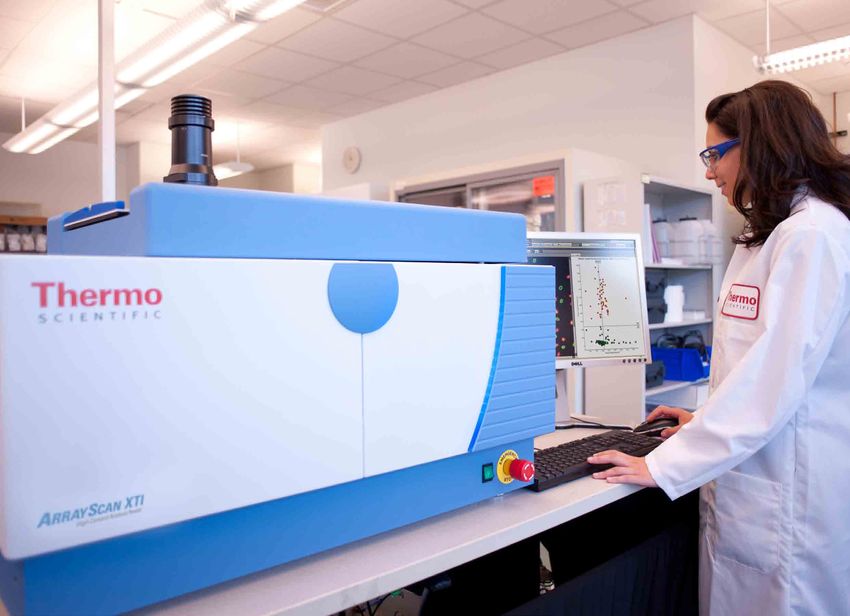
answer your most demanding
cell biology challenges
infinite possibilities
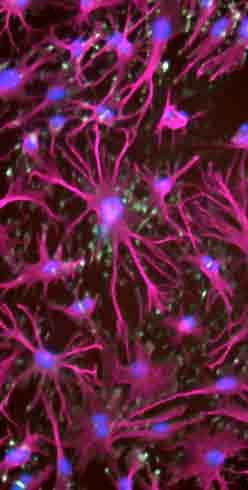
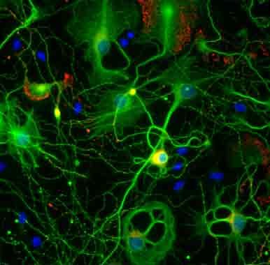
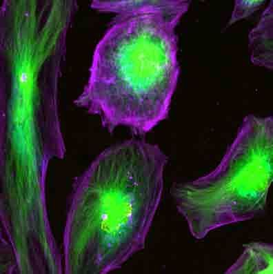
As scientists increasingly apply high content analysis to stem cell characterization, multi-dimensional cell
models, primary cultures and tissues, the challenges posed by these applications demand new types of
imaging techniques. The Thermo Scientific™ ArrayScan™ XTI HCA Infinity Configuration delivers the latest
advances in multi-dimensional high content imaging and analysis – without sacrificing your productivity.
• Modular and flexible design for your most demanding high-content assays
• Integrated variable pin-hole confocal technology with innovative LED illumination
• Solid-state seven-color LED illumination, live cell and label free capabilities
as standard
• Best-in-class image analysis software
• Open standard, scalable image and data management software
• The high-content reader at the core of hundreds of published papers
Visit our website at thermoscientific.com/infinity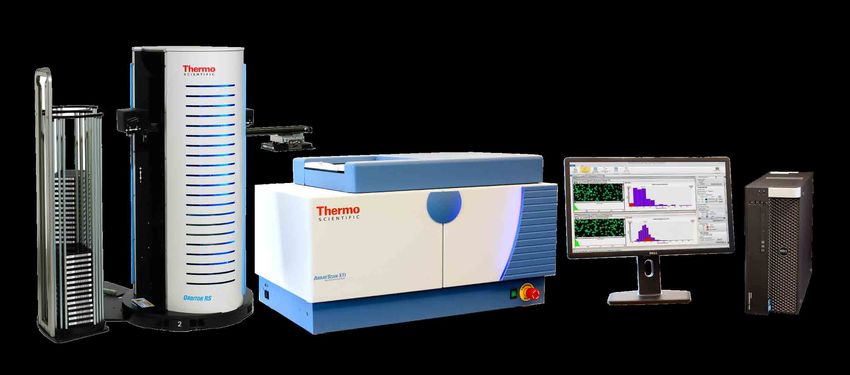
take high content analysis
                                  into new areas of cell biology
                                                                                                                definite answers

Developed by the inventors of high-content analysis using over a decade of unequalled experience, the
ArrayScan XTI HCA Infinity Configuration offers the broadest set of capabilities for the large-scale study of
cell biology (cellomics) in a single, modular platform. From high-content assay development, through basic
cell biology research to systems biology and drug discovery and toxicology, the ArrayScan XTI HCA Infinity
Configuration has been designed to deliver robust, biologically relevant answers, with minimal effort and
with the fastest “image-to-answer” on the market.

            Developing higher-content assays?                                   More advanced biology?
            • Get up and running quickly with hundreds                          • Live cell and label free capabilities allow
              of pre-built, validated image analysis protocols                    pharmacokinetics, cell motility and extended
            • Assay development is easy with our interactive                      toxicity studies to be performed
              image analysis software – no expertise required                   • Advanced widefield and confocal optics
                                                                                  allow imaging of complex 3D cell structures,
                                                                                  cell aggregations and tissues

            Worried by all those images and data?                               New to high-content analysis?
            • Our data management software is based on                          • Our industry-leading training program allows
              open standards and does not require complex,                        you to quickly master the basics and become
              expensive computer hardware                                         productive in your research
            • Comprehensive visual analytics analyze the data                   • Support for your high-content biology from
              and generate reports so you can communicate                         our Center of Excellence and Field Application
              your answers quickly and effectively                                Scientists
                                                                                • Global support from our experienced service
                                                                                  and support team

                To order or request additional information, call USA 1.800.432.4091 • Asia +81 3 5826 1659 • Europe +32 (0)53 85 71 84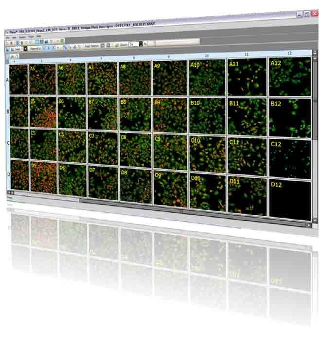
all your high content needs
in one modular integrated platform
integrating multi-dimensional imaging capabilities in one tool
1. Live Cell Kinetics 3. Optics by Carl Zeiss™
The live cell module enables the Acquiring high-quality images is the
dynamics of intracellular molecular first step in high-content analysis.
interactions and cell motion to be At the core of the instrument is the
studied. In addition, the system fully automated Zeiss™ Axiovert
features the Zeiss™ Definite Z1 microscope that offers a range
Focus module to maintain cells in of objectives and other options for
optimal focus during long live cell maximum imaging flexibility.
experiments.
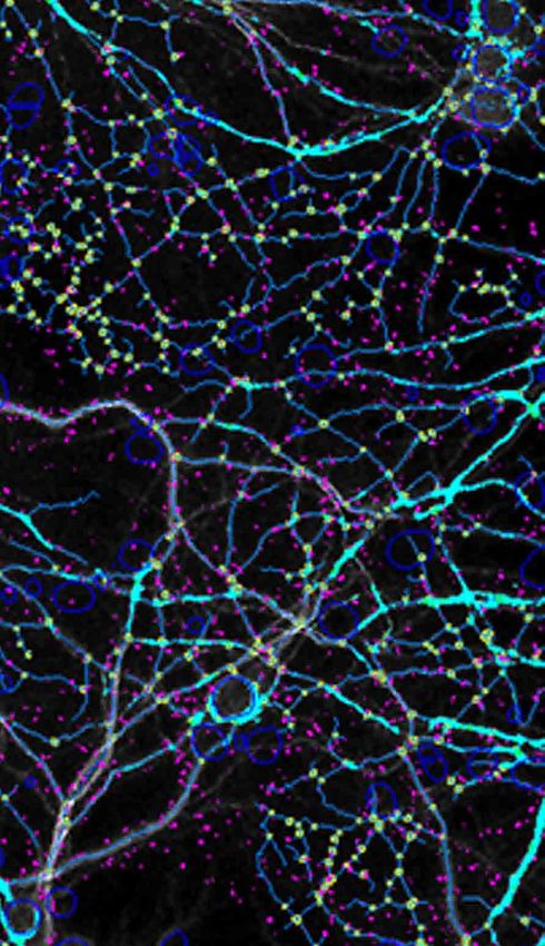
2. Confocal 4. Label Free Imaging
The integrated confocal module The images collected by the Thermo
provides maximum flexibility for all Scientific™ Brightfield Module can
your biology and cell-based assay be used to quantitate morphological
needs. The latest in high-speed features of cells, or as a way to gain
Nipkow spinning disk technology an extra channel for fluorescence,
and a variable pinhole, coupled increasing assay flexibility and
with a unique seven-color LED light significantly expanding assay
source allows stem cell colonies, capability.
tissues and 3D microenvironments
to be imaged with ease.
1
4 3
2
Visit our website at thermoscientific.com/infinity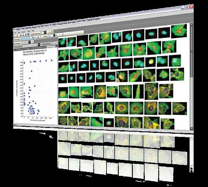
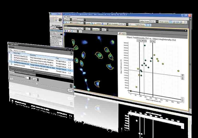
5. Powerful, Flexible Image Analysis                                      7. Orbitor RS Robot
      Software                                                                  Designed for use in the lab, the
      Designed to make single-cell                                              Thermo Scientific™ Orbitor™ RS
      measurements with precision and                                           Microplate Mover increases and
      efficiency, Thermo Scientific™                                            expands the throughput capacity
      BioApplications are the engine of                                         of your current reader.
      high-content analysis and coupled
      with the Thermo Scientific™ HCS
      Studio™ Cell Analysis Software
      provide an intuitive, interactive step-
      by-step way to quickly build and
      optimize hundreds of image-based
      assays.

      6. Image and Data Management                                                8. LED Light Engine
      Realizing the full potential of high-                                       High content analysis starts with a
      content analysis requires effective                                         high-quality image which, in turn,
      handling of massive amounts of                                              depends on optimal illumination.
      images and data. Open standard,                                             The Thermo Scientific™ seven-
      fast and secure, the Thermo                                                 color LED Light Engine is an
      Scientific™ Store™ Express Image                                            ultra-stable, long-life, solid-state
      and Database Management Software                                            illumination source that delivers
      provides an out-of-the-box solution                                         the most biologically relevant,
      allowing you to immediately search,                                         statistically robust data about your
      access, analyze and re-analyze all                                          cells, in the fastest time possible.
      your images and data no matter
      the source. For a more scalable
      solution, choose the optional Thermo
      Scientific™ Store™ SE Image and
      Database Management Software.

7

                                                                                           5

                                                              8
                                                                                                              6

    To order or request additional information, call USA 1.800.432.4091 • Asia +81 3 5826 1659 • Europe +32 (0)53 85 71 84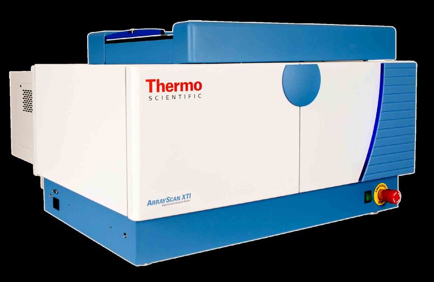
thermoscientific.com/highcontent
©2014 Thermo Fisher Scientific Inc. All rights reserved. Zeiss and Carl Zeiss are trademarks of Carl Zeiss AG Corporation. All other trademarks are the property of
Thermo Fisher Scientific Inc. and its subsidiaries. Specifications, terms and pricing are subject to change. Not all products are available in all countries. Please consult
your local sales representative for details.

USA +1 800 432 4091 info.cellomics@thermofisher.com
Asia +81 3 5826 1659 info.cellomics.asia@thermofisher.com
Europe +32 (0)53 85 71 84 info.cellomics.eu@thermofisher.com

C-BR_ASIN0313
You can also read