KOL KNOCKOUT CATARACT EDITION: Surgeons Battling for the Best Outcomes - Cataract & Refractive Surgery Today
←
→
Page content transcription
If your browser does not render page correctly, please read the page content below

Supplement to                                                                                               September/October 2021

       KOL KNOCKOUT ™
                                                                                          FACULTY:
                                                                                          Uday Devgan, MD, FACS, FRCS
                                                                                          Zaina Al-Mohtaseb, MD

       CATARACT EDITION:                                                                  Ravi D. Goel, MD
                                                                                          David A. Goldman, MD

       Surgeons Battling for
                                                                                          Himani Goyal, MD
                                                                                          John A. Hovanesian, MD

       the Best Outcomes                                                                  Robert F. Melendez, MD, MBA
                                                                                          Denise M. Visco, MD, MBA

                                                                                                        Provided by

     A continuing medical education activity provided by Evolve Medical Education LLC.
     This activity is supported by an unrestricted educational grant from Alcon Vision.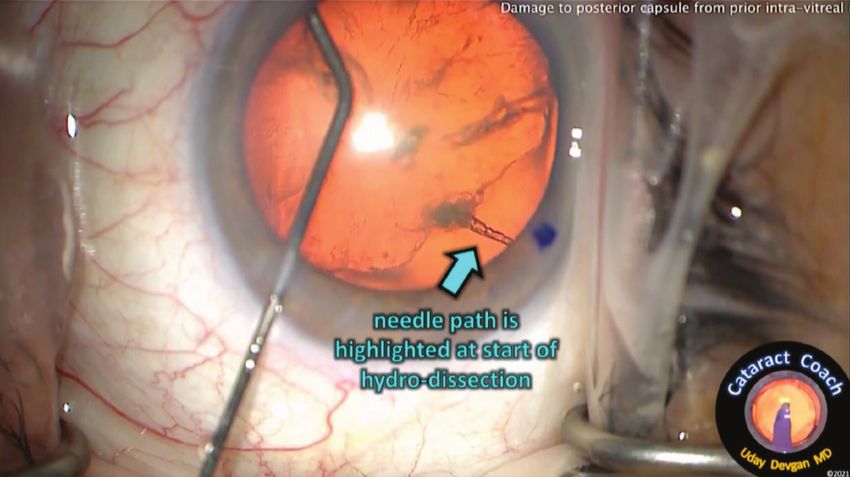
KOL Knockout — Cataract Edition:                  TM                                                                                    Release Date: September 2021
                                                                                                                                           Expiration Date: October 2022

   Surgeons Battling for the Best Outcomes
   FACULTY MEMBERS

         UDAY DEVGAN, MD,                  ZAINA AL-MOHTASEB, MD                         RAVI D. GOEL, MD                 DAVID A. GOLDMAN, MD
            FACS, FRCS                     Associate Professor of Ophthalmology        Comprehensive Ophthalmologist                Goldman Eye
                  Program Chair            Associate Residency Program Director           Regional Eye Associates              Palm Beach Gardens, FL
              Devgan Eye Surgery                Baylor College of Medicine                     Cherry Hill, NJ
          Professor of Ophthalmology                    Houston, TX                              Instructor
            Jules Stein Eye Institute                                                        Wills Eye Hospital
             Chief of Ophthalmology                                                          Philadelphia, PA
          Olive View-UCLA Med Center
                 Los Angeles, CA

         HIMANI GOYAL, MD                  JOHN A. HOVANESIAN, MD                     ROBERT F. MELENDEZ,                     DENISE M. VISCO,
          Clinical Assistant Professor                 Clinical Faculty                    MD, MBA                               MD, MBA
              New York University                 Jules Stein Eye Institute                    Founder and CEO               Medical Director and Founder
         Grossman School of Medicine         University of California Los Angeles            Juliette Eye Institute                  Eyes of York
                  New York, NY                     Harvard Eye Associates                Clinical Associate Professor                  York, PA
                                                       Laguna Hills, CA                   University of New Mexico
                                                                                               Albuquerque, NM

   CONTENT SOURCE                                                                   LEARNING OBJECTIVES
     This continuing medical education (CME) activity captures con-                   Upon completion of this activity, the participant should be able to:
   tent from a three live virtual symposia.                                           • Summarize the technology of femtosecond laser-assisted
                                                                                        cataract surgery
   ACTIVITY DESCRIPTION                                                               • Explain how to incorporate femtosecond laser technology
      This supplement summarizes content from a series of three                         into cataract surgery techniques
   unique, live virtual symposia hosted by Uday Devgan, MD, FACS,                     • Establish when to incorporate intraoperative aberrometry to
   FRCS. The game show-style quiz competition with real-time                            provide better refractive outcomes for cataract patients
   audience voting featured cataract-focused case studies and                         • Evaluate patient lifestyles and expectations to improve
   discussions regarding patient care and surgical approaches among                     intraocular lens choice
   seven key opinion leaders/contestants. The archived video series can               • Compare current and emerging technologies in advanced
   be viewed here: https://evolvemeded.com/course-group/kol-knockout-                   technology intraocular lenses
   cataract-edition-aberrometry-femtosecond-lasers-advanced-iols.
                                                                                    GRANTOR STATEMENT
   TARGET AUDIENCE                                                                     This activity is supported by an unrestricted educational grant
      This certified CME activity is designed for ophthalmologists.                 from Alcon Vision.

2 SUPPLEMENT TO CATARACT & REFRACTIVE SURGERY TODAY / MILLENNIALEYE | SEPTEMBER/OCTOBER 2021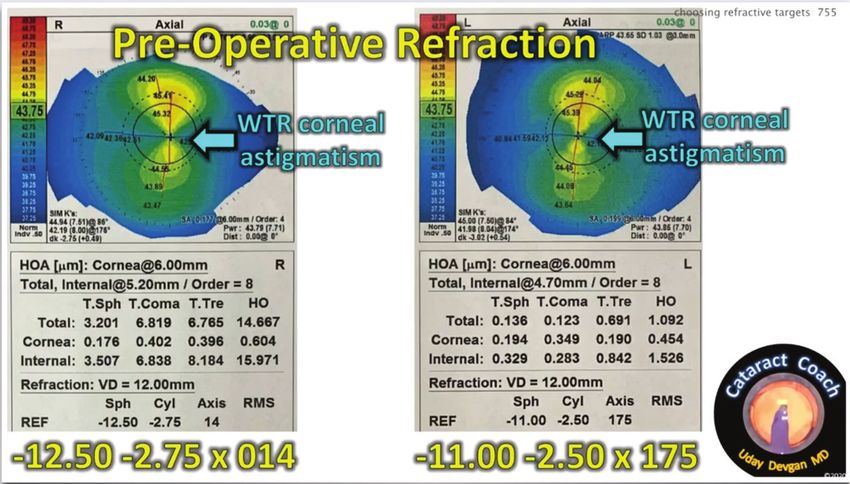
ACCREDITATION STATEMENT                                                       in the form of Advisory Board: 1-800-Doctors, Aerie Pharmaceuticals,
  Evolve Medical Education LLC (Evolve) is accredited by the                  Abbott Medical Optics, Blephex, Cord, Eyedetec, Glaukos, Guardian
Accreditation Council for Continuing Medical Education (ACCME)                Health Sciences, Ingenoeye, IOP/Katena, Ivantis, Kala Pharmaceuticals,
to provide continuing medical education for physicians.                       MDBackline, Ocular Therapeutix, Omeros, ReVision Optics, Shire,
                                                                              Sight Sciences, TearLab, Tearfilm Innovations, Valeant, and Veracity.
CREDIT DESIGNATION STATEMENT                                                  Consultant: 1-800-Doctors, Acufocus, Aerie Pharmaceuticals, Alcon
   Evolve Medical Education designates this enduring material for             Vision, Allegro Ophthalmics, Allergan, Abbott Medical Optics,
a maximum of 1.5 AMA PRA Category 1 Credit™. Physicians should                BlephEx, Eyedetec, Glaukos, Guardian Health Sciences, IOP/Katena,
claim only the credit commensurate with the extent of their                   Ivantis, Kala Pharmaceuticals, Novartis, Ocular Therapeutix, Omeros,
participation in the activity.                                                ReVision Optics, Sarentis Ophthalmics, Sensimed, Shire, TearLab,
                                                                              Tearfilm Innovations, Valeant, and Veracity. Grant/Research: Aerie
TO OBTAIN CREDIT                                                              Pharmaceuticals, Abbott Medical Optics, Alcon Vision, Allergan,
   To obtain credit for this activity, you must read the activity in          Cloudbreak Therapeutics, Cord, Eydetec, Glaukos, Ingenoeye, IOP/
its entirety and complete the Pretest/Posttest/Activity Evaluation/           Katena, Novartis, Ocular Therapeutix, Omeros, ReVision Optics,
Satisfaction Measures Form, which consists of a series of multiple-choice     Shire, Tearfilm Innovations, and Valeant. Stock/Shareholder/Equity:
questions. To answer these questions online and receive real-time             Alcon Vision, Alicia Surgery Center, Allegro Ophthalmics, Allergan,
results, please go to https://evolvemeded.com/course/2039-supp. Upon          BlephEx, Eyedetec, Glaukos, Guardian Health Sciences, Harvard Eye
completing the activity and self-assessment test, you may print a CME         Associates, Harvard Hearing, Ingenoeye, MDBackline, Novartis, Ocular
credit letter awarding 1.5 AMA PRA Category 1 Credits™. Alternatively,        Therapeutix, RxSight, Sarentis Ophthalmics, Sight Sciences, and
please complete the Posttest/Activity Evaluation/Satisfaction Form            Tearfilm Innovations. Royalties: SlackBooks and TLC: The Laser Center.
and mail or fax to Evolve Medical Education LLC, 353 West Lancaster
Avenue, Second Floor, Wayne, PA 19087; Fax: (215) 933-3950.                      Robert F. Melendez, MD, MBA, has had a financial agreement
                                                                              or affiliation during the past year with the following commercial
DISCLOSURE POLICY                                                             interests in the form of Speaker: Alcon Vision.
  It is the policy of Evolve that faculty and other individuals who
are in the position to control the content of this activity disclose             Denise M. Visco, MD, MBA, has had a financial agreement
any real or apparent conflicts of interest relating to the topics of this     or affiliation during the past year with the following commercial
educational activity. Evolve has full policies in place that will identify    interests in the form of Consultant: Bruder, Carl Ziess Meditec,
and resolve all conflicts of interest prior to this educational activity.     Cassini, Lensar, Omeros, and Sun Pharmaceutical Industries. Grant/
                                                                              Research Support: Carl Ziess Meditec, Lensar, Omeros, Oyster Point
   The following faculty/staff members have the following financial           Pharma, and Sun Pharmaceutical Industries. Speaker’s Bureau: Carl
relationships with commercial interests:                                      Ziess Meditec, Lensar, Omeros, and Sun Pharmaceutical Industries.
                                                                              Stock/Shareholder: Eyevance.
   Uday Devgan, MD, FACS, FRCS, has had a financial agreement or
affiliation during the past year with the following commercial interests in   EDITORIAL SUPPORT DISCLOSURES
the form of Speaker’s Bureau: Bausch + Lomb and Novartis. Shareholder:          The Evolve staff and planners have no financial relationships with
Advanced Euclidean Solutions, CataractCoach.com, and LensGen.                 commercial interests. Michelle Dalton, writer, and Nisha Mukherjee, MD,
                                                                              peer reviewer, have no financial relationships with commercial interests.
   Zaina Al-Mohtaseb, MD, has had a financial agreement or
affiliation during the past year with the following commercial                OFF-LABEL STATEMENT
interests in the form of Consultant: Alcon Vision, Bausch + Lomb,                This educational activity may contain discussion of published and/
CorneaGen, and Ocular Therapeutix.                                            or investigational uses of agents that are not indicated by the FDA. The
                                                                              opinions expressed in the educational activity are those of the faculty.
  Ravi D. Goel, MD, has had no financial agreement or affiliation             Please refer to the official prescribing information for each product for
during the past year.                                                         discussion of approved indications, contraindications, and warnings.

   David A. Goldman, MD, has had a financial agreement or                     DISCLAIMER
affiliation during the past year with the following commercial                  The views and opinions expressed in this educational activity are
interests in the form of Consultant: Alcon Vision, Allergan, Bausch +         those of the faculty and do not necessarily represent the views of Evolve,
Lomb, and Novartis. Speaker's Bureau: Allergan and Eyevance. Stock/           Cataract & Refractive Surgery Today, MillennialEYE, or Alcon Vision.
Shareholder: Alcon Vision, Allergan, Johnson & Johnson Vision, Kala
Pharmaceuticals, Ocular Therapeutix, Stroma, and Tarsus.                      DIGITAL EDITION
                                                                                This supplement is part of a larger curriculum that includes
  Himani Goyal, MD, has had a financial agreement or affiliation              archived versions of three live virtual symposia. Go to https://evolve-
during the past year with the following commercial interests in               meded.com/course-group/kol-knockout-cataract-edition-aberrom-
the form of Consultant: Carl Zeiss Meditec. Grant/Research Support:           etry-femtosecond-lasers-advanced-iols to view
Research to Prevent Blindness.                                                the full series.
                                                                                To view the online version of the supple-
   John A. Hovanesian, MD, has had a financial agreement or                   ment, please go to https://evolvemeded.com/
affiliation during the past year with the following commercial interests      course/2039-supp.

                                                              SEPTEMBER/OCTOBER 2021 | SUPPLEMENT TO CATARACT & REFRACTIVE SURGERY TODAY / MILLENNIALEYE 3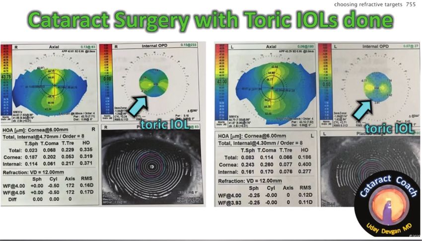
PRETEST QUESTIONS
                 PLEASE COMPLETE PRIOR TO ACCESSING THE MATERIAL AND SUBMIT WITH POSTTEST/
                         ACTIVITY EVALUATION/SATISFACTION MEASURES FOR CME CREDIT.

1. Please rate your confidence in your ability to incorporate intraoperative          6. A 55-year-old high hyperope presents to your cataract surgery clinic
   aberrometry to provide better refractive outcomes for cataract patients                for evaluation. She has been reliant on +8.50 D glasses for distance and
   (based on a scale of 1 to 5, with 1 being not at all confident and 5 being             +11.00 D glasses for reading for the past 10 years. She now desires spectacle
   extremely confident).                                                                  independence. Which of the following statement about her postoperative
       a. 1                                                                               vision is TRUE?
       b. 2                                                                                   a. She may notice more image magnification after surgery, so objects may
       c. 3                                                                                       seem larger after surgery.
       d. 4                                                                                   b. She may notice less image magnification after surgery, so objects may
       e. 5                                                                                       seem smaller than before surgery.
                                                                                              c. Image magnification after surgery will not change.
2. Which of the following is an important step in approaching an intumescent                 d. She may notice fluctuating image magnification after surgery.
   white cataract?
      a. Needle decompression of liquefied cortical material prior to starting the    7. Which of the following statements about astigmatic correction after cataract
          capsulorhexis                                                                   surgery is TRUE?
      b. Underfilling the anterior chamber with viscoelastic                                 a. The presence of astigmatism can increase depth of focus, and fully
      c. Performing the capsulorhexis prior to filling the anterior chamber with                correcting astigmatism may collapse the depth of field after surgery.
          viscoelastic                                                                       b. T he presence of astigmatism can reduce depth of focus, and fully
      d. Performing the capsulorhexis with only forceps                                          correcting astigmatism may increase the depth of field after surgery.
                                                                                             c. T
                                                                                                 he presence of astigmatism can increase depth of focus, and fully
3. Which of the following steps is an important when posterior capsular                        correcting astigmatism may further increase depth of field after surgery.
   rupture is suspected?                                                                     d. The presence of astigmatism can reduce depth of focus, and fully
      a. Turning off irrigation                                                                  correcting astigmatism may further reduce the depth of field after surgery.
      b. Filling the anterior chamber with viscoelastic while maintaining irrigation
      c. Switching to the irrigation/aspiration device                                 8. Which of the following patients would benefit most from an intraocular lens
      d. Enlarging the incision to 3 mm                                                   (IOL) with diffractive rings, such as trifocal, bifocal, multifocal, and some
                                                                                          extended depth of field designs?
                                                                                             a. A patient with low-angle alpha and low-angle kappa
4. A n 81-year-old male presents to your clinic with hand motion vision. On                 b. A patient with high-angle alpha and low-angle kappa
   exam, you notice a milky white intumescent cataract. You schedule him for                 c. A patient with low-angle alpha and high-angle kappa
   cataract surgery the next day. You pierce the center of the anterior capsule              d. A patient with high-angle alpha and high-angle kappa
   with a needle and decompress some liquefied cortex. What is a method you
   can use to ensure the posterior liquefied cortex is decompressed prior to           9. Which patient would be the most appropriate to receive multifocal IOL
   initiating the capsulorhexis?                                                          surgery?
       a. Pierce the lens again with the needle and continue advancing the needle           a. A 30-year-old -2.00 D patient with a clear lens
           posteriorly through the lens.                                                     b. A 50-year-old -1.50 D patient with 1+ NS
       b. Use the irrigation aspiration device to aspirate more cortex                       c. A 70-year-old patient with refractive error of +5.00 D -2.00 x 180
       c. Rock the nucleus back and forth with your needle and reaspirate with                  with 2+ NS
           your needle                                                                       d. A 30-year-old +1.00 D patient with a clear lens
       d. Hydrodissect with balanced salt solution prior to initiating the
           capsulorhexis                                                               10. Which of the following patient(s) is/are an ideal trifocal IOL candidate?
                                                                                           Select all that apply.
5. A patient with a history of frequent intravitreal injections presents to your            a. Emmetropic patient with a clear lens
   clinic for preoperative cataract surgery screening. You notice a focal opacity            b. Emmetropic patient with a cataract
   in the inferotemporal quadrant of the lens. You suspect posterior capsular                c. Hyperopic patient with a clear lens
   compromise. During cataract surgery, what step would most likely lead to                  d. Hyperopic patient with a cataract
   further compromise of the posterior capsule?
       a. Clear corneal incision
       b. Hydrodissection
       c. Phacoemulsification of the lens
       d. Irrigation/aspiration of nuclear material

    4 SUPPLEMENT TO CATARACT & REFRACTIVE SURGERY TODAY / MILLENNIALEYE | SEPTEMBER/OCTOBER 2021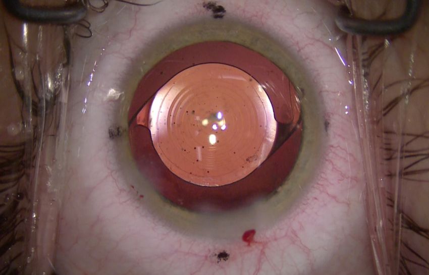
KOL KNOCKOUTTM — CATARACT EDITION:
                                                                                                                                       Surgeons Battling for the Best Outcomes

KOL KnockoutTM — Cataract Edition:
Surgeons Battling for the Best Outcomes
Cataract surgery is one of the most commonly performed surgeries worldwide, and by 2050, the number of people in the United States with
cataracts is expected to reach 50 million (up from 24.4 million in 2010).1 The rapidly evolving field has seen substantial improvements in
preoperative biometry, intraoperative aberrometry, enhanced or extended depth of focus (EDOF) intraocular lenses (IOLs), new IOL calculators,
femtosecond lasers, presbyopia-correcting IOLs and corneal inlays.
Captured from a series of three live, virtual “knock out rounds,” we’ve put together case studies to evaluate some of these innovations, but more
importantly, to determine what steps we would take in real-world, real-time scenarios. We hope you not only enjoy these case presentations, but
can put our discussion into use in your clinics tomorrow.
                                                                                                  — Uday Devgan, MD, FACS, FRCS, Program Chair

ROUND 1 | CASE 1: ARGENTINIAN FLAG                                                     we can ascertain the capsule has been decompressed, the next
           AND LENS CHOICES                                                             step is to make the incision and complete the rhexis. This is my
   Uday Devgan, MD, FACS, FRCS: Our first case is an                                    approach to minimize the risk of that Argentinian flag sign.
intumescent white cataract. We know these types of cataracts
tend to present with a thin and fragile anterior capsule that
makes performing phacoemulsification tricky.2 This is particularly                      Q        DR. DEVGAN: What are your approaches? What
                                                                                                 instruments or tools you'd use to do a capsulorhexis in
true when we’re creating the continuous circular capsulorhexis                          an intumescent white cataract to prevent Argentinian flag sign?
(CCC), because puncturing the anterior capsule results in a                                Ravi Goel, MD: I like your approach, but in these cases I prefer to
decrease in anterior capsular chamber pressure, which then                              make two side port incisions and ensure the intraocular pressure
causes the remaining intralenticular to anteriorly displace the                         in the eye is above 30 mm Hg. Next, I stain with trypan blue 0.06%.
lens, leading to a radial extension of the capsular tear; this is oth-                  Next, I use the Seibel capsulorhexis forceps to do the rhexis under
erwise known as the Argentinian flag sign for the blue/white/blue                       high pressure through two side port incisions. I can make a small
color pattern.2-5                                                                       capsulorhexis initially, and then perform the double capsulorhexis
   In our case, we used a 27-gauge needle to decompress the                             technique.6,7 I love that I don't have a main incision until I finish
cataract. We used trypan blue dye to stain the capsule. After                           the capsulorhexis. That allows me to rock the nucleus a bit, which
using ophthalmic viscoelastic (OVD) to pressurize the anterior                          can help remove some of the cortex and that milky substance.
chamber, we can tell the bag is also pressurized with intumescent
fluid. See Figure 1.                                                                      Dr. Devgan: Great technique. Figure 2 illustrates what you
   What’s important to note here is that we have not yet created                        mean by rocking the nucleus to release more of the liquefied
the main incision. In Figure 1, we’ve gone right to the center of                       cortex. Dr. Goel, do you use two port incisions and then a
the capsule to try to aspirate and release some of the fluid. Once                      25-gauge forceps?

Figure 1. An intumescent cataract with needle insertion to decompress.                  Figure 2. Intumescent white cataract and rocking the nucleus.

                                                                         SEPTEMBER/OCTOBER 2021 | SUPPLEMENT TO CATARACT & REFRACTIVE SURGERY TODAY / MILLENNIALEYE 5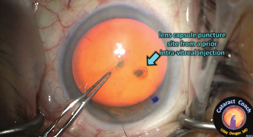
KOL KNOCKOUTTM — CATARACT EDITION:
  Surgeons Battling for the Best Outcomes

      Dr. Goel: Yes, the specialized microcapsulorhexis forceps.

     Dr. Devgan: Dr. Al-Mohtaseb, what's your approach in
   these cases?

       Zaina Al-Mohtaseb, MD: I definitely like using a 27- or 25-gauge
   half-inch needle to decompress prior to creating the main wound.
   I like putting in a Healon5 viscoadaptive (Johnson & Johnson
   Vision), which also helps with performing the rhexis. Once I
   decompress, I create my main wound. The key for me is to ensure
   you’ve removed not just the central liquefied cortex, but also the
   cortex that might be in the periphery of the lens. You must be
   very careful during this step. I remove a good amount and then              Figure 3. Trifocal lens implantation in a patient with white cataract.
   press down on the lens nucleus to make sure the fluid drains. It is
   important to remember the rhexis will tend to run out because
   of the liquefied cortex in the periphery or behind the lens that is         stage, sometimes within 30 days. I’ve planned on using the
   not removed with decompression. If it's a soft cataract instead of          femtosecond laser on this patient, as colleagues have told me
   a dense white cataract, I use bimanual irrigation aspiration8-10 to         they’ve had success using the laser for these cases. There is also
   remove all the cortical material.                                           support in the literature.3,11,12

     Dr. Devgan: That’s another great technique.                                  Dr. Al-Mohtaseb: It’s reasonable to use the femtosecond laser
   Dr. Melendez, what is your approach for these intumescent                   in these cases. I’ve had cases in which the lens is too intumescent
   white cataracts?                                                            and liquefied cortex comes out, which can happen if the laser
                                                                               takes a little longer to perform the capsulorhexis. You just need
       Robert Melendez, MD, MBA: With the case you have presented              to remember it won’t be continuous; you’ll need to remove the
   so far, there is only one difficult step. Overall, the case itself is not   capsule with care and assume there will be tags. Still, I think it's a
   difficult. It is important, however, that you keep positive pressure        very reasonable approach.
   on that anterior capsule. I stain the anterior capsule with trypan
   blue, then I use OVD. Once there is an adequate amount of OVD                 Dr. Goel: I think using a femtosecond laser is a reasonable
   in the eye to keep positive pressure on the anterior capsule, I’ll          option. I recall a case you recently posted, Dr. Devgan, in which
   puncture the anterior capsule with a cystotome. I also make two             you went in with a needle and it went immediately to the
   side port incisions. I initially make a tiny puncture, but I keep it        Argentinian Flag sign. I think you could still have the potential
   very small and circular. In my experience, I’ve found by doing that,        of that occurring, even with a femtosecond laser, but I believe
   if it does tear, it will do so circumferentially as opposed to tearing      using a femtosecond laser to control the situation is a reason-
   straight out in most cases.                                                 able approach.
       Then, I'll go in with the 27-gauge cannula through another
   side port and start to evacuate more of the liquefied cortex.                  Dr. Devgan: My advice for those of us who want to use the
   Oftentimes, you'll see it ooze and go to one of the paracentesis            femtosecond laser is to still stain with trypan blue. Once you’re
   ports. At that point, I’ll usually add more OVD, and go back in             in the eye, don’t presume the capsule is (essentially) free-floating.
   with the 27-gauge to aspirate more for the liquefied cortex.                You’ll need to grab the capsule and make the tearing motions
       Dr. Al-Mohtaseb made a good point about removing some                   because there may still be a few attachments that you'll then be
   of the peripheral liquefied cortex to decompress that high                  able to complete.
   pressurized lens before adding more viscoelastic. Then, we move                Continuing with the case, the rest of which is fairly
   forward with the capsulotomy.                                               straightforward and uneventful. The nucleus can be chopped and
                                                                               aspirated rather quickly. What does make this case a bit more
     Dr. Devgan: In these cases, would you consider using a                    interesting is the choice of lens. We opted to insert a trifocal IOL
   femtosecond laser to create the rhexis before you even enter                in this patient. I added triamcinolone in the anterior chamber to
   the eye?                                                                    quiet any inflammation. The patient had about 0.50 D of astig-
                                                                               matism, so we also performed limbal relaxing incisions (LRIs). The
     Dr. Melendez: I have a case coming up that is very similar—an             literature shows that newer trifocal lenses reduce photic phenom-
   intumescent cataract. Patients at a higher risk for intumescent             enon compared to the previous bifocal designs, and they deliver
   cataracts are those with a 4+ white to posterior subcapsular                high levels of patient satisfaction.13-19 Figure 3 shows the end of
   cataract, as those can progress very rapidly to an intumescent              the procedure.

6 SUPPLEMENT TO CATARACT & REFRACTIVE SURGERY TODAY / MILLENNIALEYE | SEPTEMBER/OCTOBER 2021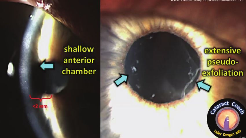
KOL KNOCKOUTTM — CATARACT EDITION:
                                                                                                                       Surgeons Battling for the Best Outcomes

Q          DR. DEVGAN: What are your thoughts about implanting
           a premium lens in these patients? Can these patients
                                                                          I think you have to make sure the ocular surface and the rest
                                                                        of the ophthalmic exam is normal, especially in the contralateral
achieve an accurate refractive outcome even if they’ve started off      eye. This patient probably should have a B-scan before surgery to
with white cataracts? How do you do the lens calculations?              ensure nothing else was going on.
   Dr. Al-Mohtaseb: Those are all very good questions. If there are       I rely on ORA in these types of cases, also in postrefractive cases,
no issues with the patient’s ocular surface, and we have good axial     as the literature also supports that.21
length measurements, I think a premium lens is a viable option.
The newer optical biometers, such as the IOLMaster 700 (Carl Zeiss         Dr. Devgan: In this case, the patient was thrilled. She ended up
Meditec) or the Lenstar 900 (Haag-Streit) can image very dense          just about plano—to go from a white cataract to clean and crisp
lenses much better than earlier iterations. White cataracts are         vision with a trifocal lens and with minimal complaints about
still very difficult to predict and plan for. I definitely recommend    nighttime glare or halos made this one of my favorite cases.
doing an A-scan immersion, making sure your gates are correct on
the immersion, and that you’re comfortable with the axial length
measurement. Compare it to the other eye to reconfirm.                  ROUND 1 | C ASE 2: TREATING PATIENTS WITH
   It’s also about managing expectations. I tell these patients we                 ZONULAR LAXITY
may be off our refractive mark, but we can rectify it with refractive      Dr. Devgan: Our next case is a patient with severe zonular
surgery if necessary. So, yes, I think premium lenses are something     laxity. Figure 4 shows the preoperative images, which again
to consider even in white cataracts.                                    reinforces Dr. Al-Mohtaseb’s point about why the preoperative
                                                                        exam is so crucial.
   Dr. Devgan: Dr. Melendez, how do you do your IOL calculations           In this patient, there was a very shallow anterior chamber and
in these patients?                                                      very small pupil (visible on the left side of Figure 4). This is a very
                                                                        tiny eye; the IOL power for postoperative emmetropia was +34 D.
   Dr. Melendez: First and foremost, there must be a careful            The right side of Figure 4 shows that same eye under maximum
consent with the patient so they understand the potential risk of       pharmacologic dilation.
a tear that may lead to having a different lens implanted. We’re           The capsulorhexis was a little difficult, a little tough. There
doing an internal study at our clinic comparing the IOLMaster           was a little laxity on the lens capsule and it was difficult to get
700 with the Argos Swept-source Optical Coherence Tomography            started; I was unable to poke it with forceps and had to use a
Biometer (Alcon) in measuring dense cataracts like this one.            sharp needle instead. I was able to make a 5-mm capsulorhexis;
   I recently had a white intumescent cataract, very similar to this    my forceps have marks so I can measure it directly. The
case. The IOLMaster 700 could not capture the image, but the            capsulorhexis is nice and round and centered. For the time
Argos with ERV setting was able to capture the measurements.            being, there was enough capsular support or zonular support to
I also have the intraoperative ORA System (Alcon). This system          hold the capsule. But I could rock the entire nucleus and have
uses preoperative biometry measurements and intraoperative              the capsulorhexis shift.
interferometry to measure an aphakic eye’s refractive power for
appropriate IOL selection,20,21 which I’ve found particularly useful
in these types of surgeries.                                            Q        DR. DEVGAN: How would you handle this case with very
                                                                                 weak zonules?
   But to Dr. Al-Mohtaseb’s point, using the contralateral eye             Dr. Goel: I tend to make the capsulorhexis the same way as
as a base can work, presuming the second eye is very close              I would with a white cataract—two paracenteses in the high
in measurement, and you can confirm that with axial length
measurements. With the ORA, you have measurements
intraoperatively as well to help with the best IOL calculation.

  Dr. Goel: I would start by looking for an older spectacle
prescription before the patient developed the intumescent
cataract. I'd compare the refractions in both eyes. Then, I would
do axial length measurements as best I could. At our clinic, we
have the choice of an IOLMaster 700, a Lenstar, or an A-scan.
  For me, this is a case in which I’d use the ORA intraoperatively,
because you noted there was a small amount of astigmatism as
well. The literature has shown using intraoperative aberrometry
increased the proportion of eyes with postoperative refractive
astigmatism of less than 0.50 D and reduced the mean
postoperative refractive astigmatism at 1 month.22                      Figure 4. Preoperative images of a shallow anterior chamber.

                                                         SEPTEMBER/OCTOBER 2021 | SUPPLEMENT TO CATARACT & REFRACTIVE SURGERY TODAY / MILLENNIALEYE 7
KOL KNOCKOUTTM — CATARACT EDITION:
  Surgeons Battling for the Best Outcomes

   hyperopes. I do this because I’m afraid when the anterior chamber           area of zonular weakness. I like to use Mackool hooks overlying
   depth is 2.5 mm or less that it may be too shallow. Having the two          this area. In these cases, usually the pupils are small; so I usually
   side port incisions and making the capsulorhexis with the MSC               use iris hooks to do the iris retraction, a little bit farther out in
   forceps has worked for me.                                                  that area of zonular weakness. I use Mackool hooks and go in
      I also consider using Mackool hooks with a Chang Modified                with a manual reverse cannula. I'll basically hold circumferentially
   Retractor, which is a capsular tension retractor. I would also use          as opposed to just tangentially because that can disinsert the bag
   a capsular tension ring (CTR) as late as possible in the procedure,         even more if you pull directly to the center. Circumferentially is
   but as soon as you need it. In this case, I would have tried to insert      key in a circular fashion.
   the ring as soon as I saw the jiggling.
                                                                                 Dr. Devgan: I absolutely agree. Dr. Al-Mohtaseb, please share
      Dr. Melendez: Knowing the patient has pseudoexfoliation                  your thoughts.
   should raise your suspicion that you’re going to have a loose bag.
   We already know that pseudoexfoliation and zonular laxity are                  Dr. Al-Mohtaseb: I like to insert CTRs manually, but taking
   associated with an increased risk of vitreous loss23 and with late          out the cortex can be difficult after putting a CTR in these cases.
   IOL dislocation.24 In this case, when you tried to puncture the             Prior to placing my CTR, I go in with OVD and viscodissect the
   lens, there was a good amount of wrinkling which increased when             cortex. To place the CTR, I go in through a paracentesis so there's
   you did puncture it. To me, that implies the bag was already very           less movement than going in from the wound. I use a Sinskey
   loose right out of the gate. Based on your description, it seems as         hook through the wound to hold the CTR as I'm threading it in,
   though the zonular laxity may have been limited to one area. In             so it doesn't push against the anterior capsule. I don't put the
   these cases, I use the Mackool hooks, but I also believe exposure           Sinskey hook in the eyelit. Once the CTR is in, I rotate my hand
   is key. We should not be afraid to insert a couple of iris hooks if         clockwise to allow for the CTR to be fully placed in the bag. I feel
   that pupil comes down. You really want good exposure to observe             I have more control when I do this manually.
   the edge of the bag in the event that there is any disinsertion of
   the bag.                                                                      Dr. Goel: I’ve used an injector, but I agree with Dr.
                                                                               Al-Mohtaseb. I have seen videos of using the Sinskey hook in the
      Dr. Al-Mohtaseb: First, I ensure the pupil is well dilated. Then,        eyelid to introduce the CTR in a controlled fashion. I absolutely
   to perform the capsulorhexis, I either use capsular hooks or I place        agree that the irrigation/aspiration (I/A) has to be super delicate
   capsular tension ring segments and hook the eyelit with an iris             because of the zonular weakness. For maximum stability, I also
   hook. I believe there is more stability when I use the CTR segments         go circumferentially.
   than when I use four large capsular hooks. This is particularly true
   in cases of really dense lenses. Avoiding further zonular loss by              Dr. Devgan: Fantastic pearls. I agree with your advice, and
   being careful with rotation and hydrodissection is important and            I placed the CTR with an injector and went circumferentially.
   so is chopping the lens.                                                    We switched to a bimanual approach for the I/A, which
                                                                               made clean up much easier. But this patient is also 87. What’s
     Dr. Devgan: Another suggestion is to use iris hooks to stabilize          your lens choice in these cases? Single-piece IOL in the bag? In
   the capsule procedure once you debulk as much of the lens as                the sulcus?
   possible, just like Dr. Goel mentioned.
     Preoperative discussions with these patients is of the upmost               Dr. Melendez: We have numerous options. I’ll use a single
   importance. I may joke with them that their surgery is going to             piece if there is less than 3 clock hours of zonular weakness. Your
   take a week off my life and they should be really sweet to me no            case may have up to 4 clock hours, and I would recommend
   matter the outcome. But in all seriousness, it is important to con-         using a three-piece IOL, doing a reverse capture and putting the
   vey to the patient this is a very difficult surgical procedure and          haptics in the sulcus. Then, I would push the lens in the bag.
   there are no guarantees of the outcome.
     Returning to the case: I almost never do stop and chop, but I                Dr. Devgan: Fantastic pearl. Dr. Goel, what do you think?
   did a groove to debulk the lens centrally in this case because there
   was so little working room. In my hands, this is easier than having            Dr. Goel: No one would fault you in this case if you used either
   two chopped halves; with a groove, each half is more like 40 to             a single-piece or a three-piece IOL. I think with a CTR you could
   45% due to the removed lens material from grooving, so it’s just            use a single-piece. A CTR could help you very carefully place a
   smaller. The nucleus comes out fairly easily, and I move to cortex          three-piece IOL into the sulcus, as Dr. Melendez described, with
   removal. At this point, I’ve decided to insert a CTR.                       the haptics and the sulcus and the optic captured in the bag.
                                                                               Certainly, I would calculate for both. I'd calculate an AC IOL, too,
   Q      DR. DEVGAN: In your hands, when would you place a CTR
          and how do you place one (manually, with an injector)?
                                                                               but you have to take each of these cases as they come. I don’t
                                                                               know that I’d generalize or exclude one type of lens or where to
     Dr. Melendez: I typically don't use CTRs. I try to locate the             place it.

8 SUPPLEMENT TO CATARACT & REFRACTIVE SURGERY TODAY / MILLENNIALEYE | SEPTEMBER/OCTOBER 2021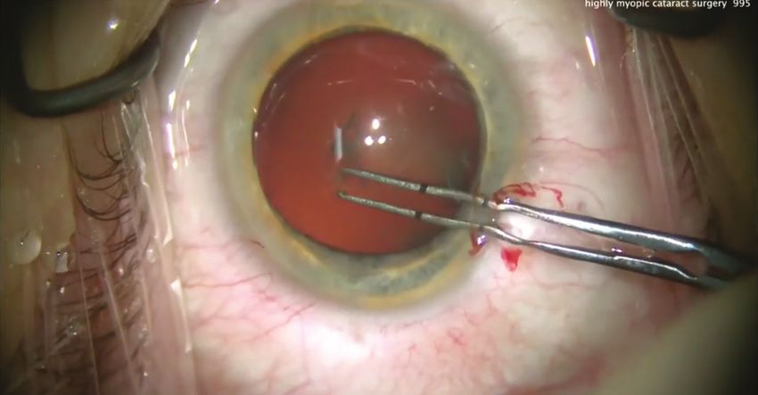
KOL KNOCKOUTTM — CATARACT EDITION:
                                                                                                                Surgeons Battling for the Best Outcomes

   Dr. Al-Mohtaseb: I would use a single-piece IOL with the CTR.               Second, I also recommend looking for zonular loss, which
I would put it in the bag. If I feel there is a lot of zonular loss or if   can happen in these eyes more frequently than we might think.
it was a much younger patient, then I would suture CTR Ahmed                Zonular weakness coupled with high myopia can lead to late
segments to the sclera. Otherwise, I can always hook the CTR if it          IOL dislocation.25
does dislocate later. I like putting in the one-piece IOL with a CTR.          Third, as soon as you enter with the phacoemulsification,
                                                                            reverse pupillary block is very uncomfortable for the patient and
  Dr. Devgan: To continue with our case, I chose a three-piece              it deepens everything into the eye. I prefer to go in with a second
lens. I inserted the haptics into the sulcus and captured the               instrument, lift up the iris and then go in with my phaco. In my
optic by button-holing it behind the capsulorhexis. Hopefully,              hands, that helps reduce the risk of reverse pupillary block from
that will give this patient good long-term stability. I'm happy to          occurring. We need to remember that reverse pupillary block can
tell you the patient did pretty well. But this was a very stressful         occur even during I/A. That is a good enough reason to change
case. I really cannot emphasize enough how important it is to               how I approach these cases.
talk with the patient in the preoperative period.                              Finally (as I do in all cases), I try to avoid a posterior capsule tear
  I do want to reiterate that these are not hypothetical cases—they         because of the known risk of retinal detachment being so much
are from my own clinic and certainly stressful. I think the patients        higher in highly myopic cases.26-28
appreciate it when you tell them ahead of time how difficult it's
going to be. Then, they're more accepting of a wider outcome range.            David A. Goldman, MD: I agree with Dr. Al-Mohtaseb’s
                                                                            comments. I will add that if you get a reverse block, one option is
ROUND 2 | CASE 3: HIGH MYOPE                                               to lift the iris, or lower the bottle height. If possible, I recommend
  Dr. Devgan: Our next case is a high myope. This 60-year-old               using an acrylic lens because these patients are at a higher risk for
patient has an axial length of greater than 30 mm, he’s -15.00 D            retinal pathologies.27,29
and after a nuclear sclerotic shift, he’s -20.00 D in his refraction.          When you’re counseling these patients, explain that the risk
These cases present with several issues; among them is lens                 of retinal detachment is influenced by the patient’s posterior
calculations. Figure 5 shows how I’m “cheating” when I’m making             vitreous detachment (PVD) status, gender, and age. There is a
the capsulorhexis. I’m using forceps that are marked on the tip             significant year-over-year risk of retinal detachment and risk with
so I can actually measure out 5 mm. This patient has a huge                 cataract surgery. I tell these patients they must follow-up with a
eye—13 mm white-to-white and a large, dilated pupil.                        retinal specialist for the rest of their lives.
  One pearl here: Don’t use the pupil as guidance in creating
the capsulorhexis in these cases, otherwise you’ll end up with an             Dr. Devgan: Dr. Goyal, are you sending these cases to your
oversized rhexis that won’t hold the IOL in place.                          retina specialist before surgery?
  Another issue that can present itself intraoperatively with highly
myopic eyes and long axial lengths is reverse pupillary block.                Himani Goyal, MD: I definitely do. I want to make sure there
                                                                            are no retinal issues to consider. I’m not good at scleral depression,
Q        DR. DEVGAN: In these cases of high myopes and long
         axial lengths, what do you do differently intraoperatively?
                                                                            and I can’t see everything that’s there. With these cases, I feel the
                                                                            retina specialist brings more to the table than I can alone. The
   Dr. Al-Mohtaseb: In these cases, the first thing to remember is          retina specialist will let me know if there is anything we need
that you’re going to be much deeper in the eye. You must hold               to do prophylactically to the eye, but it also allows the patient
your hand position very differently. You're almost operating like           to meet the retina specialist who will likely be treating them if
a retina surgeon. You have to be very careful when you're doing             something does happen down the road.
your rhexis, for all the reasons Dr. Devgan explained.
                                                                             Dr. Devgan: Do you send them back to the retina specialist a
                                                                            month later for a postoperative evaluation?

                                                                              Dr. Goyal: Generally I do. Obviously, if something happened
                                                                            intraoperatively, I’d refer right away.
                                                                              In terms of what I do differently intraoperatively, Drs.
                                                                            Goldman and Al-Mohtaseb covered the topic eloquently. You
                                                                            need to decrease the pressure if you’re using a phaco device
                                                                            with that, or decreasing the bottle height can achieve the same
                                                                            thing, which is bringing things a little closer to you. I wasn’t
                                                                            aware that could also help stop the reverse pupillary block.
                                                                            That’s a really good pearl I'm going to try with my next few
                                                                            cases with my resident.
Figure 5. Marked forceps help with capsulorhexis planning.

                                                             SEPTEMBER/OCTOBER 2021 | SUPPLEMENT TO CATARACT & REFRACTIVE SURGERY TODAY / MILLENNIALEYE 9
KOL KNOCKOUTTM — CATARACT EDITION:
  Surgeons Battling for the Best Outcomes

      Dr. Devgan: If you use your chopper to barely tent the iris, that        refraction and using Barrett or SRK/T IOL formulas, these patients
   helps break the pressure from the anterior chamber and posterior            end up remaining myopic. I don't want to adjust for that because
   chamber—not the vitreous cavity, but the two chambers. This                 myopic eyes like to be myopic.
   is also called lens-iris diaphragm retropulsion syndrome (LIDRS),
   which Dr. Robert Osher first described nearly 20 years ago.30                 Dr. Devgan: Good points. Patients with those much higher
      Returning to the case, it was easy to remove the cataract. I try         myopic levels may not be able to achieve 20/20 VA.
   to avoid barotrauma (or pressure trauma), as I do not want the                In this case, we used a three-piece IOL, and I agree with
   anterior chamber to collapse and then deepen. To minimize that              Dr. Goyal that the Barrett formula and Ladas Superformula AI are
   risk, I will very carefully come out of the eye, switch to the I/A          particularly useful for these eyes. You may find the capsular bag to
   probe to keep the entire anterior chamber inflated and avoid put-           be large enough that you end up with a wrinkle on the posterior
   ting traction on the vitreous base.                                         capsule. My advice is to ensure you inflate it enough so the lens
                                                                               can be inserted and positioned without that wrinkle.
   Q         DR. DEVGAN: Is there a role here for intraoperative
             aberrometry? And what do you aim for postoperatively?
   How does that change (or does it?) if the patient wants to be               Q          DR. DEVGAN: How do you handle patient expectations?
                                                                                          Particularly if they wanted to have 20/20 VA?
   plano, or expects to be 20/20?                                                 Dr. Goldman: It’s no different with these patients than
      Dr. Al-Mohtaseb: It’s reasonable to not aim exactly plano but            high hyperopes or post-LASIK. We talk to them a bit more
   a little minus. The key is you definitely don't want to make the            extensively beforehand to explain why their surgery is more dif-
   patient hyperopic because they're going to be miserable. Someone            ficult. I’ve found doing that changes it from a complication to an
   with that level of myopia is probably not removing their glasses            expectation. But if they’re really unhappy at -1.00 D from their
   to read, which is another factor to consider. I aim for close to            -20.00 D, we can correct it with PRK.
   plano but on the myopic side. I do think aberrometry is helpful
   in the extremes of these axial lengths. It's another data point to             Dr. Al-Mohtaseb: You have to listen to the patient, regardless
   help with your preoperative measurements that you've taken. In              of how much we realize going from a -20.00 D to -1.00 D is a
   addition, if you are using the Holladay formula, you want to make           difficult surgery. It goes back to what Dr. Goldman said–proper
   sure to adjust your axial length.31                                         preoperative counseling will help manage postoperative
                                                                               expectations. I advise these patients to give it some time, but I will
     Dr. Devgan: Dr. Goldman, how important is effective length                refer them to the optometrist or contact lens specialist for a trial
   position (ELP)? If the lens power is zero, what role does ELP play?         before I do refractive surgery to make sure there potential VA is
                                                                               20/20 with refraction.
     Dr. Goldman: It’s not as crucial a factor as having a hyperope
   with those higher-powered lenses. But it’s still a factor. As               ROUND 2 | C ASE 4: UNDERCORRECTION IN
   Dr. Al-Mohtaseb has said, you want to aim to err on the                                CONTACT LENS PATIENTS
   myopic side. I don't do intraoperative aberrometry for these                  Dr. Devgan: Our next case is also a high myope (see Figure 6
   kinds of cases. For someone who needs a toric lens, I’ll use                for the preoperative imaging). The surgery itself was uneventful,
   intraoperative aberrometry to ensure everything is aligned along            and the patient had toric lenses implanted. At slit lamp there
   the right axis. But because the ELP is so dynamic during ORA                was nothing unusual. He’s is truly plano in both eyes. But
   measurements, I don’t rely on the sphere measurements as much               he’s unhappy, and here’s why: he had been undercorrected
   post-implantation of the lens.                                              in contact lenses and had been -1.00 D OU while using them.
                                                                               He’s on the computer all day for work, and he’s used to
     Dr. Devgan: Dr. Goyal, what are your thoughts and thought                 being -1.00 D, and now I’ve made him plano (see Figure 7 for
   process for choosing a postoperative refractive target. Do you like         postoperative outcomes).
   aberrometry in these eyes?

      Dr. Goyal: It’s a mix of what everyone else has said. First, it will     Q         DR. DEVGAN: Dr. Goyal, how would you handle this case?
                                                                                         Do you perform hyperopic LASIK?
   depend on patient age. Equally important is what the patient                  Dr. Goyal: This is a case in which you must have specific patient
   does, if they’re currently using bifocals or progressives, and if           input: When is his vision good for him? When is he not happy
   they’re looking to have their presbyopia addressed as well. Usually,        with it? If he reads and does a lot of near work, this outcome
   these patients are taking off their glasses to read or they’re happy        should be good, and he will only need glasses for distance. So, no, I
   with bifocals. I tell them if they’re really after plano, they’re not       would not go back in.
   ever going to be able to “take out” their contact lenses and will
   likely always need a pair of readers.                                         Dr. Goldman: I’d let it settle and see if he adapts. If he continues
      In my hands, over time I’ve realized highly myopic eyes remain           to complain, I might consider a piggyback lens. They’re really easy
   more myopia. Even though I’m aiming for that same target                    to implant, and you can add a three-piece IOL in the sulcus; it’s

10 SUPPLEMENT TO CATARACT & REFRACTIVE SURGERY TODAY / MILLENNIALEYE | SEPTEMBER/OCTOBER 2021
KOL KNOCKOUTTM — CATARACT EDITION:
Surgeons Battling for the Best Outcomes
Figure 6. Preoperative imaging. Figure 7. Postoperative outcomes.
a very quick and easy procedure. These are probably the hardest comorbidities such as dry eye.32-34 Prior refractive surgery is a
patients to treat. I’ve had multiple patients complain that they soft contraindication.
want distance vision with their cataract surgery and when I give
them that, they’re upset because they could “already see up Denise Visco, MD, MBA: The first red flag for me is the patient's
close.” We now include a statement on our informed consent that personality. As the surgeon, we need to ensure the patient can
acknowledges the need for reading glasses. handle the side effects of the multifocal IOLs. Dr. Hovanesian
With this case, you could do hyperopic LASIK, but I’d lean more alluded to some of the other factors I consider, which I lump
toward a piggyback lens. into three categories. There’s the optics of the eye (cornea and
retina). What’s the angle kappa? Have they undergone previous
Dr. Al-Mohtaseb: These patients assume they can read laser vision correction? Then the cornea itself, is it healthy? Is the
without correction. You need to make this part of the preopera- eye dry? Do they have a pterygium? Do they have a Salzmann's
tive conversation to determine what type of reading they do and nodule? Then I consider the retina: whether they have macular
how often. degeneration, or an epiretinal membrane. The healthier the
For this case, I’d be very conservative and would try to avoid eye, the better the end result after cataract surgery. But if their
performing hyperopic LASIK or go back in for an IOL exchange. personality suggests they’re not going to be able to tolerate some
I’d try to talk him into wearing a pair of glasses or contacts for dysphotopsias, then they're not going to be a good candidate, no
reading, as he may be more comfortable with that. matter how healthy their eye may be.
Dr. Devgan: We suggested wearing contact lenses again for his
computer work, and he told us he was fine wearing +1 readers. So Q DR. DEVGAN: How important is it that you nail your
refractive target and how do you ensure that?
in this case, we had a good outcome and played it conservatively Dr. Goldman: It’s particularly crucial to nail the target with
without performing any kind of enhancement. the multifocal lenses because the patient is expecting spectacle
ROUND 3 | C ASE 5: IDENTIFYING THE IDEAL
TRIFOCAL CANDIDATE
Dr. Devgan: In this case, we had already removed the cataract
and we were left with a nice looking rhexis. The lens was a 27.50 D
PanOptix toric lens. I’m really happy the manufacturers are now
giving us all the new technology in a toric platform as well. Figure 8
shows a PanOptix toric lens in the bag with a nice red reflex. That’s
been a huge help to me to be able to hit the refractive targets.
Q DR. DEVGAN: Who is your ideal trifocal or multifocal
patient?
John A. Hovanesian, MD: Trifocal lenses really have been a
game-changing technology because of the additional spectacle
independence. Fully 83% of my patients are completely
spectacle-free with bilateral PanOptix. But there are caveats:
patients must have healthy eyes without any significant Figure 8. PanOptix toric lens in the capsular bag.
SEPTEMBER/OCTOBER 2021 | SUPPLEMENT TO CATARACT & REFRACTIVE SURGERY TODAY / MILLENNIALEYE 11KOL KNOCKOUTTM — CATARACT EDITION:
Surgeons Battling for the Best Outcomes
independence. I prefer to read the biometry scans myself, to I advocate becoming adept in. If you have good blades, an LRI
ensure they look appropriate and then I compare the two eyes. can be done right at the slit lamp. Numerous studies have found
Prior LASIK does not necessarily make the patient contraindicated, that for each degree of rotation, there’s a 3.3% loss of efficacy
because those are also the patients who are more motivated to with a toric lens. So, 30° of misalignment neutralized the effect
have the technologically advanced lenses. They’ve been without altogether.35-39
glasses and they want to stay that way. I do discuss the possibility I agree with Dr. Visco’s point that you need to pick the right
of a second surgery if there are any refractive surprises, but we candidates, although the newest generation lenses have larger
have access to excimer lasers in our offices, so I can do a PRK central optics so they can be a bit more forgiving for things like
enhancement if necessary. angle alpha and angle kappa.
In healthy eyes, the significant contraindication for me is a
patient who voices concerns about any kind of halo. When Dr. Hovanesian: We use ORA and we recently showed that
PanOptix was first introduced, I was concerned about halos, when we use intraoperative aberrometry, about 60% of the time
but no one was developing them or complaining, so I stopped we actually increase the toric correction we're using because the
discussing it. Once in a rare blue moon, a patient will mention ORA indicates there’s more astigmatism intraoperatively than
rings. My discussion now sounds something like, “You may see preoperatively.40 When we’ve used the higher toric correction, our
some rings around lights with this lens, if that's going to be a end results have been closer to sphere, so I put a real value in ORA
problem, let's try a different technology.” To your original point for toric cases. Purkinje images are useful to ensure alignment
though, hitting the target is critical. If cataract surgeons want to as well.
use these lenses, they must have the most upgraded biometry. As far as angle kappa, some recent studies suggest that angle
kappa or angle alpha may not be as important as higher order
Dr. Devgan: Let’s continue with the case. I like to aim for aberrations in screening patients.41-44 If higher order aberrations
plano if I can, but if there's any doubt, I'd rather err on the side (HOAs) are greater than 0.5 µm in the central 6 mm of the
of slightly plus than slightly minus. In this case, the iris prolapsed cornea, that is a concern for any of the multifocal lenses. My
out of the eye, despite having a good incision. So that was a brief advice is to be sure to know how to look for HOAs in the central
complication. The other thing to remember about these cases is 6 mm of the cornea.
the lens has to be dialed into the proper toric axis.
ROUND 3 | C ASE 6: WHEN TO OPERATE … OR NOT
Q DR. DEVGAN: What techniques do you employ to line
up these lenses? How important is it to have those rings Q DR. DEVGAN: The next case features are preoperative
refraction is +5.00 -2.00 x 175°. How does your treatment
in the patient’s visual axis? How does your technique change if regimen differ if the patient is 30 or 50 or 70 years old? Would
there’s a large angle alpha or kappa? Does anyone use LASIK be an option here for the 30-year-old?
Purkinje images? Dr. Visco: I would try to avoid surgery in the 30-year-old, as I
Dr. Visco: I'm fortunate that I have a Lensar (Lensar), so I use think they’re a bit young. If the 30-year-old has a healthy cornea
the IntelliAxis-L. My capsulorhexis is created with a femtosecond and they’re contact lens intolerant, I’d do a refractive lensectomy,
laser that corrects for cyclotorsion, so I don't need to mark but I’d prefer not to do LASIK. Of course, there’s no question I’d
the patient. And I also have Callisto (Carl Zeiss Meditec) to do surgery for the 50-year-old and 70-year-old.
double-check the axis. Any amount of astigmatism should be
corrected. Patients with multifocals are very exquisitely sensitive Dr. Hovanesian: For the 30-year-old, I’d recommend toric
to residual cylinder—it affects their result and will need to be contacts lenses. There’s no conversation I’ve ever had with
corrected if it’s not corrected at the time of the initial surgery. If a 30-year-old that helped them to understand what losing
they have a large angle kappa, I avoid the lens. Pushing the lens to accommodation means. At some level, I’ve always been
the nasal side of the bag doesn’t work. We need to deliver results underwhelmed with refractive lens exchanges (RLE) on patients
for our patients, not merely so-so outcomes. that young. Unfortunately, we don’t have good hyperopic
phakic lenses.
Dr. Goldman: In the video of this case, there was a bit of an For the 50-year-old, it's a very reasonable option, although
iris prolapse, but Dr. Devgan immediately “burped” out some they, too, need a discussion because they have reasonable
of the viscoelastic while the iris was back inside the eye. For less accommodation, even at age 50. They’ve begun to lose it but
experienced surgeons, that’s a key point to remember. If you usually there's some cataract and that’s a trade-off worth making.
don’t perform that step to remove some of the viscoelastic, And in the 70-year-old, I wouldn’t think twice about proceeding.
the iris is going to keep coming back out your wound.
Decompressing the chamber is going to save you from having Dr. Goldman: I'd consider laser vision correction in the
that recurrent iris prolapse. 30-year-old if their cycloplegic is equal to the manifest and they
Because the lens can still rotate after it’s been implanted, the have thick corneas and everything else looks normal. They’d
ability to touch up patients in your office with an LRI is something need to be counseled that there is going to be some regression
12 SUPPLEMENT TO CATARACT & REFRACTIVE SURGERY TODAY / MILLENNIALEYE | SEPTEMBER/OCTOBER 2021KOL KNOCKOUTTM — CATARACT EDITION:
Surgeons Battling for the Best Outcomes
Dr. Hovanesian: There’s an expectation on the part of some
50-year-olds that they're going to be 22 again. So be overly careful
about managing patient expectations. None of the multifocals
are perfect, and the Vivity lens (Alcon), while probably the safest
multifocal we’ve ever had, may not provide a greater range of
vision than a phakic 50-year-old already has naturally. The range
of the Vivity may be about 1.50 D, which is likely about the
natural level of accommodation in that patient.
Dr. Devgan: Great advice. Figure 9 shows the +5.00 -2.00 x 175°
70-year-old having a Vivity lens implanted.
Figure 9. Lens implanted in a 70-year-old.
Dr. Visco: One thing I think is important is the quality of
the lens material in RLE in younger patients. Alcon has been
introducing lenses on the Clareon platform, which does not
produce glistenings. That’s an important aspect to consider with
these lenses. Other lenses that are really great for RLE include
the Tecnis Synergy (Johnson & Johnson Vision) and the ZKB00
(Johnson & Johnson Vision), which is an intermediate reading lens.
In my hands, I can get J2 vision with minimal (if any) glare or halos
with the ZKB00. Again, you need to set the expectation with these
patients: They won’t be able to thread a needle, but they will be
able to read large newsprint.
ROUND 3 | C ASE 7: DAMAGED LENS CAPSULE
Figure 10. Lens capsule punctured from previous intravitreal injection. FOLLOWING INTRAVITREAL INJECTION
Dr. Devgan: In this case, the lens capsule has been punctured
from a previous intravitreal injection. Figure 10 shows the
puncture. The needle path is clear, but this was a resident case
and they didn’t see it. Figure 11 shows the needle path at the
start of the hydrodissection. The resident thinks there’s just a
little defect.
Dr. Visco: I thought the capsulorhexis was so nice and it
was just about 5 mm. I thought the surgeon must have had a
premonition for the reverse optic capture that they were going to
have to put through the capsule.
Q DR. DEVGAN: Absolutely. With the increasing numbers of
patients with retinal diseases who are receiving intravit-
Figure 11. Needle path at start of hydrodissection. real injections, we’re going to be seeing more of these cases with
capsule puncture. If you see this, what are your suggestions? How
over time. We explain that’s just what happens with hyperopia do you treat this patient differently?
and astigmatism. But living in a world with both hyperopia and Dr. Goldman: There's a really good chance there’s going to
astigmatism can be pretty miserable. Hyperopes in general are be no capsule behind there or it’s going to open up. For these
happier with most things we can do for them. cases, I like to viscodissect the entire nucleus out of the bag with
I’ve become more bullish on RLE on 50-year-olds. In fact, a low-flow settings; I'll lower the bottle height. I'll take the lens out
colleague recently had a similar patient, 50-year-old who had RLE of the anterior chamber—I’ve found these tend to be soft lenses,
with PanOptix, who's a -2.00 sphere preoperative. That's probably so you're not using a lot of big emulsification energy. I'll keep
the worst patient to do it on and she was outstandingly satisfied applying viscoelastic as needed, to keep that vitreous back. But
with the outcome. Compared to the multifocals we had a genera- the key with these cases is to ensure you’ve removed the whole
tion ago, we've really come along way and I'm a lot more comfort- nucleus. If you end up having to do a little vitrectomy in order to
able and I've lowered the age at which I'll recommend RLE. remove the cortex once the nucleus is out, that’s fine. You’ve left
SEPTEMBER/OCTOBER 2021 | SUPPLEMENT TO CATARACT & REFRACTIVE SURGERY TODAY / MILLENNIALEYE 13You can also read