Two cases of phacoemulsification in the presence of a small pupil using an iris expander - DergiPark
←
→
Page content transcription
If your browser does not render page correctly, please read the page content below
Turkish Journal of Veterinary and Animal Sciences Turk J Vet Anim Sci
(2019) 43: 159-166
http://journals.tubitak.gov.tr/veterinary/
© TÜBİTAK
Case Report doi:10.3906/vet-1808-62
Two cases of phacoemulsification in the presence of a small pupil using an iris expander
1 1, 2 2
Ha-Eun LEE , Joon-Young KIM *, Da-Eun LEE , Jin-Gu KANG
1
Veterinary Medical Teaching Hospital, Konkuk University, Seoul, Republic of Korea
2
College of Veterinary Medicine, Konkuk University, Seoul, Republic of Korea
Received: 21.08.2018 Accepted/Published Online: 25.01.2019 Final Version: 12.02.2019
Abstract: A small pupil and poor pupil dilation are well-known risk factors faced by veterinary ophthalmologists during and after cataract
surgery. We here describe two cases of phacoemulsification performed using an iris expander (Visitec I-Ring Pupil Expander, Beaver-
Visitec International, USA) for treating cataracts in dogs with small pupils. Using this technique, we performed cataract extraction
effectively in dogs with insufficiently dilated pupils. This case report provides useful information on the use of an iris expander for
phacoemulsification in eyes with a small pupil, which can be applied in veterinary settings.
Key words: Canine, cataract extraction, I-Ring pupil expander, poor pupil dilation
1. Introduction However, reports on small pupil management with an iris
Some dogs who present for cataract removal have eyes expander are rare in the veterinary literature. To the best of
with small pupils that dilate poorly. Poor pupil dilation our knowledge, because iris manipulation in dogs induces
and fibrillary material in the pupillary axis can present more serious inflammation than in humans, the reports of
significant obstacles to safe phacoemulsification of a small pupil management in dogs are rare.
cataract (1). Poor pupil dilation is caused by aging, The purpose of this case report was to present an
synechiae, previous trauma or surgery, diabetes, uveitis, account of treatment of cataract surgery in the eyes of dogs
chronic miotic therapy, or pseudoexfoliation (2). Small with small pupils using an iris expander.
pupils complicate the phacoemulsification procedure
and increase the risk of zonular dehiscence, capsular 2. Case history
rupture, vitreous loss, dropped nucleus, tear of the iris 2.1. Case 1
sphincter, bleeding, and rupture of the posterior lens A 7-year-old castrated male Poodle was referred with a left
capsule (2,3). The surgeon can ignore the pupil size and eye cataract and a history of glaucoma. The local animal
perform small-incision surgery maneuvers through an hospital reported limited vision in the left eye and transient
unenlarged incision; however, this may result in the above high intraocular pressure (42 mmHg), and administered
complications (2,4). antiglaucoma eye drops (Cosopt, 2% dorzolamide and
Small pupil enlargement techniques can essentially 0.5% timolol, MSD, Riom, France) before referring the
be divided into 4 categories: viscomydriasis, surgical case to our institution.
(papillary membrane removal, multiple partial On initial ophthalmic examination, this eye showed
sphincterotomies, etc.), iris stretching (iris retractors, etc.), a positive dazzle reflex and menace response. The direct
and iris expanders (5). Pharmacological therapy, by means and indirect pupillary light reflex (PLR) test revealed a
of nonsteroidal eye drops or strong mydriatics, may be slow positive response. Severe conjunctival hyperemia
ineffective in cases with posterior synechiae (2,4). Surgical and episcleral congestion were present. Tear production
or iris stretching methods may be associated with bleeding, was normal in the Schirmer tear test (STT) (25 mm/
permanent loss of iris sphincter function, and abnormal min), although intraocular pressure (IOP) (TONO-Pen
pupil shape postoperatively (5). Iris expanders represent VET, Reichert Technologies, Depew, NY, USA) was low
a more effective option for maintaining mydriasis as well (6 mmHg). Slit lamp biomicroscopy (Hawk Eye, Dioptrix,
as protecting the pupillary margin during surgery than Toulouse, France) indicated severe ciliary flush and
traditional iris retractors (6) in human ophthalmology. mild corneal edema (Figure 1a). Additionally, posterior
* Correspondence: canvet@hanafos.com
159
This work is licensed under a Creative Commons Attribution 4.0 International License.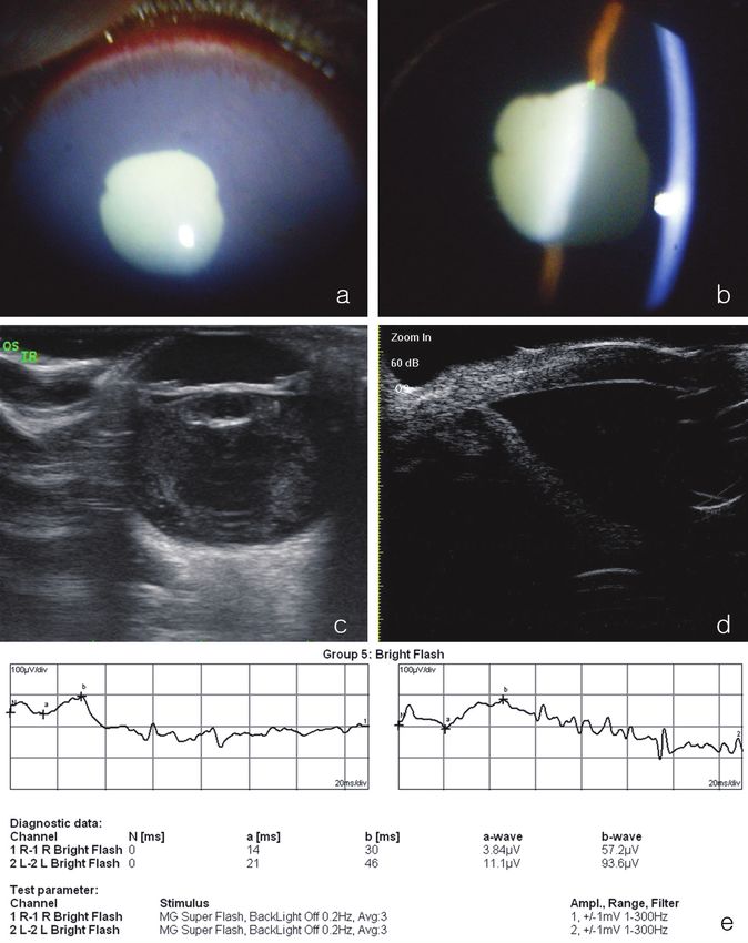
LEE et al. / Turk J Vet Anim Sci
synechiae were detected at 10 o’clock and 2 o’clock (Figure was introduced to engage the I-Ring pupil expander
1a). The color of the iris was very dark and aqueous (Figure 2b). Phacoemulsification was performed using a
flare was detected (Figures 1a and 1b). The lens showed mini-capsulorhexis technique, because severe capsular
a hypermature cataract (Figures 1a and 1b). Posterior fibrosis hindered the use of capsulectomy (7). The nucleus
synechiae were also revealed. Ultrasonography revealed did not seem to be particularly hard, because the patient
severe vitreous degeneration and decreased lens size was only 7 years old. Instead, the progression of cataracts
(Figure 1c). An almost-closed ciliary cleft was found on was rapid, and the lens material rapidly liquefied, leading
an ultrasound biomicroscopy examination (UBM) (MD- to phacolytic uveitis. Thus, because the lens capsule
320WD1101; MEDA Co., Ltd.; Tianjin, China) (Figure was not stable during surgery, phacoemulsification was
1d). No abnormalities in retinal function were noted on performed with low ultrasound power and low bottle
electroretinography examination (ERG) without sedation height. An intraocular lens (IOL) was not implanted due to
or anesthesia with a bright flash system (10 cd s/m2, the ruptured posterior capsule. After performing anterior
mini-Ganzfeld photopic test; RETI port, Roland Consult) vitrectomy to remove the dislocated vitreous, the residual
(Figure 1e). The diagnosis was hypermature cataract with viscoelastic agent was removed carefully. Subsequently,
phacolytic uveitis. Furthermore, secondary glaucoma the I-Ring pupil expander was gently disengaged using a
was also suspected because of the collapsed ciliary cleft. manipulator and completely drawn into the inserter from
Cataract extraction and trabeculectomy were selected for the primary incision. The corneal incision was closed
treatment. Because of transient intraocular hypertension with a 9-0 polyglactin 910 suture (Vicryl, Ethicon LLC,
and the narrowed ciliary cleft, we could not use mydriatics Somerville, NJ, USA).
prior to surgery. Modified filtering surgery with Ologen collagen matrix
Immediately before the surgery, atropine (Isopto was performed following phacoemulsification using the
Atropine, Alcon NV, Hünenberg, Switzerland), tropicamide method described by Lee et al. (8).
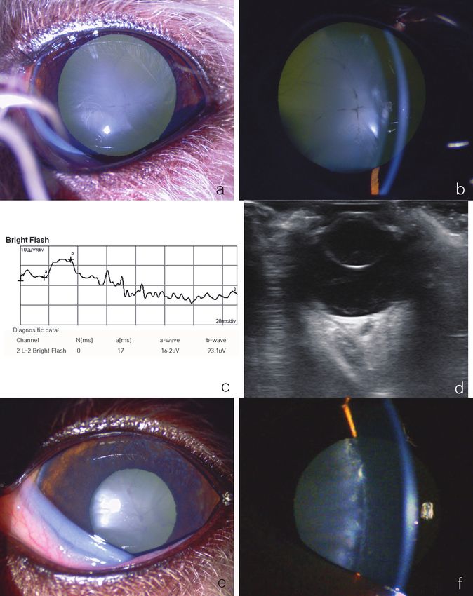
with phenylephrine (Mydrin P, Santen Pharmaceutical 2.2. Case 2
Co. Ltd., Osaka, Japan), prednisolone acetate (Pred A 2-year-old female Bichon Frise was referred with a left
Forte, Allergan, Irvine, CA, USA), flurbiprofen sodium eye cataract that had been detected 1 month before.
(flurbiprofen sodium, Bausch & Lomb, Tampa, FL, USA), During initial ophthalmic examination, the affected
and ofloxacin (Tarivid, Santen Pharmaceutical Co. Ltd.) eye showed a positive dazzle reflex, although the menace
eye drops were administered every 30 min for 2 h; however, response was negative. The PLR, STT (21 mm/min), and
the pupil did not fully dilate because of severe posterior IOP (22 mmHg) were normal. Slit lamp biomicroscopy
synechiae. We therefore prepared an iris expander (Visitec indicated an immature cataract (Figures 3a and 3b). Except
I-Ring Pupil Expander, Beaver-Visitec International, for the cataract, no abnormality was detected in this eye.
Waltham, MA, USA). Upon bright-flash ERG examination, retinal function was
Cefazolin (30 mg/kg intravenously; Safdin, Daehan observed to be normal (Figure 3c). Ultrasound imaging
New Pharm Co., Ltd., Seoul, Korea) was administered as a showed the intumescence of the lens, but there was no
prophylactic antibiotic. Propofol (6 mg/kg intravenously; evidence of lens luxation (Figure 3d). We diagnosed the
Provive 1%, Myung-moon Pharm. Co., Ltd., Seoul, dog with an immature cataract, and at the owner’s request,
Korea) was used to induce anesthesia. Anesthesia was the animal underwent cataract surgery 8 days later.
maintained using isoflurane (Isoflurane; Choongwae Co., Prednisolone (PDS) eye drops (twice a day) and systemic
Ltd., Seoul, Korea). The patient was positioned in dorsal PDS (0.5 mg/kg, twice a day) were administered for 5 days
recumbency. Atracurium besilate (Atra, Hana Pharm before the operation.
Co. Ltd., Gyeonggi, Korea), a neuromuscular blocking Immediately prior to the operation, the affected
agent, was intravenously injected to facilitate the globe eye showed miosis. The IOP was 12 mmHg. Moderate
positioning and reduce the external force arising from conjunctival hyperemia, mild episcleral congestion, and
the extraocular muscles. The ophthalmic surgical site was nictitating membrane protrusion were also detected
routinely prepared. (Figure 3e). The iris color was darker than that at initial
A 3-mm clear corneal incision was made. Intracameral examination, the anterior chamber was deeper, and mild
sodium hyaluronate 1.4% (Healon GV, AMO, Uppsala, aqueous flare was detected (Figure 3f). Prior to surgery, we
Sweden) was injected into the anterior chamber. administered atropine, tropicamide with phenylephrine,
Subsequently, a second 1-mm incision was made at an prednisolone acetate, flurbiprofen sodium, and ofloxacin
angle of 70° from the first incision. Synechiolysis was eye drops every 30 min for 2 h. However, the pupil did
performed with an iris spatula. An I-Ring pupil expander, not dilate sufficiently for cataract extraction. We therefore
in an inserter, was introduced into the anterior chamber prepared an I-Ring pupil expander. The cause of this severe
through the primary incision (Figure 2a). A manipulator miosis was considered to be lens-induced uveitis.
160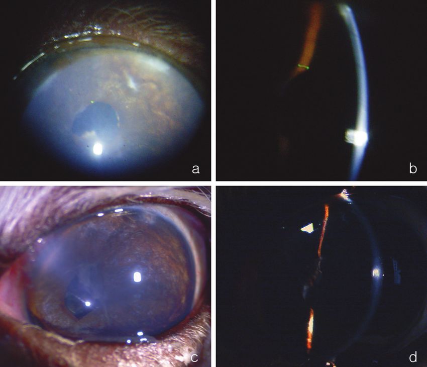
LEE et al. / Turk J Vet Anim Sci

Figure 1. Initial ophthalmic examination of the left eye in Case 1. a, b) Slit-lamp biomicroscopic examination; severe ciliary flush, mild
corneal edema, and an aqueous flare are shown. c) Ultrasonography examination; increased lens opacity, decreased lens volume, and
severe vitreous degeneration are visible. d) Ultrasound biomicroscopic examination showed a narrow ciliary cleft. e) Electroretinography
examination showed normal cone cell function in the left eye.

                                                                                                                                     161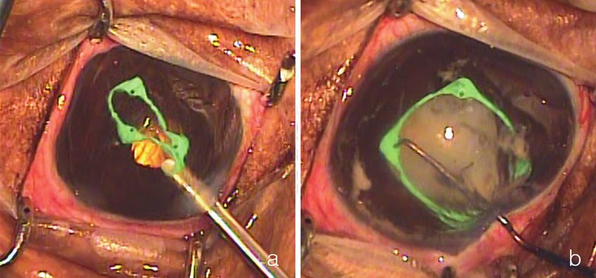
LEE et al. / Turk J Vet Anim Sci

Figure 2. Intraoperative pictures of Case 1. a) An I-Ring pupil expander, in an inserter, was introduced into the anterior chamber
through the primary incision. b) A manipulator was introduced to engage the I-Ring pupil expander.

    Routine ophthalmic anesthesia was performed as for             tropicamide with phenylephrine, prednisolone acetate,
Case 1. The patient was positioned in dorsal recumbency.           flurbiprofen sodium, and ofloxacin eye drops were applied
Atracurium besilate was intravenously injected to facilitate       four times a day. The frequency of application was gradually
globe positioning and to reduce external forces from the           decreased over 2 months. Although the IOP remained low
extraocular muscles. For asepsis, the ophthalmic surgical          (6 mmHg) for 1 month postoperatively, ocular hypertension
site was routinely prepared.                                       developed later. Glaucoma was initially well controlled
    A 3-mm clear corneal incision was made. The                    with antiglaucoma eye drops (Cosopt [three times a day],
anterior lens capsule was stained with Trypan Blue                 Xalatan [twice a day, latanoprost], Pfizer Manufacturing
(Optithech Tissue Blue, Tarun Enterprises, Allahabad,              Belgium, Puurs, Belgium). Corneal opacity did not clear
India). Subsequently, Healon GV was injected into                  up completely, but continuously improved (Figures 5a and
the anterior chamber. An I-Ring pupil expander, in an              5b). Ciliary flush also disappeared. Although the vision in
inserter, was introduced into the anterior chamber. The            this eye was maintained for 250 days postoperatively, it was
I-Ring pupil expander was placed in the pupil using a              later lost because the retina detached due to uncontrolled
manipulator. Capsulorhexis was performed with a 26-G               glaucoma.
needle and continuous curvilinear capsulorhexis forceps.           3.1.2. Case 2
Subsequently, a second 1-mm incision was made at an                The menace response and dazzle reflex were found to
angle of 70° from the first incision. Cataract extraction was      be normal immediately after surgery. Postoperative
performed using a routine phacoemulsification technique            medications were the same as in Case 1. Severe flare,
(Figure 4a). After an IOL (an-lens MD4-13, an-vision Inc.,         moderate corneal edema, and moderate conjunctival
Salt Lake City, UT, USA) was implanted (Figure 4b), the            hyperemia continued for 10 days and then disappeared.
I-Ring pupil expander was gently removed, as described in          Posterior synechiae were present from the 2 o’clock to 5
case 1. The viscoelastic agent was removed with balanced           o’clock position and the pupil did not dilate well during
salt solution. The corneal incision was closed with a 9-0          50 days of follow-up (Figure 5c). Most of the flare had
polyglactin 910 suture.                                            disappeared (Figure 5d). IOP (18 mmHg) and vision were
                                                                   well maintained.
3. Results and discussion
                                                                   3.2. Discussion
3.1. Surgical outcomes and follow-up                               A small pupil is a well-known risk factor for numerous
3.1.1. Case 1                                                      complications during and after cataract surgery (9).
After the operation, the menace response and the dazzle            Various surgical methods have been reported for dilating
reflex were immediately normal. For 10 days after surgery,         pupils that show poor dilation (9). Since pharmacological

162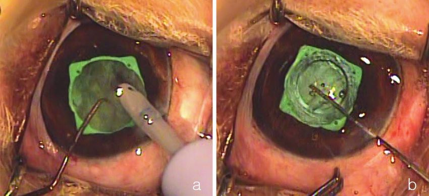
LEE et al. / Turk J Vet Anim Sci

Figure 3. Initial (a, b, c, d) and 8 days later (e, f) ophthalmic examination of left eye (Case 2). a, b) Slit lamp biomicroscopy indicated
an immature cataract. c) Electroretinography examination showed normal cone cell function in left eye. d) Ultrasound revealed
intumescence of the lens, but there was no evidence of lens luxation. e) Eight days later, lens opacity had deteriorated and the anterior
chamber had deepened. The pupil did not dilate well even though we used mydriatics (atropine, phenylephrine, and tropicamide). f)
Aqueous flare detected in the anterior chamber 8 days later.

                                                                                                                                      163
LEE et al. / Turk J Vet Anim Sci
Figure 4. Intraoperative pictures of Case 2. a) Cataract extraction was performed with a routine phacoemulsification technique. b) An
intraocular lens was implanted through the I-Ring pupil expander.
approaches to dilating the pupil before or during cataract terms of pupil diameter during surgery, the I-Ring has
operation cannot guarantee the result, a mechanical pupil some advantages over the Malyugin ring. First, the I-Ring
dilator should be used at the time of phacoemulsification pupil expander can be more safely manipulated via four
(9). Iris expanders have become increasingly popular, positioning holes and acts as a barrier against iris aspiration
because they allow easy mechanical dilation of the pupil during phacoemulsification, in 360 degrees (10).
(9,10). In veterinary medicine, the difficulty with small However, iris expanders also have limitations in
pupil cases is resolved by means of nonsteroidal eye veterinary ophthalmology. First, the canine mean pupil
drops, strong mydriatics, and viscomydriasis (4), although diameter is wider than that of humans. The diameter of
with these methods, pupil dilation remains insufficient expanders should therefore be wider than those developed
for phacoemulsification in some cases. In cases of small for humans, so that the pupil size will be sufficiently wide for
pupils with posterior synechiae, iris cutting methods, iris safe operation during canine phacoemulsification. Second,
stretching methods, or iris retainer methods are needed iris expanders cannot adjust the pupil size according to
to dilate the pupil sufficiently (2). Iris cutting methods the preference of the surgeon (3). Third, expanders are
and iris stretching methods have similar disadvantages, in only single-use; thus, to reduce costs, reusable expanders
that they require a certain level of experience and carry should be developed. Although the I-Ring pupil expander
a risk of sphincter rupture (2). Among the iris retainer can protect the iris during surgery, the contact between
methods, iris expanders compensate for the deficiencies the expander and the iris can induce uveitis, which
of other two methods (2,9). Representative iris expanders results in posterior synechiae. In this present study, severe
are the diamond-shaped Malyugin ring (MicroSurgical posterior synechiae were detected after surgery in both
Technology Inc., Redmond, WA, USA) and the circular cases. It is not clear whether these synechiae originated
I-Ring pupil expander (10). Both expanders have the from preoperative uveitis or contact of the pupil expander
advantage of easy insertion and removal via an injector, as with the iris. Severe uveitis occurred in both cases after
compared with the traditional iris retainer, and decrease operation and may have resulted in the severe synechiae.
the risk of sphincter rupture because the pupil is not In Case 1, we used combined cataract and glaucoma
stretched excessively. surgery, which may have caused severe postoperative
While the Malyugin ring is inserted through a 2.2-mm uveitis and a risk of hypotonic IOP. In this case, combined
single port and the mean pupil diameter during surgery surgery was indicated because of the narrowed ciliary cleft
is 6.25 mm, the I-Ring pupil expander, which was used that was detected by UBM examination and the transient
during our surgery, is inserted through a 2.5-mm single ocular hypertension that was reported. We did not
port and provides a mean pupil diameter of 6.3 mm during choose to use endolaser cyclophotocoagulation because
surgery (10). Although there is no major advantage in of the severe preoperative uveitis. Filtering surgery was
164LEE et al. / Turk J Vet Anim Sci
Figure 5. Postoperative pictures. a, b) Slit lamp examination of Case 1, 65 days postoperatively. c, d) Slit lamp examination of Case 2,
50 days postoperatively.
performed as rapidly as possible after cataract surgery to technique has some limitations; for instance, it can lead
minimize the probability of hypotonic IOP. to the development of synechiae, and it can dilate the
In conclusion, it is useful and effective to use the pupil only to 6.3 mm in canine cataract patients. As no
I-Ring pupil expander to manage small pupils that cannot previous reports of using the I-Ring pupil expander to
be sufficiently dilated by medication during cataract manage small pupils in veterinary medicine are available,
extraction. Although mechanical iris retraction and we believe that these case reports will provide insight into
expansion are not recommended because manipulations a useful technique for cataract extraction in cases with an
of the canine iris can result in serious inflammation, small- insufficiently dilated pupil, and the information provided
pupil enlargement techniques are occasionally required; can be applied in veterinary settings in the future.
in such cases, the I-Ring pupil expander is useful. This
165LEE et al. / Turk J Vet Anim Sci
References
1. Smith GT, Liu CS. Flexible iris hooks for phacoemulsification 7. Gimbel HV. Principles of nuclear phacoemulsification.
in patients with iridoschisis. J Cataract Refr Surg 2000; 26: In: Steinert RF, editor. Cataract Surgery, Techniques,
1277-1280. Complications, and Management. 2nd ed. Philadelphia, PA,
USA: Saunders; 2004. pp. 153-181.
2. Kershner RM. Management of the small pupil for clear corneal
cataract surgery. J Cataract Refr Surg 2002; 28: 1826-1831. 8. Lee SJ, Kim JY, Jeong SW. Modified trabeculectomy, using
Ologen collagen matrix implants, for treating glaucoma in 4
3. Akman A, Yilmaz G, Oto S, Akova YA. Comparison of various
dogs. Vet Ophthalmol 2016; 19: E23.
pupil dilatation methods for phacoemulsification in eyes with
a small pupil secondary to pseudoexfoliation. Ophthalmology 9. Malyugin B. Cataract surgery in small pupils. Indian J
2004; 111: 1693-1698. Ophthalmol 2017; 65: 1323-1328.
4. Wilkie DA, Colitz CMH. Surgery of the lens. In: Gelatt KN, 10. Tian JJ, Garcia GA, Karanjia R, Lu KL. Comparison of 2 pupil
Gilger BC, Kern TJ, editors. Veterinary Ophthalmology. 5th ed. expansion devices for small-pupil cataract surgery. J Cataract
Ames, IA, USA: John Wiley & Sons, Inc.; 2013. pp. 1234-1286. Refr Surg 2016; 42: 1235-1237.
5. Kim ES, Han SB, Lee SJ, Kim M. Cataract surgery through the
small pupil. Clin Interv Aging 2016; 11: 1387-1389.
6. Fine IH, Packer M, Hoffman RS. Phacoemulsification in the
presence of a small pupil. In: Steinert RF, editor. Cataract
Surgery, Techniques, Complications, and Management. 2nd
ed. Philadelphia, PA, USA: Saunders; 2004. pp. 211-221.
166You can also read