The erbium laser: the Star Wars of dentistry
←
→
Page content transcription
If your browser does not render page correctly, please read the page content below
Clinical excellence

The erbium laser:
the Star Wars of dentistry
Fred S. Margolis looks at dentistry’s new weapon against tooth decay, the erbium laser

Dentistry has a new weapon in the fight against tooth decay.         Hard tissue laser dentistry includes the use of the laser for
This ‘light saber’ of dentistry is the erbium laser.              Class I through Class VI preparation of carious teeth. The
   The dental laser is the latest modern innovation for the       main advantages of the erbium laser for this use are the
21st Century. Erbium lasers have proven safe and effective        following:
for the removal of tooth decay and cavity preparation, in         • No anesthesia in the majority of patients due to the
addition to many soft tissue and hard tissue surgical             numbing effect of the laser
procedures.                                                       • No waiting for the patient to be anesthetised in the
   The FDA approved the erbium laser for marketing in the         majority of patients
United States in 1997. It offers an alternative to the high-      • No concern about the patient biting their lip, cheek, or
speed drill, eliminating fear and patient discomfort for both     tongue
adults and children. The laser is revolutionising dental care –   • More pleasant experience, due to not being anesthetised.
just as it has in many other areas of our lives. With the            The erbium laser can be used for soft tissue surgery in
erbium laser, the dentist can provide a new method of dental      many ways. These include:
care that can often be performed without local anaesthesia        • Gingivectomy (Figure 5)
(Figure 1).                                                       • Frenectomy (labial and lingual)
   Most patients find laser procedures remarkably                 • Gingivoplasty
comfortable. So comfortable, in fact, that in many cases no       • Exposure of teeth to aid tooth eruption
anesthesia is required (Figure 2). People who have                • Operculectomy
experienced laser treatment for cavity preparation report         • Gingival removal to expose areas for restorations
feeling nothing more than the touch of the handpiece and          • Aphthous ulcers (Figure 7)
an occasional slight sensation of warmth. Teenage patients        • Pulp therapy
report a ‘tingling’ feeling. Unfortunately, conventional          • Abnormal gingival architecture associated with orthodontic
drilling must still be used for the removal of previous metal     movement
restorations. The erbium laser has been used to prepare           • Excision of soft tissue tumors, including fibromas, lipomas,
crowns and veneers without the aid of conventional rotary         etc (Convissar, 2000).
instruments (Nash, 2002).
   The dental laser often eliminates the unpleasant
after-effects associated with many dental procedures –            The types of erbium lasers
soreness, bleeding, inflammation, sutures and numbness. It        Paghdiwala, in 1988, tested the ability of the erbium:YAG
also creates no known after-effects of its own. ‘The advantage    laser to ablate dental hard tissues. He prepared holes in
of laser surgery is the minimisation of intra-operative           enamel and dentine with low energy. Without water cooling,
haemorrhage and a decrease in post-operative pain                 the cavities exhibited no cracks and little or no charring.
symptoms. Carbon dioxide and Nd:YAG lasers have been                 The erbium:YAG (2.94 µm) laser was approved for market-
used effectively for soft tissue oral surgery procedures. Argon   ing by the US Food and Drug Administration in May 1997.
lasers have also been used in oral surgical procedures. One of    This laser wavelength was shown to produce precise ablation
the main advantages of laser surgery conventional excision        of sound and carious dentin and enamel with a thermal
with scalpel is the reported lessening of post-operative pain     penetration of shallow proportions. The erbium chromium:
and the ability to excise or ablate with less bleeding’ (Rizoiu   YSGG (2.78um) is made of erbium, chromium, yttrium,
et al, 1996) (Figure 3).                                          scandium, gallium and garnet, and has the same properties
                                                                  as the erbium:YAG laser (Convissar, 2000) (Figure 2).

Clinical uses for the erbium laser
The erbium laser has various uses, which can be divided into      Mechanism of action on hard tissue
hard and soft tissue procedures for dentistry.                    According to Hadley et al, the mechanism of action on the 4

14                                                                                                           Private Dentistry May 2009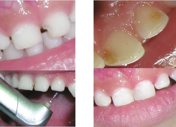
Figure 2: Left photo: Er,Cr;YSGG Waterlase MD. Note: Illumination.
Right photo: Class II preparation with no anaesthetic and no handpiece
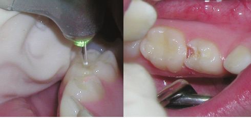
Figure 1: Class III Preparation with the Waterlase MD
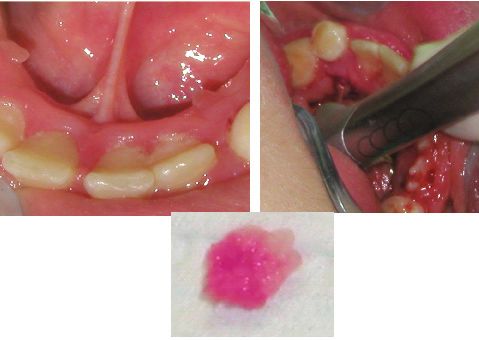
Figure 4: Ankyloglossia in a 15-year-old male
Figure 3: Waterlase MD preparing primary first molar for vital
pulpotomy
hard tissues of the human body (i.e. enamel, dentin, photoaccoustic effect that develops is characteristic of a short
cementum and bone), is that the Erbium laser ‘delivers interaction time (100 microseconds) and a high energy
photons into an air/water spray matrix with resultant density. The incident laser energy is absorbed in a thin
microexplosive forces on water droplets. This process is surface layer. Water, hydroxyapatite, and collagen have an
hypothesised to contribute significantly to the mechanism of affinity for this laser energy. The water spray of the laser
hard-tissue cutting.’ (Hadley et al, 2000) (Figure 2) handpiece accelerates this effect. Water-mediated explosive
‘Both the Er:YAG and the Er;Cr:YSGG can be categorised tissue removal has been shown to be the most efficient way
as having photomechanical effects. Laser light that is highly of removing tissue, while transferring minimal heat to the
energetic and is short pulsed causes fast heating of the dental remaining tooth.’ (Miserendino and Pick, 1995)
tissue in a small area. A fast shockwave is created when the Scanning electron microscopy (SEM) has shown that the
energy dissipates explosively as a volumetric expansion of erbium laser ‘makes clean cuts through enamel and dentin
the water occurs. This is called cavitation. Water molecules without creating a significant smear layer’ (Hadley, 2000).
in the target area are superheated, explode, and thus ablate The mechanism of action of the erbium laser is that: ‘Water
the tooth structure and caries. A bactericidal effect, typical of that is bound to the crystalline structures of the tooth
laser/tissue interaction, also occurs. The shockwaves that absorbs the laser light readily and easily. The vapourisation
occur are due to a rapid photovapourisation of water, of the water within the mineral substrate causes a massive
producing a volumetric change of state of the liquid within volume expansion, and this expansion causes the surround-
the tooth. This change creates high pressure, removing and ing material to literally explode away.’ (Coluzzi, 2000)
destroying selective areas of adjacent tissue. The The erbium laser is slower in cutting through enamel 4
16 Private Dentistry May 2009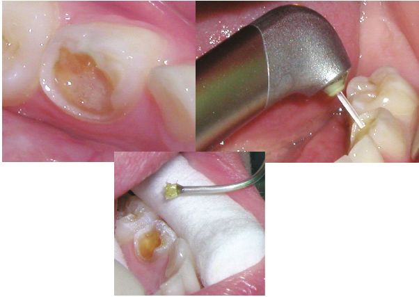
Figure 5: Left: Waterlase MD. Right: Immediate post-surgery. Note: No
bleeding
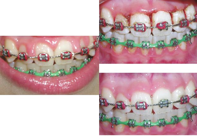
Figure 6: Left: 12 year old female with gingival hyperplasia. Right
upper: Immediate gingivectomy (Waterlase MD). Lower right: One
month post-surgery
Figure 7: Aphthous ulcer before and after treatment with Waterlase MD Figure 8: Excision of fibroma with Waterlase MD
than dentine. This is due to the fact that there is more water photographs were used to conclude that erbium:YAG laser
in dentine than enamel and more water in carious dentine, preparation of dentine left a surface for strong bonding or
so the ablation of each of these tissues occurs at a varying composite material. Frentzen reported the surface
rate. Erbium lasers have been shown to cut hard dental morphology of enamel remained rough after erbium:YAG
tissues with efficacy and depth that corresponds to the preparation. The laser treatment ‘allowed additional etching,
increasing power setting and use of a water spray (Eversole resulting in a microretentive pattern’ (Convissar, 2000)
and Rizoiu, 1995). (Figure 9).
Etching of the enamel surface Pulpal tolerance
Lased tooth surfaces have been evaluated for their ability to No odontoblastic alterations have been noted, nor is there
form adhesion with various bonding agents; shear and any inflammatory response in the pulp chamber beneath the
tensile strength assays have been used to compare bonding preparation (Eversole and Rizoiu, 1995). Histopathologic
to lased and acid-etched enamel and dentinal surfaces studies in animals and humans have shown that pulpal
(Hadley, 2000). The etching of enamel and dentine by the tissues underlying deep cavity preparations made with an
erbium laser has been shown to be ‘facilitated or even erbium laser do not undergo pathological changes. It was
improved over acid etching techniques’ (Eversole and Rizoiu, shown, utilising rats teeth, that fibroblast proliferation is
1995). In a study performed by Visuri et al in 1996, the laser observed sooner and more frequently in the specimens
sampled dental surfaces had improved bond strengths when treated with the erbium:YAG laser than those prepared with
compared to acid-etched and handpiece controls. SEM the high-speed drill (Takamori, 2000). 4
18 Private Dentistry May 2009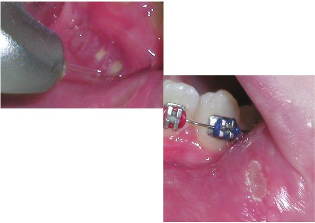
Figure 9: Female, age 12: maxillary frenectomy and gingivoplasty with
                                                                erbium laser. Upper right: Immediate post-surgery. Lower left: One
                                                                month post-surgery

   Also, Rizoiu et al (1996) found that the laser powered       effectively remove cements and composites with ablation
Er;Cr:YSGG, when used for preparation of carious lesions,       efficiency similar to that of healthy tooth structure. Previ-
had ‘no apparent adverse thermal effect as measured in the      ously placed dental sealants can also be removed with the
pulp space’ (Keller and Hibst, 1997).                           laser. Dental restorative preparations are possible and while
   Coluzzi (2000) states that ‘…laboratory studies indicate     not as precise as with a bur, these preparations can be
that the pulpal temperature of the treated tooth may actually   improved by limited hand instrumentation, which should
decrease by five degrees centigrade during laser treatment.’    allow for successful placement and retention of dental and
   The efficiency of ablation by the Erbium:YAG laser has       restorative materials (Coluzzi, 2000) (Figure 2).
been explained as a ‘thermally induced mechanical process…
the incident Er:YAG laser radiation is absorbed in a thin
surface layer, causing sudden heating and vapourisation of      Comfort of the patient: handpiece vs. erbium laser
the water. A high steam pressure then leads to                  The non-contact laser preparation seems to be comfortable
microexplosions with erupting particles with a crater           to the patients, whereas drilling may cause pain sensations
corresponding morphology.                                       due to various causes such as vibration, pressure, heat, and
   ‘Because the tissue is not vapourised completely but only    noise. In an article by Hadley et al (2000), intra-operative
disintegrated into fragments, the radiant energy is converted   discomfort levels indicated a higher prevalence of discomfort
efficiently into the ablation that alters the morphological     among the air turbine/bur-treated teeth than among the
structure of the tissue. No melting process takes place that    erbium laser treated teeth.
might lead to considerable heat damage to the surrounding          Keller and Hibst (1997) reported on two clinical studies. In
tissues.’ (Miserendino and Pick 1995)                           the first study, 67 teeth of 33 patients were prepared with an
                                                                erbium:YAG laser. Buccal preparations were used for this
                                                                study. In all 67 treatments, no pain was reported in 24 teeth
Precision of the erbium laser in cutting tooth                  and minimal pain in 38 teeth. In 29 of 41 teeth with deep
structure                                                       caries, only minimal discomfort was reported. In a second
Keller (1997) showed that the erbium laser can be applied to    study, the pain of the laser versus mechanical drilling was
both primary and secondary carious lesions. It can also         compared. Only 6% of the patients required local             4
20                                                                                                              Private Dentistry May 2009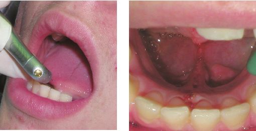
anaesthesia with laser preparation, compared to 11% of the         Two days post-operatively, the wounds are closed in the
patients with drilled cavity preparations. 83% of the patients     epithelial parts. Eight days post-operatively, the epithelial
indicated that the bur was more uncomfortable than the             would healing is complete (Figure 8). The subepithelial
laser; 88% of the patients indicated a preference of the           fibrous tissue is not totally repaired at this time. This corre-
Erbium:YAG laser. ‘The pain perception during laser treat-         sponds to normal wound healing after surgical incision or
ment was reduced; the pain was described as only like short        excision by scalpels. In contrast, the carbon dioxide laser
needle sticks.’ (Miserendino and Pick, 1995)                       cuts show a delay in wound healing of two to three days
                                                                   because of the extended thermal damage zones (Miserendino
                                                                   and Pick, 1995). PD
Other effects of the laser on the tooth
Caries prevention
Hicks (1993) reported the effect on caries-like lesions and        References
progression in enamel after the use of the argon laser. The        Coluzzi, D.J. (2000) An Overview of Laser Wavelengths Used
resulting surfaces were shown to have a lowered pH from 5.5        in Dentistry. Chapter in: The Dental Clinics of North America.
to 4.78. This hardened enamel was four times more resistant        Philadelphia: W.B. Saunders,
to acid dissolution. The increase in resistance caused a           Convissar, R.A.(2000) The Dental Clinics of North America.
significant reduction in the depth of the carious lesion.          Philadelphia: W.B. Saunders
Theorising, the enamel micropores may trap the ions                Eversole, L.R. and Rizoiu, I.M.( December 1995) Preliminary
released (calcium, phosphate, fluoride) that become                Investigations on the Utility of an Erbium, Chromium YSGG
dissolved during the formation of caries. Enamel that has          Laser. CDA Journal, 41-47
been lased has a greater attraction for the calcium phosphate      Hadley, J.,et al. (2000) A Laser-Powered Hydrokinetic System.
and fluoride ions, with the result of a reprecipitation of the     J ADA; 131: 777-785
mineral phase. Therefore, irradiation by laser may be              Hicks, M.J., et al. (1993) Caries-like lesion initiation and
important in caries prevention in enamel that is sound.            progression in sound enamel following argon laser irradia-
                                                                   tion: An in-vitro study. ASDC J Dent Child. 60: 201-206
Histologic effects                                                 Keller, U., Hibst, R. (1997) Effects of Er:YAG Laser in Caries
Effects observed by both the lased and the control groups          Treatment: A Clinical Pilot Study. Lasers in Surgery and
showed no significant differences in the quantitative effects      Medicine 20: 32-38
on the odontoblasts, predentine, and dentine, which showed         Miserendino, L.J., Pick, R.M.(1995)Lasers in Dentistry.
mild changes in both groups. When the conventional drill           Chicago. Quintessence International
was used, the histologic variations were larger for the pulpal     Nash, R. (2002) Crown and Veneer Preparation Using the
tissue, odontoblasts, and predentine. The immediate and            Er,Cr:USGG Waterlase Hard and Soft Tissue Laser.
long-term effects of the erbium laser for caries removal,          Contemporary Esthetics and Restorative Practice, October: 80-
preparation of the cavity, and etching of the enamel have          86
shown no significant differences.                                  Rizoiu, I.M., et al. (1996) Effects of an erbium, chromium:
   These variations suggest to the author that the dental drill    yttrium, scandium, gallium,garnet laser on mucocutaneous
may be more harmful to the tooth than the erbium laser. The        soft tissues. Oral Surg Oral Med Oral Pathol Oral Radiol. 82:
localised areas of healing seen within the pulp adjacent to        386-395
the cavity preparations should be considered a normal              Takamori, K. (2000) A Histopathological and Immunohisto-
physiological response. There was no significant damage to         chemical Study of Dental Pulp and Pulpal Nerve Fibers in
the pulp on histological examination of teeth in which it          Rats After the Cavity Preparation Using Er:YAG Laser. J Endod.
appeared radiographically that the laser cavity preparation        26: 2
reached the pulp. ‘The laser may have a potential bactericidal     Visuri, S.R., et al.(1996) Shear Strength of composite bonded
and sealing effect on the pulp when exposed.’ (Convissar,          to Er:YAG laser-prepared dentin. J Dent Res 75: 1, 599-605
2000) (Figure 4)

Mechanism of action on soft tissues
The erbium:YAG laser has indications in soft tissue surgery if     Comments to pd@fmc.co.uk
no coagulation effect is desired, i.e. removal of hyperplastic
gingival tissue, periodontal surgery, and ablation of large        Fred S. Margolis, DDS, FICD, FACD is adjunct clinical assistant
benign lesions of the oral mucosa or the skin, without             professor for paediatric dentistry at University of Illinois College
closing the wound by sutures, according to Keller and Hibst        of Dentistry in Chicago, Illinois, USA. He is also clinical
(1997) (Figure 9). After focused Er:YAG laser irradiation, small   instructor at Loyola University’s Oral Health Centre in
and deep cuts with partial bleeding are seen. Shallow and          Maywood, Illinois, USA.
large lesions are produced by defocused irradiation. In all
cases, only a minimal damage zone of carbonisation occurs.

22                                                                                                               Private Dentistry May 2009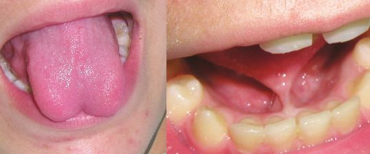
You can also read