Modification of a haematoxylin, eosin, and natural saffron staining method for the detection of connective tissue
←
→
Page content transcription
If your browser does not render page correctly, please read the page content below
J Vet Res 65, 2021
DOI:10.2478/jvetres-2021-0008
Modification of a haematoxylin, eosin,
and natural saffron staining method
for the detection of connective tissue
Cassandra Ceccopieri, Joanna Skonieczna, Jan P. Madej
Division of Histology and Embryology, Department of Biostructure and Animal Physiology,
Wrocław University of Environmental and Life Sciences, 50-375 Wrocław, Poland
ceccopieri@yahoo.com
Received: August 10, 2020 Accepted: January 12, 2021
Abstract
Introduction: The aim of our study was to optimise an existing staining procedure: haematoxylin-eosin saffron (HES). The
method follows the classical haematoxylin and eosin protocol with the addition of a staining step using natural saffron to better
identify the collagen fibres. Material and Methods: The saffron solution was obtained by dissolving ground saffron stigmas in
absolute alcohol. In order to test the HES method for its staining ability on four main types of collagen (I, II, III, and IV), specific
tissues (skin, tooth, cartilage, aorta, spleen, and penis) were chosen. Results: The procedure showed a sharp differentiation between
muscle, stained red or pink, and connective tissue, stained bright yellow or orange. Conclusion: HES allows the diagnosis of
reticulin fibrosis undetected in HE and in previous saffron staining procedures. HES represents an advantageous alternative to HE
staining giving highly reproducible results with high diagnostic value.
Keywords: saffron, collagen fibres, multichromatic staining, connective tissue.
Introduction component of the extracellular matrix and is, like most
of the extracellular fibres, an acidophilic structure (6).
Saffron is a spice obtained from the threads of The acidity of its water-soluble carotenoids makes
Crocus sativus’ flowers. Originating in the Middle East, saffron a suitable dye for collagen fibres.
saffron has been used for centuries as a spice and The function of collagen is to confer strength and
colouring agent for food and clothes (13). The main support for the organs. It can be found in many parts of
bioactive compounds of saffron are picrocrocin, the body and occurs in different types, of which almost
responsible for the bitter taste; safranal, emitting the 30 are known (7). The most frequently found are the
scent; and the carotenoid pigments crocetin and crocin, fibrillar types (I, II, III, V, XI, XXIV, and XXVII) that
which give the typical vivid crimson colour (1). Crocins, are characterised by extensive cross-linking which
safranal, and picrocrocin have been proved to have provides mechanical strength for high-stress tissues like
a cytotoxic effect on human cancer cells (11). skin, cartilage, and bone (5).
In 1719 saffron was used by Leeuwenhoek to stain When secreted in excess, collagen may lead to
animal tissue sections and simplify the microscopic tissue hardening and overgrowth causing a pathological
analysis. In the 20th century, Conn applied it to detect change called fibrosis. Fibrosis is usually a side effect of
liver damage and obtain differential staining for the chronic inflammatory reactions triggered by different
glandular cells of the stomach (9). Saffron was then kinds of stimuli like physical or chemical insults,
employed to distinguish collagen fibres in several infections, hypersensitivity reactions, autoimmune
multichromatic stains such as a variant of Masson’s responses, and radiation (14).
trichrome using haematin, phloxine, and saffron (3) or In 1997 Edston and Gröntoft (4) introduced the first
modified Russell-Movat pentachrome (10). method adding a saffron staining step to the classical
Collagen is the primary structural element of the haematoxylin and eosin substitute erythrosin B protocol.
dermis of vertebrates and it makes up to 30% of total The procedure gives impressive results in differentiating
body protein in mammals (7). It represents the main the dermis from the muscular tissue underneath it in skin
© 2021 C. Ceccopieri et al. This is an open access article distributed under the Creative Commons Attribution-
NonCommercial-NoDerivs license (http://creativecommons.org/licenses/by-nc-nd/3.0/)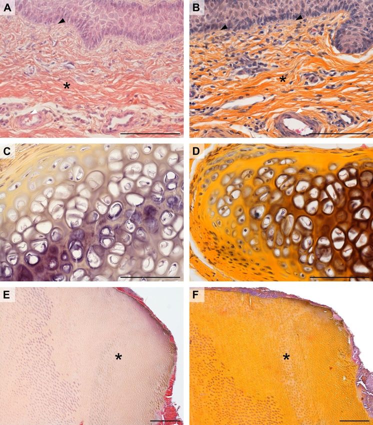
C. Ceccopieri et al./J Vet Res/65 (2021)
samples, but it fails to clearly distinguish muscle from HES procedure. The cut sections were mounted on
connective tissue in several other organs like blood glass slides, deparaffinised in xylene, and rehydrated in
vessels, the spleen, and the penis. Furthermore, in the a degressive series of ethanol solutions. The samples
case of tooth samples, the aforementioned method does were incubated in haematoxylin solution for 4 min, then
not stain the collagen present in the dentin. rinsed in distilled water and immersed in acid alcohol for
We introduce an improved version of the method 1 min. The slides were then washed in running tap water
presented by Edston and Gröntoft succinctly described for 10 min, incubated in eosin solution for 4 min and
as haematoxylin-eosin saffron (HES). It consists of rinsed in distilled water for a few seconds. Subsequently,
a trichromatic staining procedure for routine use in the specimens were briefly immersed in 96% ethanol
histological laboratories that facilitates the identification and incubated in the saffron alcohol solution (pre-
of collagen fibres and accelerates pathological warmed to 35–40°C for 10 min) for 15 min; the slides
investigation of connective tissue in a wide range of were examined microscopically after 5 min incubation
organs. in the saffron solution following a fast rinse in distilled
water as is recommended. The slides were then
immersed in two changes of absolute alcohol for 5 min
Material and Methods and mounted with cover slides.
Tissues. To evaluate the HES method for its ability
to stain the four main types of collagen (I, II, III, and IV), Results
skin, tooth, cartilage, aorta, spleen, and penis tissues
were selected. The tissue samples (stored in paraffin The procedure sharply differentiated between
blocks) were retrieved from the archive of the Division muscle, stained red or pink, and connective tissue
of Histology and Embryology of Wrocław University of stained bright yellow or orange.
Environmental and Life Sciences. The organs had HES for routine stainings. The artery wall
previously been fixed in a 4% buffered formaldehyde (mouflon aorta) slides exemplify how HES (Fig. 1B)
solution, washed in running tap water, dehydrated, and gives improved contrast between connective tissue and
cleared in xylene, suitably preparing them for use in muscular tissue staining colours in comparison to the
a Shandon Citadel 1000 automatic tissue processor haematoxylin-erythrosine saffron procedure (Fig. 1A).
(Thermo Fisher, Germany) in standard mode. The A similar result was obtained in chicken spleen (Fig. 1C
embedding procedure in paraffin was performed using and D), where the HES-stained sample showed a more
a Myr Ec 500 tissue embedding centre (Especialidades visible contrast between reticular fibres (type III
Médicas Myr S.L., El Vendrell, Spain). Sections of 5 μm collagen) meshwork and the cells in the red and white
were cut with an HM 310 microtome (Microm pulp. The collagen type I-rich connective tissue of the
International, Walldorf, Germany) and placed on glass trabeculae as well as smooth muscle cells in the artery
slides (Thermo Fisher Scientific, Waltham, MA, USA). wall are also clearly visible. In rat skin (Fig. 1E and F),
Solutions. The solution of saffron in alcohol was the HES and haematoxylin-erythrosine saffron staining
prepared as follows. One gram of natural saffron stigmas procedures gave comparable results. However, the
(from a food market) was ground into a powder, saffron dye of HES allowed a higher saturation of the
dissolved in 15 mL of absolute alcohol, mixed and connective tissue stain to be achieved in the dermis.
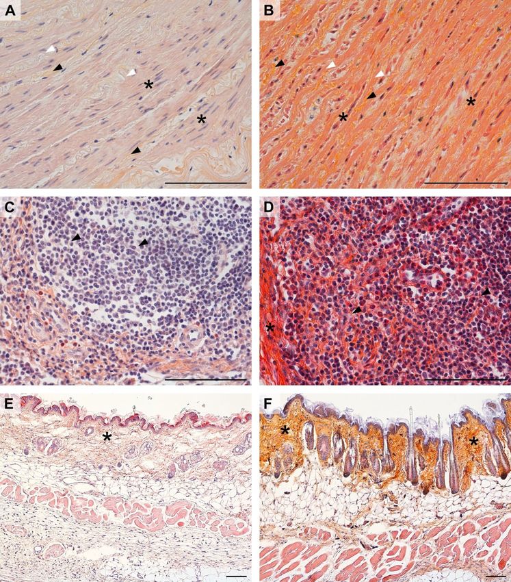
heated at 35–40℃ for 30 min on a heated stirrer. The In Fig. 2A, the saffron dye of the haematoxylin-
solution was then filtered and stored at room temperature erythrosine saffron procedure failed to stain the
for further use. To obtain the eosin staining solution, 10 g connective tissue in the pig penis, which only took a dark
of eosin powder (Avantor Performance Materials, red colour from the erythrosine B dye. In contrast, the
Gliwice, Poland) was dissolved in 1L of distilled water HES-dyed specimen (Fig. 2B) showed a clear distinction
and mixed for 30 min at 35–40°C on a heated stirrer. between the pink colour of the muscle cells in the tunica
Once the solution was homogeneous, 10 drops of glacial media of the artery wall given by eosin, and the yellow
acetic acid were added. The haematoxylin staining steps colour given by saffron to the collagen of the connective
were performed using the ready-to-use solution tissue of the penis. Furthermore, unlike the
according to Delafield (CAS no. MHS80; Sigma- haematoxylin-erythrosine-saffron procedure, HES
Aldrich, Darmstadt, Germany). Masson–Goldner stained the collagen in the basal membrane. In hyaline
trichrome and Reticulin silver plating according to cartilage (Fig. 2C and D), HES and haematoxylin-
Gordon and Sweet’s histological procedures were erythrosine saffron staining achieved very similar results
carried out respectively using the CAS no.1004850001 but there was still higher contrast in the HES-stained
and CAS no.1002510001 Sigma-Aldrich staining kits. sample. In rabbit tooth samples (Fig. 2E and F),
Once stained, the slides were mounted using the Euparal haematoxylin-erythrosine saffron did not stain the
ready-to-use microscopic mounting solution (CAS no. dentin. Contrary findings were made with HES, which
7356.1; Carl Roth, Karlsruhe, Germany). impregnated this sample satisfactorily.
C. Ceccopieri et al./J Vet Res/65 (2021)

     HES in diagnostic applications. HES staining is                      applied to the same specimen of cavy liver results in
also very useful in the diagnosis of reticulin fibrosis.                  lower contrast between connective and glandular
Both type I and III collagen are clearly visible (Fig. 3D),               (epithelial) tissue. Type III collagen is also more
while the structure of the cell cytoplasm is clearer than                 difficult to discern. Similar results to those of HES were
in Masson–Goldner trichrome staining (Fig. 3B).                           obtained only with silver impregnation, an elaborate and
Haematoxylin-erythrosin saffron staining (Fig. 3A)                        specific methodology designed to stain type III collagen.

Fig. 1. Comparison of staining outcomes of the haematoxylin-erythrosine saffron method (A, C, and E) and haematoxylin-eosin saffron (HES)
method (B, D, and F)
A and B – mouflon aorta; black arrowhead – collagen (mainly type I) fibres; white arrowhead – elastic fibres; asterisk – smooth muscle cells
C and D – chicken spleen; asterisk – trabeculae; arrowhead – reticular (type III collagen) fibres
E and F – rat skin; asterisk – collagen of the dermis. All scale bars =100 µm
C. Ceccopieri et al./J Vet Res/65 (2021)
Fig. 2. Comparison of staining outcomes of the haematoxylin-erythrosine saffron method (A, C, and E) and haematoxylin-eosin saffron (HES)
method (B, D, and F)
A and B – pig penis; black arrowhead – basement membrane; asterisk – collagen (mainly type I) fibres; white arrowhead – smooth muscle cells
C and D – hyaline cartilage from the trachea of the rabbit
E and F – rabbit tooth; asterisk – dentin. All scale bars = 100 µm
Discussion applied, the largest and slowest diffusing dyes, like
saffron, tend to accumulate first in the more hydrated
The procedure described in this article maintains elements of the tissues with great porosity and highly
the characteristics of the HE protocol with the advantage accessible surface area like collagen fibres.
of giving a clear distinction between collagen and muscle. Contrastingly, small acid dyes establish nonspecific
The specificity of interaction with collagen fibres is (most probably electrostatic) interactions with the amine
probably due to the chemical nature of the stain. Indeed, group of amino acids: for instance, eosin binds to free
when a combination of large and small acid dyes is NH2 groups in proteins (2).C. Ceccopieri et al./J Vet Res/65 (2021)
Fig. 3. Comparison of different staining methods in the diagnosis of reticulin fibrosis in cavy liver
A – haematoxylin-erythrosine saffron method
B – Masson–Goldner trichrome
C – silver impregnation
D – haematoxylin-eosin saffron (HES) method; asterisk – collagen (mainly type I) fibres; arrowhead – reticular (type III collagen) fibres. All scale
bars = 100 µm
When compared with previous methods erythrosin saffron methods. Similar results were
employing saffron (4), HES shows a wider spectrum of obtained only with silver impregnation, an elaborate
application allowing the collagen fibres to be procedure that requires the employment of more toxic
distinguished from the surrounding tissues with high substances.
contrast in a great variety of organ samples. Therefore, HES method represents a great alternative
Furthermore, saffron represents a non-toxic alternative to HE staining giving highly reproducible results with
to other substances. Indeed, several multichromatic high diagnostic value. This method is also easy and rapid
stains employ dyes like metanil yellow, a pH indicator to perform and requires less toxic and expensive chemicals
known for its neuro- (8) and hepatotoxicity (12) as than other techniques. Moreover, the interpretation of
a food colourant also classified as a corrosive and chronic the results of the HES method is straightforward because
hazard to aquatic life (Metanil Yellow, PubChem of its analogy to those of classical HE.
Database, CID 393558, contained for example Alcian
Blue–HE Metanil Yellow stain). Other methods, like Conflict of Interests Statement: The authors declare
Reticulin silver plating according to Gordon and that there is no conflict of interests regarding the
Sweet, apply corrosive silver nitrate (PubChem publication of this article.
Database, CID 24470), and formalin, a carcinogenic
agent (formaldehyde, PubChem Database CID 712), to Financial Disclosure Statement: The publication of the
detect collagen (type III) presence in tissue. paper was financed under the Leading Research Groups’
HES offers improved diagnostic utility, as clearly support project from the subsidy for the period 2020–
shown in the liver sample, where saffron allowed 2025 in the amount of 2% of the subsidy referred to
fibrosis of the reticular fibres to be revealed. Whereas Art. 387 (3) of the Law of 20 July 2018 on Higher
fibrosis was not detected by the HE or haematoxylin- Education and Science, obtained in 2019.C. Ceccopieri et al./J Vet Res/65 (2021)
Animal Rights Statement: Not applicable. 7. Mienaltowski M.J., Birk D.E.: Structure, physiology, and
biochemistry of collagens. Adv Exp Med Biol 2014, 802, 5–29,
doi: 10.1007/978-94-007-7893-1_2.
8. Nagaraja T.N., Desiraju T.: Effects of chronic consumption of
References metanil yellow by developing and adult rats on brain regional
levels of noradrenaline, dopamine and serotonin, on acetylcholine
1. Amanpour A., Kelebek H., Selli S.: GLC/HPLC Methods for esterase activity and on operant conditioning. Food Chem Toxicol
Saffron (Crocus sativus L.). In: Bioactive Molecules in Food, 1993, 31, 41–44, doi: 10.1016/0278-6915(93)90177-z.
edited by J.M. Mérillon, K.G. Ramawat, Springer, Cham, 2019, 9. Pearse A.G.E.: History of staining 3rd edition, edited by G. Clark,
pp. 1987–2035. F.H. Kasten, Williams and Wilkins, Baltimore 1983, p. 139.
2. Bathaie S.Z., Farajzade A., Hoshyar R.: A review of the chemistry 10. Russell H.K. Jr.: A modification of Movat’s pentachrome stain.
and uses of crocins and crocetin, the carotenoid natural dyes in Arch Pathol 1972, 94, 187–191.
saffron, with particular emphasis on applications as colorants 11. Samarghandian S., Borji A.: Anticarcinogenic effect of saffron
including their use as biological stains. Biotech Histochem 2014, (Crocus sativus L.) and its ingredients. Pharmacognosy Res 2014,
89, 401–411, doi: 10.3109/10520295.2014.890741. 6, 99–107, doi: 10.4103/0974-8490.128963.
3. Bencosme S.A.: A trichrome staining method for routine use. Am 12. Saxena B., Sharma S.: Food Color Induced Hepatotoxicity in
J Clin Pathol 1954, 24, 1324–1328, doi: 10.1093/ajcp/ Swiss Albino Rats, Rattus norvegicus. Toxicol Int 2015, 22,
24.11_ts.1324. 152–157, doi: 10.4103/0971-6580.172286.
4. Edston E., Gröntoft L.: Saffron - A connective tissue counterstain 13. Schmidt T., Heitkam T., Liedtke S., Schubert V., Menzel G.:
in routine pathology. J Histotechnol 1997, 20, 123–125, doi: Adding color to a century-old enigma: multi-color chromosome
10.1179/his.1997.20.2.123. identification unravels the autotriploid nature of saffron (Crocus
5. Firestein G., Budd R., Gabriel S.E., McInnes I.B., O’Dell J.: sativus) as a hybrid of wild Crocus cartwrightianus cytotypes.
Heritable Diseases of Connective Tissue. In: Kelley and New Phytol 2019, 222, 1965–1980, doi: 10.1111/nph.15715.
Firestein’s Textbook of Rheumatology 10th edition, edited by G.S. 14. Wynn T.A.: Cellular and molecular mechanisms of fibrosis.
Firestein, R.C. Budd, S.E. Gabriel, I.B. McInnes, J.R. O’Dell, J Pathol 2008, 214, 199–210, doi: 10.1002/path.2277.
Elsevier, Philadelphia, 2016, pp.1797–1815.
6. Mescher A.L.: Junqueira’s Basic Histology: Text & Atlas, 15th edition.
edited by M. Weitz, B. Kearns, P. Boyle, McGraw-Hill Medical,
New York, 2018, pp. 96–103.You can also read