Surface-based 3D Deep Learning Framework for Segmentation of Intracranial Aneurysms from TOF-MRA Images
←
→
Page content transcription
If your browser does not render page correctly, please read the page content below

Surface-based 3D Deep Learning Framework for
                                            Segmentation of Intracranial Aneurysms from
                                                         TOF-MRA Images

                                                      Xi Yang1 , Ding Xia1,2 , Taichi Kin1 , and Takeo Igarashi1
                                                                         1
                                                                           The Universtiy of Tokyo
arXiv:2006.16161v1 [eess.IV] 29 Jun 2020

                                                                 2
                                                                     South China University of Technology

                                                 Abstract. Segmentation of intracranial aneurysms is an important task
                                                 in medical diagnosis and surgical planning. Volume-based deep learning
                                                 frameworks have been proposed for this task; however, they are not ef-
                                                 fective. In this study, we propose a surface-based deep learning frame-
                                                 work that achieves higher performance by leveraging human interven-
                                                 tion. First, the user semi-automatically generates a surface representa-
                                                 tion of the principal brain arteries model from time-of-flight magnetic res-
                                                 onance angiography images. The system then samples 3D vessel surface
                                                 fragments from the entire brain artery model and classifies the surface
                                                 fragments into those with and without aneurysms using the point-based
                                                 deep learning network (PointNet++). Next, the system applies surface
                                                 segmentation (SO-Net) to the surface fragments containing aneurysms.
                                                 We conduct a head-to-head comparison of segmentation performance by
                                                 counting voxels between the proposed surface-based framework and ex-
                                                 isting pixel-based framework, and our framework achieved a much higher
                                                 dice similarity coefficient score (72%) than the existing one (46%).

                                                 Keywords: Intracranial aneurysm segmentation · Point-based 3D deep
                                                 learning · Medical image segmentation.

                                           1   Introduction
                                           An intracranial aneurysm (IA) is a weakened or thinned part of the blood vessel
                                           in the brain that bulges like a balloon and fills up with blood. Bloated aneurysms
                                           not only compress the surrounding nerves and brain tissue, but also have a high
                                           risk of rupturing and causing blood squirt. To prevent them from rupturing, the
                                           main surgical solution is to clip their neck. Therefore, extracting the shape of
                                           aneurysms is an important part of the preoperative examination to determine
                                           the position and posture of the clips [1]. In the current practice, this process is
                                           manual, and draws the experience of experts; each case requires several minutes.
                                           Thus automating this process is a very worthwhile venture. Furthermore, with
                                           automation, we can obtain a large amount of segmented dataset, which is a
                                           venue for gaining further insights into IA through statistical analysis.
                                               To obtain images of the brain, various medical imaging techniques, such
                                           as computed tomography (CT), magnetic resonance angiography (MRA), and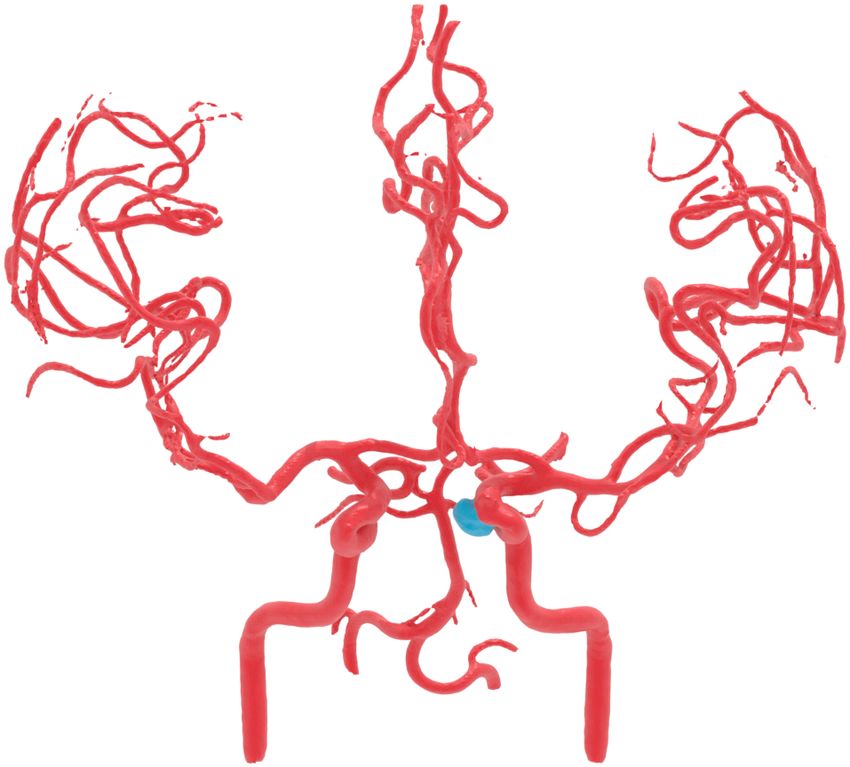
2      F. Author et al.

digital subtraction angiography (DSA), can be used. The DSA is the standard
for diagnosing intracranial IAs; however, it is invasive and time-consuming. Al-
though CT scans are efficient, it is difficult to distinguish the details of vessels
and aneurysms through CT scans. Therefore, we decided upon the MRA as a
suitable technique for preoperative examination.
     After imaging, the images must be segmented to obtain the detailed location
and shape of the aneurysm. Traditional methods used rule-based shape analy-
sis [7,9,3]; however, recently, learning-based methods are becoming popular with
the development of deep learning. For instance, deep learning networks for the
detection of IA to aid diagnosis were examined in two studies [10,14]. However,
few studies have focused on the segmentation of IAs. Podgorsak et al. [11] claimed
that segmenting IAs and the surrounding vasculature from DSA images using
the convolutional neural network is non-inferior to manually identifying the con-
tours of aneurysms. However, DSA is highly intrusive, and the applicability of
the DSA images is limited; further, they only extracted the 2D contours of the
IAs. Sichtermann et al. [13] applied a popular software based on a volume-based
neural network, DeepMedic[4], to segment IAs from MRA images. However, the
performance of their approach was suboptimal (46% in DSC).
     In this study, we proposed a novel processing pipeline for segmenting IAs by
leveraging surface-based neural networks to achieve better performance. We first
semi-automatically obtained the surface representation of the entire intracranial
blood vessel. We then collected small fragments from the entire vessel network
and performed surface-based classification on them. Finally, we applied surface-
based segmentation to the fragments that were classified as those containing
aneurysms.
     This work builds on Yang et al.’s work on an intracranial aneurysm dataset [15].
They presented a dataset for surface-based classification and segmentation, and
reported the performance of existing neural network models for each. However,
the dataset and execution are fully separated for classification and segmentation.
We present a complete processing pipeline that takes an entire intracranial vessel
network model as input, and returns IA fragments as output. We also present
the results of a head-to-head comparison between our surface-based method and
a previous volume-based method in a segmentation task performed on entire
brains [11].

2   Proposed Pipeline

Figure 1 compares our surface-based pipeline with a volume-based pipeline. In
the volume-based pipeline [13], the medical image is directly fed to a neural
network, which affixes a label to each voxel indicating whether it is part of an
aneurysm or not. In our surface-based pipeline, we first semi-automatically ob-
tained a surface representation of the principal brain arteries. We then generated
small samples along vessels from the entire model and performed surface-based
classification on them. Finally, we performed surface-based segmentation on the
samples classified as those containing aneurysms. To compare our results with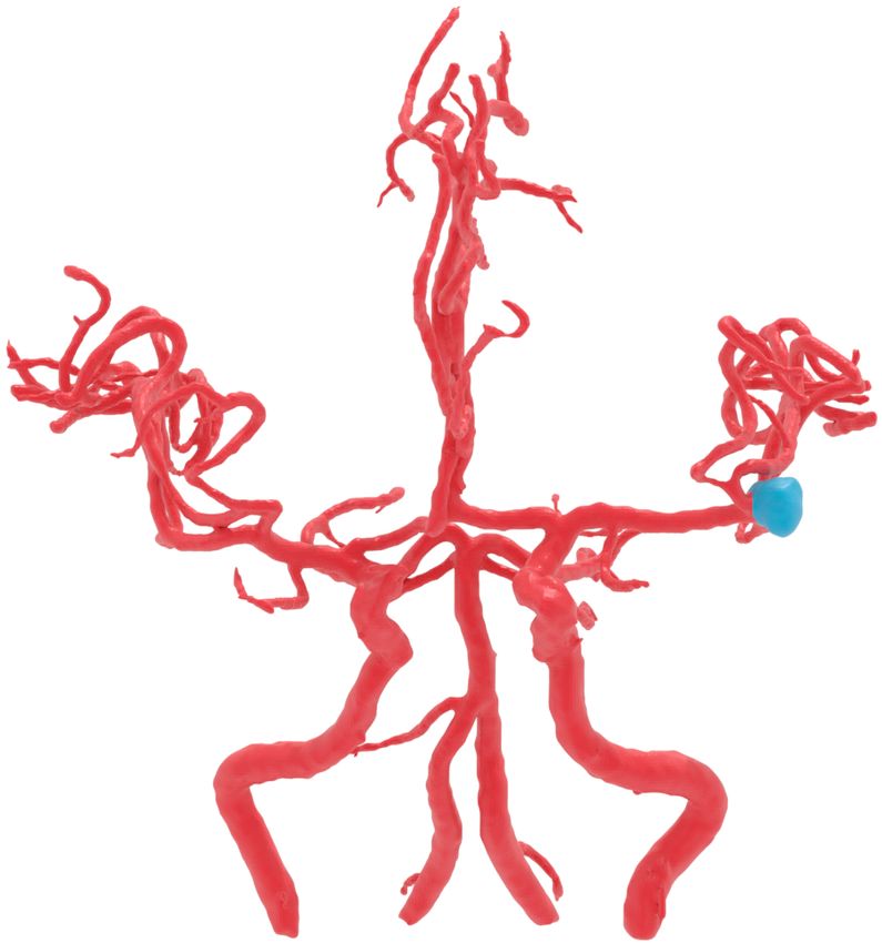
Title Suppressed Due to Excessive Length 3
Fig. 1: Comparison of proposed pipeline and volume-based pipeline.
the results yielded by volume-based methods, we voxelized the surface model of
the segmented aneurysms into volumes using winding-numbers [2].
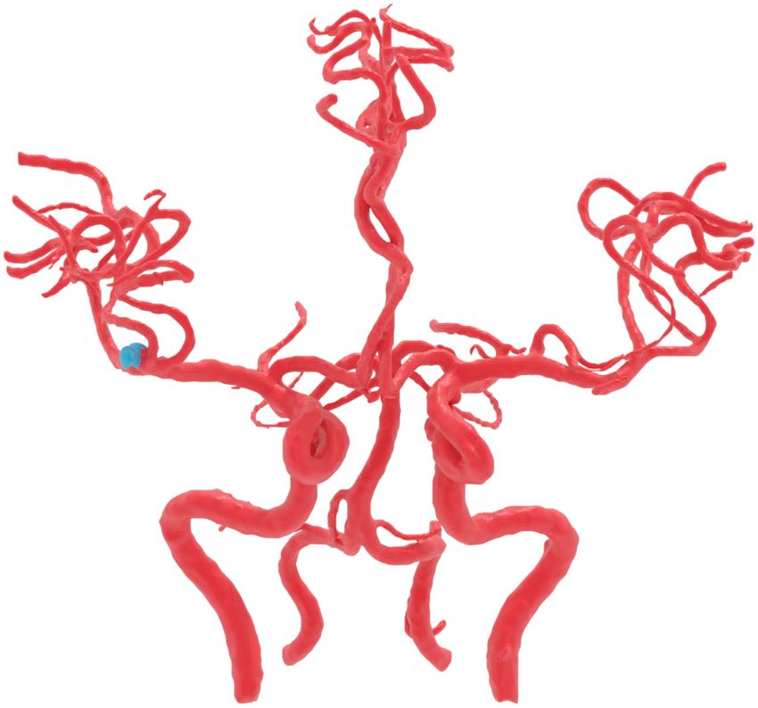
2.1 Reconstruction of Surface Models
We first obtained the surface models of the principal brain arteries of patients
from TOF-MRA image sets. We currently did this semi-automatically using a
software (Amira 2019 by Thermo Fisher Scientific, MA, USA) based on the
multi-threshold method [5]. Importantly, we focused on dealing with the sur-
rounding region of aneurysms to ensure that the complete shape of the aneurysms
is exhibited in the extracted 3D surface model, compared with the data in In-
tra [15]. In the future, we envision that this process can be mostly or fully
automated by using a network specifically designed for brain arteries.
2.2 Fragments Sampling
The aneurysmal parts are so small, compared to the entire model, that the
network we use could not segment them effectively. Therefore, we first sampled
small fragments from the entire model. We set the size of the fragments so that a
fragment roughly covers an aneurysm of typical size according to the experience
of the experts. Then, we used grid sampling to divide the 3D space of a model.
Next, from the center of each grid cell, the nearest point on the surface model
closer than a threshold (α) was selected as the starting point in each grid cell.
The grid cells that do not have nearby surface model points were ignored. Finally,
we collected the surface points around the start point whose geodesic distance
is less than a threshold (β). Note that this sampling was designed to cover the
model with some overlap; thus, uniform sampling is not a strong requirement.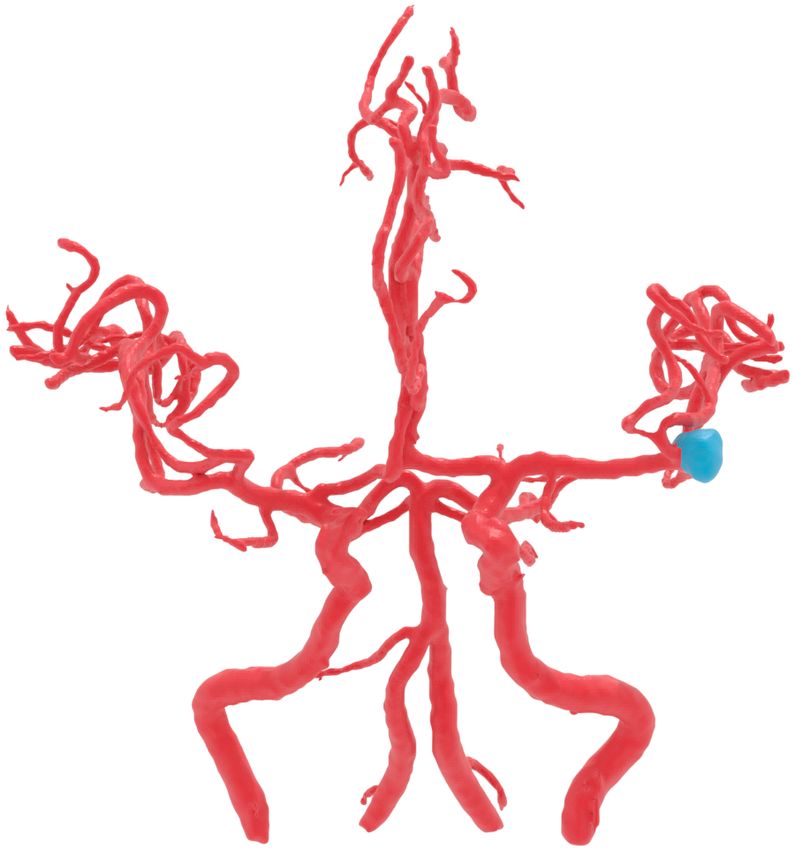
4      F. Author et al.

         Fig. 2: Details of our data-processing pipeline and algorithm.

2.3   Classification
We chose to use point-based methods rather than mesh-based methods to avoid
arduous preprocessing tasks including cleaning the models and making manifold
meshes. We used PointNet++ [12] to classify the fragments into two classes,
with or without aneurysms. The fragments with few points were ignored before
classification. However, the fragments with aneurysms were still significantly
fewer than those without aneurysms. Therefore, we used the weighted soft-max
cross-entropy loss function to train the classification network to deal with the
imbalance of the two classes. The purpose of classification was to reduce the
number of candidate fragments fed to the segmentation network and improve its
accuracy.

2.4   Segmentation
We then fed fragments with aneurysms to the segmentation network, SO-Net [8].
Only a fraction of the original points had a label after segmentation because
the point-based network uses random sampling to deal with the input mod-
els with different numbers of points. Thus, we performed segmentation with
random sampling multiple times, and assigned labels to all the points based
on voting criterion. There was the possibility of few points failing to be sam-
pled; we labeled them arteries (not aneurysms); however, they were few, and did
not significantly affect the segmentation results. We used a conditional random
field (CRF), DenseCRF [6] specifically, to refine each voting result. Finally, the
segmentation results of the individual fragments were combined to obtain the
complete segmentation result of the entire surface.
Title Suppressed Due to Excessive Length       5

2.5   Voxelization
We converted the results of our surface-based segmentation to volume to perform
a head-to-head comparison against volume-based methods. We first obtained
the query points through uniform sampling using the same interval as the MRA
images. We then computed the winding number of each query point using the
fast winding number method [2] to determine whether it is inside or outside an
aneurysm. We set the threshold of the winding number to 0.5, as suggested in
the study. One example is shown in Figure 2

3     Experiments
3.1   Dataset
We collected the TOF-MRA images sets of 103 patients with 116 aneurysms.
Each set contains at least one IA, and 512 × 512 ∼ 300 2D images sliced by
0.496mm. We annotated the aneurysmal parts on both the entire surface models
of the brain arteries and TOF-MRA images to generate the ground truth image
for classification and segmentation for neural networks. It took a total of three
experts 21 working days to perform this task. We use five-fold cross-validation
to conduct our experiments. 103 sets were shuffled, and divided into five subsets,
four subsets were used as the training data, and one was used as the test data.

3.2   Implementation details
The experiments were carried out on a PC with a GeForce RTX 2080Ti. During
data preprocessing, the normal vector of each point was estimated using the
original surface model. We also recorded the point index of the entire model as
a global ID on sampled fragments for each point to improve the efficiency of
voting. We set the sampling thresholds as α = 15, β = 1.5 ∗ α, and the samplings
in which the number of points was less than 500 were removed. We automatically
generated 7192 vessel fragments from the 103 entire models, and 392 fragments
had aneurysms inside them.
    The training hyper-parameters were set as follows. For the classification net-
work, the number of sampling points was 1024 for each fragment. The weights
of the loss function were determined according to the number of fragments. We
trained the network using 251 epochs and a batch size of 8. The classification
results were predicted by setting a discrimination threshold of 0.23. For the seg-
mentation network, the number of sampling points was 2048. We trained the
network for up to 401 epochs, with a batch size of 12. For each network, we used
the Adam optimizer with a learning rate of 10−3 .
    We also applied the method described in [13] to our dataset for comparison.
In this study, four preprocessing approaches A, B, C, and D were applied in the
paper. A has only been applied as a necessary step in DeepMedic, while B, C,
and D, were performed as additional masks for the skull-stripping of the TOF-
MRA images. B generated the masks with a fixed threshold, C used a manual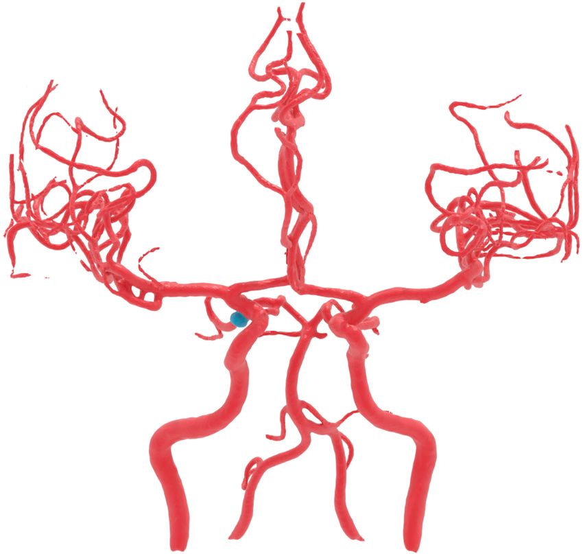
6        F. Author et al.

            Table 1: Comparison of segmentation results in DSCs (%).
                              Overall   Fold 0   Fold 1   Fold 2   Fold 3   Fold 4
       Surface-based   Mean   71.79     73.10    75.72    73.81    72.37    64.17
          (Ours)       STD     30.91    32.55    27.23    30.43    30.50    34.85
       Volume-based    Mean    45.90    56.56    47.40    38.86    45.51    41.20
      (DeepMedic B)    STD     31.00    28.77    30.25    31.57    30.88    33.22

threshold for the skull-stripping of each sample, and D added N4 bias correction
to the result of C. By analyzing the segmentation results, skull-stripping could
improve the performance; however, there was not much difference among the
results of B, C, and D. Therefore, we compared our method with B, which
has the highest reproducibility. We used BET2 to obtain the masks of skull-
stripping using the fixed threshold of 0.2 in this study. The input of the TOF-
MRA images was resized to 256×256 by down-sampling in comparison according
to the requirement of DeepMedic.

3.3    Results

About the results of our classification network, the receiver operating character-
istic (ROC) curve and confusion matrix of each classification network is shown
in Figure 3. It can be observed that all the areas of ROC curves are higher than
0.95, demonstrating that the trained classification networks are generalized. The
sensitivities of the aneurysm class of five networks are 73.63%, 81.08%, 79.49%,
86.11%, and 80.77%, respectively. This shows that we can detect the fragments
with IA precisely. By analyzing the confusion matrices, we can observe that
only a few fragments with aneurysms were misclassified because they have tiny
aneurysms or contain only a small part of the aneurysm. A 100% sensitivity
is not necessary for our classification network, because the sampled fragments
overlap, as shown in Figure 4. Some fragments without IA were not classified
correctly as well because the original data are noisy and they have a shape very
similar to a small part of aneurysms. These misclassified cases are also extremely
difficult for the segmentation network. Therefore, they do not have a significant
impact on the final segmentation result. The classification step is valuable. By
filtering out the majority of fragments that do not contain aneurysms, we can-
not only save a lot of training time but also improve the accuracy of the final
segmentation result.
     Dice similarity coefficient (DSC) was employed to evaluate the segmentation
results. The comparison of the final segmentation results is shown in Figure 5
and Table 1. The performance of the volume-based method is comparable to
the performance reported in the original paper [13]. Our surface-based method
obtained much better segmentation results than the volume-based method on
most of the data. However, few samples with tiny aneurysms are challenging for
both the volume-based method and ours.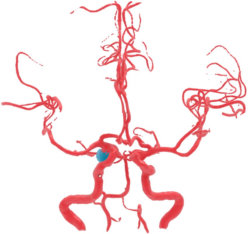
Title Suppressed Due to Excessive Length      7

Fig. 3: ROC curves and confusion matrices of five trained classification networks.

       A             B           C           D        E          F          G

Fig. 4: Fragment examples. Fragment A has a complete aneurysm. Fragments
B and C partly overlap with A, but they only have a part of the aneurysm.
Fragments D, E, F, and G are without aneurysms, but are misclassified. The
original data of D is noisy, and E, F, and G has a shape that is very similar to
a part of the aneurysm.

4   Limitation
Our current pipeline requires manual efforts by medical experts to obtain surface
models of intracranial artery networks. Thus, a possible criticism of our method
is that this process severely limits the practical value of our method. There
are three reasons why we still believe that our method has significant piratical
value. First, neurosurgeons are already constructing surface models regularly for
preoperative examination. Thus, we can assume that the surface model is already
given in our context. Second, the construction of the surface model is mostly
performed through simple thresholding [5]. An expert sets a threshold manually,
and voxels with intensity higher than the threshold are automatically extracted.
In this process, the expert is not paying attention to the details of individual
aneurysms. Aneurysms needs to be carefully segmented manually using surface
editing tools in current practice, and the automation of this process is highly
appreciated. Finally, we expect that, with the advances in deep learning methods,
surface extraction will be fully (or mostly) automatic in the future. Consequently,
the entire process will be fully automatic, which can significantly impact the field.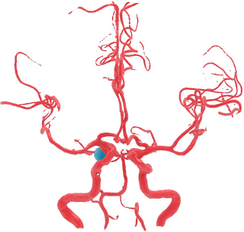
8      F. Author et al.

5   Conclusion

In this study, we proposed a new surface-based framework for the segmenta-
tion of intracranial aneurysms from TOF-MRA images. A two-steps design,
classification-segmentation, was applied to our framework using the state-of-
the-art point-based deep learning networks. We also designed sampling and
refinement methods for this IA segmentation task. The segmentation results
showed that our framework significantly outperformed to the existing volume-
based method. Surface-based methods are not yet prevalent in medical diagnosis
and surgical planning. Our results show that surface-based methods can be a reli-
able alternative to popular volume-based methods, and we hope this work impels
more efforts in this direction in other medical application domains.
Title Suppressed Due to Excessive Length         9

                  Ours                                      DeepMedic

    GT         Prediction   GT & Prediction     Input        GT         Prediction

Fig. 5: Comparison of segmentation results. Both our method and DeepMedic
yielded high segmentation accuracy on the two examples (the top two rows);
Our method yielded significantly better results than DeepMedic (the middle
three rows); Both our method and DeepMedic could not obtain the parts with IA
(the last example), the fragment with the IA was filtered out by our classification
network.
10      F. Author et al.

References

 1. Alaraj, A., Luciano, C.J., Bailey, D.P., Elsenousi, A., Roitberg, B.Z., Bernardo, A.,
    Banerjee, P.P., Charbel, F.T.: Virtual reality cerebral aneurysm clipping simulation
    with real-time haptic feedback. Operative Neurosurgery 11(1), 52–58 (2015)
 2. Barill, G., Dickson, N.G., Schmidt, R., Levin, D.I., Jacobson, A.: Fast winding
    numbers for soups and clouds. ACM Transactions on Graphics (TOG) 37(4), 1–12
    (2018)
 3. Dakua, S.P., Abinahed, J., Al-Ansari, A.: A pca-based approach for brain aneurysm
    segmentation. Multidimensional Systems and Signal Processing 29(1), 257–277
    (2018)
 4. Kamnitsas, K., Ledig, C., Newcombe, V.F., Simpson, J.P., Kane, A.D., Menon,
    D.K., Rueckert, D., Glocker, B.: Efficient multi-scale 3d cnn with fully connected
    crf for accurate brain lesion segmentation. Medical image analysis 36, 61–78 (2017)
 5. Kin, T., Nakatomi, H., Shojima, M., Tanaka, M., Ino, K., Mori, H., Kunimatsu, A.,
    Oyama, H., Saito, N.: A new strategic neurosurgical planning tool for brainstem
    cavernous malformations using interactive computer graphics with multimodal fu-
    sion images. Journal of neurosurgery 117(1), 78–88 (2012)
 6. Krähenbühl, P., Koltun, V.: Efficient inference in fully connected crfs with gaussian
    edge potentials. In: Advances in neural information processing systems. pp. 109–
    117 (2011)
 7. Law, M.W., Chung, A.C.: Vessel and intracranial aneurysm segmentation using
    multi-range filters and local variances. In: International Conference on Medical Im-
    age Computing and Computer-Assisted Intervention. pp. 866–874. Springer (2007)
 8. Li, J., Chen, B.M., Hee Lee, G.: So-net: Self-organizing network for point cloud
    analysis. In: Proceedings of the IEEE conference on computer vision and pattern
    recognition. pp. 9397–9406 (2018)
 9. Nikravanshalmani, A., Karamimohammdi, M., Dehmeshki, J.: Segmentation and
    separation of cerebral aneurysms: A multi-phase approach. In: 2013 8th Inter-
    national Symposium on Image and Signal Processing and Analysis (ISPA). pp.
    505–510. IEEE (2013)
10. Park, A., Chute, C., Rajpurkar, P., Lou, J., Ball, R.L., Shpanskaya, K., Jabarkheel,
    R., Kim, L.H., McKenna, E., Tseng, J., et al.: Deep learning–assisted diagnosis of
    cerebral aneurysms using the headxnet model. JAMA network open 2(6), e195600–
    e195600 (2019)
11. Podgorsak, A.R., Rava, R.A., Bhurwani, M.M.S., Chandra, A.R., Davies, J.M.,
    Siddiqui, A.H., Ionita, C.N.: Automatic radiomic feature extraction using deep
    learning for angiographic parametric imaging of intracranial aneurysms. Journal
    of neurointerventional surgery (2019)
12. Qi, C.R., Yi, L., Su, H., Guibas, L.J.: Pointnet++: Deep hierarchical feature learn-
    ing on point sets in a metric space. In: Advances in neural information processing
    systems. pp. 5099–5108 (2017)
13. Sichtermann, T., Faron, A., Sijben, R., Teichert, N., Freiherr, J., Wiesmann, M.:
    Deep learning–based detection of intracranial aneurysms in 3d tof-mra. American
    Journal of Neuroradiology 40(1), 25–32 (2019)
14. Ueda, D., Yamamoto, A., Nishimori, M., Shimono, T., Doishita, S., Shimazaki, A.,
    Katayama, Y., Fukumoto, S., Choppin, A., Shimahara, Y., et al.: Deep learning
    for mr angiography: automated detection of cerebral aneurysms. Radiology 290(1),
    187–194 (2019)
Title Suppressed Due to Excessive Length 11
15. Yang, X., Xia, D., Kin, T., Igarashi, T.: Intra: 3d intracranial aneurysm dataset
for deep learning. In: The IEEE Conference on Computer Vision and Pattern
Recognition (CVPR) (2020), https://github.com/intra3d2019/IntrAYou can also read