Deep Class-specific Affinity-Guided Convolutional Network for Multimodal Unpaired Image Segmentation
←
→
Page content transcription
If your browser does not render page correctly, please read the page content below
Deep Class-specific Affinity-Guided
                                                Convolutional Network for Multimodal
                                                   Unpaired Image Segmentation

                                                 Jingkun Chen1 , Wenqi Li2 , Hongwei Li3 , and Jianguo Zhang1
                                                       1
                                                        Department of Computer Science and Engineering,
arXiv:2101.01513v1 [cs.CV] 5 Jan 2021

                                                 Southern University of Science and Technology, Shenzhen, China
                                                                    zhangjg@sustech.edu.cn
                                                                            2
                                                                              NVIDIA
                                                          3
                                                            Technical University of Munich, Germany

                                              Abstract. Multi-modal medical image segmentation plays an essential
                                              role in clinical diagnosis. It remains challenging as the input modali-
                                              ties are often not well-aligned spatially. Existing learning-based methods
                                              mainly consider sharing trainable layers across modalities and minimiz-
                                              ing visual feature discrepancies. While the problem is often formulated
                                              as joint supervised feature learning, multiple-scale features and class-
                                              specific representation have not yet been explored. In this paper, we
                                              propose an affinity-guided fully convolutional network for multimodal
                                              image segmentation. To learn effective representations, we design class-
                                              specific affinity matrices to encode the knowledge of hierarchical feature
                                              reasoning, together with the shared convolutional layers to ensure the
                                              cross-modality generalization. Our affinity matrix does not depend on
                                              spatial alignments of the visual features and thus allows us to train with
                                              unpaired, multimodal inputs. We extensively evaluated our method on
                                              two public multimodal benchmark datasets and outperform state-of-the-
                                              art methods.

                                              Keywords: Segmentation · Class-specific Affinity · Feature Transfer.

                                        1   Introduction

                                        Medical image segmentation is a key step in clinical diagnosis and treatment.
                                        Fully convolutional networks [9,1,7] have been established as powerful tools for
                                        the segmentation tasks. Benefiting from the learning capability of these models,
                                        researchers start to address more challenging and critical problems such as learn-
                                        ing from multiple imaging modalities. This is an essential task because different
                                        modalities provide complementary information and joint analysis can provide
                                        valuable insights in clinical practice.
                                            Multi-modal learning is inherently challenging for two reasons: 1) super-
                                        vised feature learning is often modality-dependent; features learned from a single
                                        modality can not easily be combined with those from other modalities; 2) joint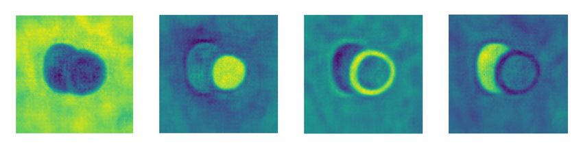
2      J. Chen et al.

learning often requires images from different modalities being spatially well-
aligned and paired; obtaining such training data is itself a costly task and often
infeasible. Fig. 1 shows sample slices from cardiac scans in different modalities.
It can be observed that although they all reveal parts of the heart anatomy, their
visual appearances vary. Segmentation networks are often sensitive to such dis-
crepancies, which has become a major obstacle for model generalization across
modalities.

Fig. 1: Left to right: slices of MR (left two) and CT (right two) cardiac scans.

    Spatial misalignment is another issue. Existing image registration methods
are often infeasible, as the spatial correspondences among modalities can be
highly complex and finding a good similarity measurement is non-trivial.
    To mitigate these issues, joint learning with unpaired data is emerging as a
promising direction [11,10,3]. MultiResUNet [4] has been proposed to improve
upon U-Net in multimodal medical image analysis. In brain image segmentation,
Nie et al. [8] trained networks independently for single modalities and then fused
the high-layer outputs for final segmentation. Yang et al. [12] used disentangled
representations to achieve CT and MR adaptation. Existing methods didn’t
take into account class-specific information, even though the features obtained
by supervised training are highly correlated with the tasks (Fig. 2).
    Our assumption is that, with the same network architecture, the underlying
anatomical features should be extracted in a similar manner across modalities.
At the same time, each network instance should have modality-specific modules
to tolerate the imaging domain gaps. With this assumption, to facilitate effective
joint feature learning, we adopt an FCN for all modalities, where the convolu-
tional kernels are shared, while the modality-specific batch feature normaliza-
tions remain local to each modality. More importantly, we extract class-specific
affinity measurements at multiple feature scales, and minimize an affinity loss
during training. Different from cross-modal feature consistency loss, our design
ensures that the networks extract modality independent features in a similar hi-
erarchical manner. Intuitively, this could be interpreted as “high-order” feature
extraction consistency compared with the feature map consistency loss. We show
that this is a more appropriate joint model regularizer that effectively guides the
anatomical feature extractions.
    In summary, our main contributions are: 1) we propose a novel unpaired mul-
timodal segmentation framework, which explicitly extracts the modality-agnostic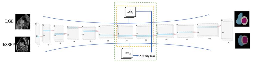
Deep Class-specific Affinity-Guided Convolutional Network for Multimodal      3

Fig. 2: Visualization of feature maps of one cardiac slice in four classes. Left to
right: the background, RV, myo and LV. The higher brightness part indicates
the region for the corresponding class; it could be observed that class-specific
feature representation is brighter than the other parts, which is highly correlated
with the ground truth mask in this class.

knowledge; 2) we introduce a joint learning strategy and a class-specific affin-
ity matrix to guide the training, which is capable of distilling between-layer
relationships at multiple scales; 3) we extensively evaluated our method on two
public multimodal benchmark datasets, and the proposed method outperforms
the state-of-the-art multimodal segmentation approaches.

2     Methodology
This section details the proposed joint training for segmentation tasks.

2.1    Multimodal Learning
We adopt an FCN as the backbone of our framework. Without any loss of gener-
ality, we present our framework in the case of training with two imaging modal-
ities. The overall architecture is illustrated in Fig. 3. The training of the system
operates on random unpaired samples from both modalities, the same set of
convolutional layers of the network are updated, while the batch normalization
layers are initialized and updated individually for each modality.

2.2    Modality-specific Batch Normalization
Using two independent sets of parameters for joint training leads to large models
and thus tends to overfit. Karani et al. [5] showed that using a domain-specific
batch normalization is effective in addressing domain gaps issue while keeping
the model compact. Here we employ the same technique for modality-specific
feature extraction. Specifically, the batch normalization layer matches the first
and the second moment of the distributions of feature x:

                                     x − E(x)
                             x∗ = γ p             +β                            (1)
                                      V ar(x) +
where γ, β are trainable parameters and are modality-dependent in our design.
 is a very small positive number to avoid dividing by zero.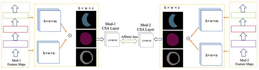
4       J. Chen et al.

Fig. 3: Architecture of the proposed class-specific affinity guided learning for mul-
timodal segmentation (CSA denotes class-specific affinity). The overall structure
of the proposed method contains two streams of FCNs with shared layers. For
the detail of the CSA module, please see Fig. 5.

2.3   Class-specific Affinity

It has been shown that feature maps in a network could reflect the saliency
of the class of interest in a multi-class setting [6] in a recent study by Levine
et al. Such a saliency map could give a robust interpretation of the reason-
ing process of model predictions. Motivated by this study, in the multi-modal
segmentation network, since all the modalities share the same tasks (e.g., multi-
class heart region segmentation), we hypothesized that the reasoning process
of model-specific channels should be similar and its feature map should reflect
class-specific saliency (i.e., interpretation of the class of interest). As shown in
Fig. 2, ideally, the region of interest in a learned feature map should be salient
and aligned well with its class-label. Therefore, for a learned feature map F (l)
of layer l and a given class c, we introduce the class-specific feature map F c (l)
defined as
                                  F c (l) = F (l) M c                            (2)
    where M c denotes the ground truth mask of size (h, w) for class c (reshape
to the size of feature map if necessary), and represents Hadamard product.
                 c
Suppose that Fm    (l) and Fnc (k) are the m-th and n-th class-specific feature maps
from layer l and k respectively, we measure their relationships using an affinity
defined by their cosine similarity, i.e.,

                                    1
                          acm,n =             c
                                       ∗ cos(Fm (l), Fnc (k))                    (3)
                                    Sc
Where Sc is the size of the region of interest in M c . Such a normalization is to
ensure that the affinity is invariant to the size of the saliency region. Suppose
that layer l and layer k have M and N number of class-specific feature maps, we
construct the between-layer affinity matrix Acm,n , where the entry at (m, n) is
Deep Class-specific Affinity-Guided Convolutional Network for Multimodal 5
acm,n . The size of Ac is M by N . Since the affinity is computed based class-specific
on feature map, we term this as the class-specific affinity (CSA) matrix.
Fig. 4: The proposed class-specific affinity guided learning layer. The dotted box
on the left shows three convolutional layers for feature extraction. The dotted
box in the middle shows the affinity computation by incorporating feature maps
at multiple scales as well as the multi-class ground truth segmentation map.
Fig. 4 shows our design of a class-specific affinity (CSA) layer. Our CSA
could be computed for each of the modality, based on which we build the CSA
module. It is worth noting that our design is based on a class-specific feature
map, and independent to the choice of modalities; therefore, our network does
not require inputs spatial alignment.
2.4 CSA Module
Suppose that we have two modalities using the same network architecture for
joint learning. We compute Ac,1 for modality-1 (e.g., CT) and Ac,2 modality-2
(e.g MR) across layer l and k. The knowledge encoded by CSA for a specific
class c could be transferred by enforcing the consistency of CSA between the
two modalities using an L2 norm. We then aggregate all of the consistencies for
all of the classes to formulate a consistency loss function as below:
C P 2
1 X 1 X c,1
LCSA = (A − Ac,2
i ) (4)
C c P i=1 i
2
where C is the number of classes. P is the total number of entries in Ac , i.e.,
M N . Normalizing by P is to ensure that the consistency is invariant to the
number of feature channels.
When minimizing the CSA loss, the affinity consistency between the two
modalities could be maximized thus ensuring joint learning. For the segmentation
loss, we use a Dice coefficient loss Ldice to ensure good segmentation at region
level, and a Lce cross-entropy loss for pixel-level segmentation. Taking all of the
three losses together, the final loss of our multi-modal learning framework for
two modalities 1 and 2 is defined as:6 J. Chen et al.
Fig. 5: The CSA guided multi-modal knowledge sharing. The CSA Layer com-
putes affinity from ”Mod-1” and ”Mod-2” respectively (according to Sec. 2.3).
The affinity loss between two modalities is computed according to Sec. 2.4.
L = α(L1dice + L2dice ) + β(L1ce + L2ce ) + λLCSA (5)
Where LCSA is the CSA transfer loss, α,β,λ are the system parameters to
weight the loss components.
3 Experiments
Datasets. We evaluated our multimodal learning method on two public datasets:
MS-CMRSeg 2019 [13] [15] and MM-WHS [14]. MS-CMRSeg 2019 contains 45
patients who had previously suffered from cardiomyopathy. Each patient has
images in three cardiac MR modalities (LGE, T2, and bSSFP) with the three
segmentation classes: left ventricles, myocardium, and right ventricles. We use
LGE and bSSFP in our experiments. For each slice, we crop the image and get
region of interests with a fixed bounding box (224×224), enclosing all the anno-
tated regions. we randomly divided the dataset into 80% for training and 20%
for testing according to the patients. MM-WHS contains the MR and CT images
whole heart from the upper abdomen to the aortic arch. The segmentation task
includes four structures: left ventricular myocardium (LVM), left atrium (LAC),
left ventricle (LVC), and ascending aorta (AA). We crop each slice with a re-
gion with a 256 × 256-pixel bounding box in the coronal plane and randomly
divided the dataset into training (80%) and test sets (20%). For preprocessing,
we use z-score normalization [2] to calibrate the intensity of each 2D slice from
all modalities in both datasets.
Implementation. Our architecture consists of nine convolutional operation
groups, one deconvolutional group and one softmax layer. Each group contains
two to four shared convolutional layers and domain-specific batch normalization
layers. We implemented the proposed method with Python-based on Tensorflow
1.14.0 library using Nvidia Quadro RTX GPU (24G). We optimize our network
with Adam optimizer with a batch size of 8. The learning rate is initialized to 1×
10−4 and decayed by 5% per 1000 iterations. Besides, we incorporated dropout
layers (drop rate of 0.75) into the network to validate the performance. In theDeep Class-specific Affinity-Guided Convolutional Network for Multimodal 7
training stage, we used three loss functions. Empirically, α, β and λ are set to 1,
1 and 0.5 respectively. Our assumption is that the higher layer features are too
closely related to the ground truth mask, so the affinity features of the migration
process are not obvious, so we choose the feature maps from the intermediate
layer. We believe that the information between remote feature maps is difficult
to express completely with affinity feature maps, so we used the feature maps
from the layers relatively close to each other in our experiment.
Comparison and Analysis. We designed the following six experimental set-
tings (single training of separate modalities (Single), Unpaired Multi-modal Seg-
mentation via Knowledge Distillation (UMMKD)[3], Joint training of two modal-
ities in shared BN model(Joint), Modality-specific batch normalization only
(MSBN), Affinity-guided learning (Affinity), and CSA guided learning (ours)).
For all settings, the network architecture and datasets are fixed so that different
methods can be compared fairly. In terms of quantitative performance measure-
ments, we adopt the volume Dice score and surface Hausdorff distance as listed
in Table 1, 2 and 3.
Table 1: Average Dice score (%) and Surface Hausdorff distance(mm) on LGE
and bSSFP, the highest performance in each class is highlighted.
LGE Dice bSSFP Dice LGE Dist. bSSFP Dist.
Method
LV myo RV LV myo RV LV myo RV LV myo RV
Single[7] 90.17 80.31 86.51 92.89 85.11 88.76 6.63 3.61 25.10 47.80 2.45 11.58
UMMKD[3] 90.41 80.48 86.99 93.38 85.69 89.91 5.00 3.46 8.66 3.00 2.24 5.39
Joint 90.22 80.91 86.61 92.74 85.19 89.12 4.00 6.08 5.12 3.16 2.45 5.00
MSBN 90.23 80.81 86.08 93.46 85.57 89.91 5.75 3.61 5.48 3.00 2.24 5.10
Affinity 90.65 81.18 87.27 93.54 85.93 89.93 5.00 3.16 9.72 3.00 2.24 5.10
Ours 91.89 83.39 87.66 93.48 85.72 90.64 4.12 3.00 5.00 3.00 2.24 4.12
1) Results on MS-CMRSeg 2019: Table 1 lists the results of three classes
cardiac segmentation. As can be observed, compared with individual training
and the other multi-modal learning, our methods showed a significant perfor-
mance gain. The overall average Dice score of three classes on two modalities
increased to 87.65% and 89.95% which is much higher than single training of sep-
arate modalities (85.66% and 88.92%) and UMMKD (85.96% and 89.66%), and
the average Hausdorff distance of three classes on LGE modality of UMMKD
decreased from 5.71mm to 4.04mm. This indicates that class-based cross-modal
knowledge transfer is effective. We have conducted the Wilcoxon signed-rank test
for the improvements of our method over UMMKD on the results based on the
patient-level predictions (the p-value is 0.010 for LGE, and 0.048 for bSSFPP),
indicating that improvements are statistically significant. It is observed that the
affinity guided learning also achieved the better result on the two modalities,
especially on the bSSFP modality, but it is still lower than the CSA module, as
the CSA module learned to share the class-based semantic knowledge.8 J. Chen et al.
2) Results on MM-WHS: The proposed method was also used to segment six
classes cardiac MRI and CT, and achieves promising segmentation performance
on this relatively large dataset (Table 2 and 3), with an average Dice score of
79.27% for LVM, 87.46% for LAC, and 81.42% for AA on CT modality; 88.89%
for LVM, 92.53% for LVC, and 96.07% for AA. Hausdorff distance of our method
on the two modalities is also lower in most of the classes. The differences when
comparing with the models trained in MS-CMRSeg 2019 were the number of
samples for training and the weight of the CSA loss increased to 0.5. Overall,
it can be seen that the proposed methods (ours) outperforms both the single
modality model and the multi-modal method, which confirms their effectiveness.
We tested the CSA learning between the more distant layers and the closer layers,
and we found that the performance within the closer layers is better. The CSA
knowledge between the closer layers can be better learned and also easier to be
migrated.
Table 2: Average Dice score (%) on MRI and CT.
CT MRI
Method
LVM LAC LVC AA LVM LAC LVC AA
Single[7] 78.17 85.84 93.07 81.08 87.56 90.29 91.66 94.26
UMMKD[3] 78.73 83.47 93.29 81.41 87.89 91.68 91.88 95.32
Joint 77.92 83.96 93.51 80.08 84.03 88.41 90.92 94.67
MSBN 78.82 86.07 94.40 80.57 87.57 92.30 91.88 96.02
Affinity 78.63 87.13 94.35 79.49 85.31 91.06 91.84 92.24
Ours 79.27 87.46 94.22 81.42 88.89 91.18 92.53 96.07
Table 3: Surface Hausdorff distance(mm) on CT and MRI.
CT MRI
Method
LVM LAC LVC AA LVM LAC LVC AA
Single[7] 5.00 10.63 5.00 13.38 8.50 15.03 4.47 6.00
UMMKD[3] 5.83 14.40 6.01 13.97 10.05 8.94 4.47 3.61
Joint 5.39 14.32 4.24 13.30 6.40 40.17 9.84 4.47
MSBN 5.00 10.63 4.24 13.93 4.12 9.06 4.47 3.00
Affinity 6.00 10.63 4.89 15.13 10.05 12.08 5.00 67.09
Ours 5.39 10.30 4.24 15.80 4.12 9.49 4.12 2.83
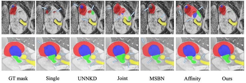
Visualization. Fig. 6 shows the predicted masks from the six methods. It could
be seen that our methods improve the performance of only using single-modal
method and the other multi-modal method, especially for the LGE modality.
These observations are consistent with those shown in the Table 1, 2 and 3.Deep Class-specific Affinity-Guided Convolutional Network for Multimodal 9
Fig. 6: Segmentation maps on CT (top) and MRI (bottom). Left to right: Ground
Truth mask, Single, UMMKD, Joint, MSBN, Affinity and CSA.
4 Conclusion
We propose a new framework for unpaired multi-modal segmentation, and intro-
duce class-specific affinity measurements to regularize the jointly model training.
We have derived the formulations and experimented with spatially 2D feature
maps. As future work, the same concepts could be extended to spatially 3D
cases. The results based on the proposed class-specific affinity loss are encourag-
ing. Further quantitative analysis of the feature maps and investigating model
interpretability is also an interesting future direction.
References
1. Badrinarayanan, V., Kendall, A., Cipolla, R.: Segnet: A deep convolutional
encoder-decoder architecture for image segmentation. IEEE transactions on pat-
tern analysis and machine intelligence 39(12), 2481–2495 (2017)
2. Chen, J., Li, H., Zhang, J., Menze, B.: Adversarial convolutional networks with
weak domain-transfer for multi-sequence cardiac mr images segmentation. In: Inter-
national Workshop on Statistical Atlases and Computational Models of the Heart.
pp. 317–325. Springer (2019)
3. Dou, Q., Liu, Q., Heng, P.A., Glocker, B.: Unpaired multi-modal segmentation via
knowledge distillation. TMI (2020)
4. Ibtehaz, N., Rahman, M.S.: Multiresunet: Rethinking the u-net architecture for
multimodal biomedical image segmentation. Neural Networks 121, 74–87 (2020)
5. Karani, N., Chaitanya, K., Baumgartner, C., Konukoglu, E.: A lifelong learning
approach to brain mr segmentation across scanners and protocols. In: International
Conference on Medical Image Computing and Computer-Assisted Intervention. pp.
476–484. Springer (2018)
6. Levine, A., Singla, S., Feizi, S.: Certifiably robust interpretation in deep learning.
arXiv preprint arXiv:1905.12105 (2019)
7. Long, J., Shelhamer, E., Darrell, T.: Fully convolutional networks for semantic
segmentation. In: Proceedings of the IEEE conference on computer vision and
pattern recognition. pp. 3431–3440 (2015)10 J. Chen et al.
8. Nie, D., Wang, L., Gao, Y., Shen, D.: Fully convolutional networks for multi-
modality isointense infant brain image segmentation. In: 2016 IEEE 13Th interna-
tional symposium on biomedical imaging (ISBI). pp. 1342–1345. IEEE (2016)
9. Ronneberger, O., Fischer, P., Brox, T.: U-net: Convolutional networks for biomedi-
cal image segmentation. In: International Conference on Medical image computing
and computer-assisted intervention. pp. 234–241. Springer (2015)
10. Valindria, V.V., Pawlowski, N., Rajchl, M., Lavdas, I., Aboagye, E.O., Rockall,
A.G., Rueckert, D., Glocker, B.: Multi-modal learning from unpaired images: Ap-
plication to multi-organ segmentation in ct and mri. In: 2018 IEEE Winter Con-
ference on Applications of Computer Vision (WACV). pp. 547–556. IEEE (2018)
11. Wolterink, J.M., Dinkla, A.M., Savenije, M.H., Seevinck, P.R., van den Berg, C.A.,
Išgum, I.: Deep mr to ct synthesis using unpaired data. In: International workshop
on simulation and synthesis in medical imaging. pp. 14–23. Springer (2017)
12. Yang, J., Dvornek, N.C., Zhang, F., Chapiro, J., Lin, M., Duncan, J.S.: Unsuper-
vised domain adaptation via disentangled representations: Application to cross-
modality liver segmentation. In: International Conference on Medical Image Com-
puting and Computer-Assisted Intervention. pp. 255–263. Springer (2019)
13. Zhuang, X.: Multivariate mixture model for cardiac segmentation from multi-
sequence mri. In: International Conference on Medical Image Computing and
Computer-Assisted Intervention. pp. 581–588. Springer (2016)
14. Zhuang, X., Li, L., Payer, C., Štern, D., Urschler, M., Heinrich, M.P., Oster, J.,
Wang, C., Smedby, Ö., Bian, C., et al.: Evaluation of algorithms for multi-modality
whole heart segmentation: an open-access grand challenge. Medical image analysis
58, 101537 (2019)
15. Zhuang, X., Xu, J., Luo, X., Chen, C., Ouyang, C., Rueckert, D., Campello, V.M.,
Lekadir, K., Vesal, S., RaviKumar, N., et al.: Cardiac segmentation on late gadolin-
ium enhancement mri: A benchmark study from multi-sequence cardiac mr seg-
mentation challenge. arXiv preprint arXiv:2006.12434 (2020)You can also read