Effect of Alendronate on Healing of Bone Defect and Gingival Tissue in Osteopenic Rats
←
→
Page content transcription
If your browser does not render page correctly, please read the page content below
Int. J. Morphol.,
38(3):683-688, 2020.

              Effect of Alendronate on Healing of Bone Defect
                   and Gingival Tissue in Osteopenic Rats

                            Efecto del Alendronato en la Reparación de Defectos Óseos
                                   y Tejido Gingival en Ratas con Osteopenia

                 José Henrique Santana Quinto1; Eder Alberto Sigua-Rodriguez1,2; Maria Raquel Marçal Natali3;
                         Sergio Olate4,5; Roberto Kenji Nakamura Cuman6 & Gustavo Jacobucci Farah1

QUINTO, J. H. S.; SIGUA-RODRIGUEZ, E. A.; NATALI, M. R. M.; OLATE, S.; CUMAN, R. K. N. & FARAH, G. J. Effect of
alendronate on healing of bone defect and gingival tissue in osteopenic rats. Int. J. Morphol., 38(3):683-688, 2020.

         SUMMARY: The aim was to evaluate bone repair and gingival tissue repair in osteopenic rats. Fifteen female wistar rats were
included; in all of them ovariectomy was realized to induce osteopenia; after 45 days, the animals were submitted to 2 surgical techinques
1) dental extraction of the upper central incisor with no socket preservation and 2) 5 mm cranial defect in the calvarium; 5 rats were
included in the control group (G1) withput alendronate application; in the group 2 (G2) was used subcutenous alendronate (0.5 mg/kg)
once for three weeks and then was realizd the both surgical techniques. In group 3 (G3), after ovariectomy was realized the both dental
extraction and the calvarium defect and after that was realized the alendronate protocol. In each group, after six week was realized
euthanasia and descriptive histological analysis of the surgical areas involved. In bone formation of the 5 mm cranial defect was observed
with good progression in the 3 experimental models and no modification in quality of bone repair was observed. For the gingival tissue
in the extraction socket, no differences were observed between G1 and G3. On other hand, in G2 a thinner and reduced gingival epithelium
was found. Our results showed that alendronate was not an obstacle for bone repair; deficiencies in re-epithelialization of oral mucosa
show the impact of alendronate before dental extraction.

        KEY WORDS: Biphosphonate; Osteonecrosis; Osteoporosis.

INTRODUCTION

       Bisphosphonates (BPs) are routinely drugs to treat                 2009). The most common clinical finding is related to
osteoporosis, involving close to 200 million people in the                ulcerated mucosa area with localized bone necrotic exposure.
world (Agis et al., 2010). BPs are drugs responsible for                  The signs and symptoms as well as the etiology and treatment
reducing bone resorption by their metabolized nitrogen                    of this disease have been not well revised.
compounds be toxic to osteoclasts, affecting their survival
and function (Li et al., 1997; Landesberg et al., 2008;                           On other hand, it is well documented in the literature
Brozoski et al., 2012; Kumar & Sinha, 2013). They may                     the effect of BPs on the bone, however, there are few studies
have approximately 10 years -of half life and their continued             evaluating the toxicity of BPs on oral mucosa (Ruggiero
use may also trigger osteonecrosis of the jaws with incidence             & Woo, 2008). The available studies show that these drugs
on population ranges from 0.7 to 12 % (Cooper et al., 2010;               are responsible for reducing the proliferation of epithelial
Mozzati et al., 2013).                                                    and fibroblastic cells with a limited in healing of soft tissue
                                                                          and more bone exposure to the oral microflora (Lin & Lane,
       Osteonecrosis induced by BPs is characterized for                  2004).Therefore the aim of this research was to investigate
the involvement of the maxillomandibular complex with                     the effect of alendronate in hard and soft tissue repair in
preference in mandible related to subjects not exposed to                 an in vivo osteopenic model developed with
radiotherapy on the head and neck region (Ohashi et al.,                  ovariectomized rats.
1
  Residency Program in Oral and Maxillofacial Surgery, State University of Maringá, Paraná, Brazil.
2
  Centro de Investigaciones del Colegio Odontológico (CICO), Institución Universitaria Colegios de Colombia, Bogotá, Colombia.
3
  Department of Physiological Science, State University of Maringá, Paraná, Brazil.
4
  Division of Oral, Facial and Maxillofacial Surgery, Universidad de La Frontera, Temuco, Chile.
5
  Center of Excelence in Morphologycal and Surgical Studies (CEMyQ), Universidad de La Frontera, Temuco, Chile.
6
  Department of Pharmacology, State University of Maringá, Paraná, Brazil.

                                                                                                                                    683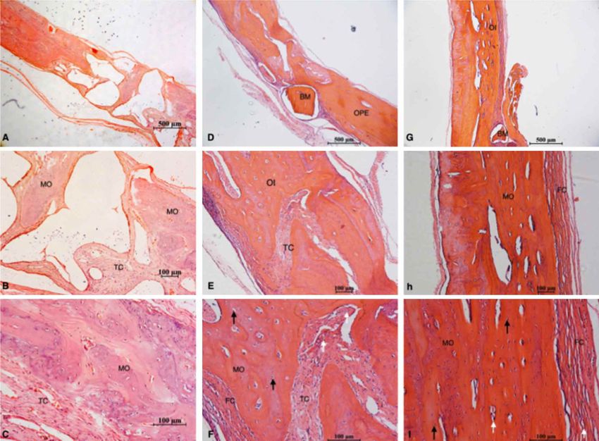
QUINTO, J. H. S.; SIGUA-RODRIGUEZ, E. A.; NATALI, M. R. M.; OLATE, S.; CUMAN, R. K. N. & FARAH, G. J. Effect of alendronate on healing of bone defect and gingival tissue in
                                                          osteopenic rats. Int. J. Morphol., 38(3):683-688, 2020.

MATERIAL AND METHOD                                                                       5 osteopenic animals treated with dental extraction and the
                                                                                          calvarium defects grafted with xenogenic biomaterial
                                                                                          (Bionnovation®) and collagen membrane (Baummer®) over
        The procedure involving the use of animals was                                    the defect; 3 week after surgery, was applied subcutaneous
approved by the Ethics Committee on Animal                                                alendronate (0.5 mg / kg) once a week for 3 weeks (Fig. 1).
Experimentation (CEEA) of the State University of Maringa
(Protocol No 057/2014). Fifteen female Wistar rats (Rattus                                        All the surgical procedure was performed at
norvegicus) with 60 days of age, weighing approximately                                   Laboratory of the Department of Pharmacology and
300 g, were placed in cages containing three animals, covered                             Therapeutics at State University of Maringá on the same
with sawdust and kept in standard vivarium conditions with                                afternoon, following intraperitoneal anesthesia with anesthetic
clear light / dark cycle of 12 hours at controlled temperatures                           solution of xylazine (10 mg / kg) and ketamine (100 mg / kg).
(25 ° C) with water and food ad libitum (NuvitalL®).                                      All fifteen animals were operated with the same protocol.

       In this methodology, the first surgical step was the                                      Euthanasia of the animals was performed with triple
ovariectomy to induce the osteopenia. This surgery was                                    anesthetic dose of xylazine and ketamine, six week after the
realized by vet surgeon with experience in this technique                                 last intervention in each group, using a routine
and after forty-five days, the bone mineral density decreased                             pharmacological protocol. Samples of calvarium and the area
as reported previously (Scheper et al., 2009) and at this                                 involved in the extraction socket were collected and process
moment the sample was divided in three differents groups.                                 with 10 % formalina solution for 72 h; after that, samples
The surgical model included two surgical defect in each                                   were decalcified with acid nitric 5 % for 21 days and then
animal, being 1) dental extraction of the upper right central                             embedded in paraffin. For histological analysis, 6 µm slice
incisor with no socket preservation and 2) the creation of 5                              were made, stained with haematoxylin and eosin. Histology
mm cranial defect on the calvarium realized by a 5 mm                                     was descriptive for all the samples.
trephine using low speed (Yano et al., 2014).

        Control Group (G1) included five animals with no                                  RESULTS
use of alendronate, submitted to dental extraction and 5 mm
cranial defect filled with blood clot. Group 2 (G2) included
5 osteopenic animals treated with subcutaneous alendronate                                       Calvaria 5 mm Defect: In G1 was observed low
(0.5 mg / kg) once a week for 3 weeks; after drug treatment,                              incremental growth lines, a low level of osteoclasts,
the animals were subjected to dental extraction and the same                              osteoblasts and osteocytes, low level of bone formation and
5 mm cranial defect on the calvarium filled with xenogenic                                a greater presence of connective tissue, mainly in the center
biomaterial (Bionnovation®) and collagen membrane                                         of the defect (Fig. 2 a-c). In the same line, was observed a
(Baummer®) on the grafted defect. Group 3 (G3) included                                   minor bone formation than G2 and G3 (Fig. 2 d-f).

       Fig. 1. Sample included in this research; each group, with 5 animals each, included a protocol to participate as control group
       (G1), and experimental groups (G2 and G3)

684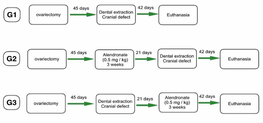
QUINTO, J. H. S.; SIGUA-RODRIGUEZ, E. A.; NATALI, M. R. M.; OLATE, S.; CUMAN, R. K. N. & FARAH, G. J. Effect of alendronate on healing of bone defect and gingival tissue in
osteopenic rats. Int. J. Morphol., 38(3):683-688, 2020.
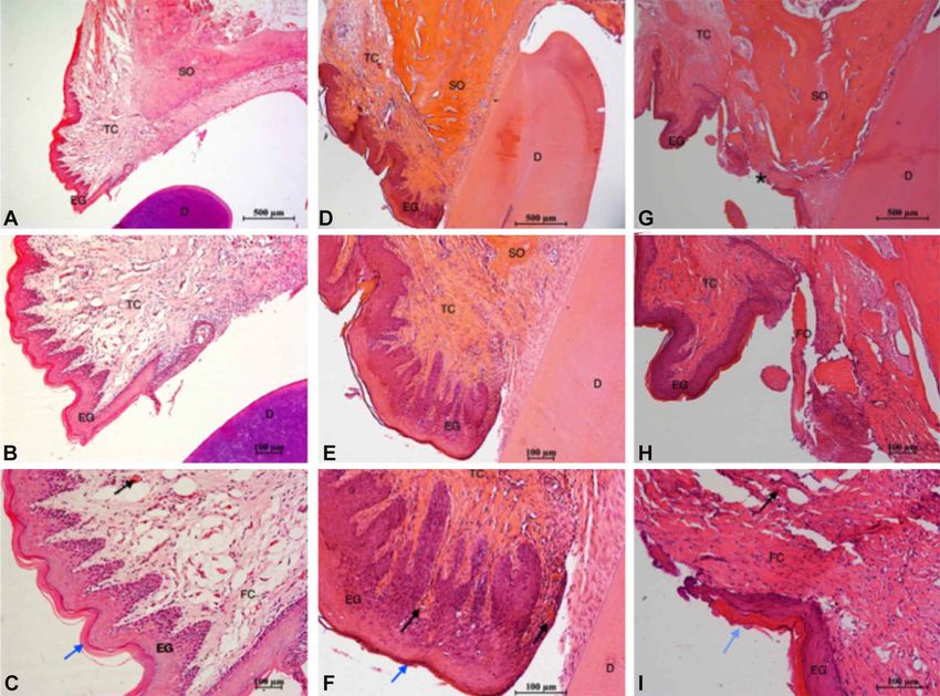
In G3 was observed osteocytes surrounded by epithelial ridges deep and well defined (Fig. 3 a-c) and in
acidophilic bone matrix close to some osteogenic and G3, a regular description as G1 was observed (Fig. 2 d-f).
fibroblastic cells; some particles of biomaterial was observed, G2 showed differences when compared to G1 and G3,
however, a large amount of bone tissue with advanced presenting a gingival epithelium fragile, reduced cell and
remodeling was present, showing macrophages in connective areas with poor definition and areas without epithelium (Fig.
tissue, bone channels and a large number abundant blood 2 g-i).
vessels (Fig. 2 d-f).
In G2 showed bone matrix regions with different DISCUSSION
degrees of acidofilias, featuring a woven bone, presence of
blood vessels, osteogenic cells (osteoblasts, osteoclasts,
osteocytes) and a connective tissue with abundant collagen For gingival tissue analysis, in G3, using three-week
fibers and fibroblasts (Fig. 2 g-i). time for tissue healing before receiving treatment with
alendronate, was observed similar condition to the control
Dental extraction with no socket preservation: As a group (G1). In clinical evaluation, Marx et al. (2005)
summary, in all the samples was observed gingival recommend a dental assessment before the patients starting
epithelium, connective tissue, collagen fibers, blood vessels treatment with BFs, to eliminate sources of infection; if
and bone septum. In G1 was observed a stratified squamous surgical procedure is needed, it is recommended wait healing
epithelium gum, keratinized, thicker with granular layer and for 30 days, and only after this period start treatment with
Fig. 2. Histological sections obtained 6 weeks after the creation of 5 mm cranial defects. G1 (A, B, C), G2 (D, E, F) and G3 (G, H, I).
Histological analysis was observed shiwong bone matrix (MO), connective tissue (TC), preexisting bone (OPE), immature bone (OI),
bundles of collagen fibers (FC), bio-materials (BM), osteocytes (black arrows) and blood vessels (white arrows) (hematoxylin and eosin,
4X, 10X and 20X).
685QUINTO, J. H. S.; SIGUA-RODRIGUEZ, E. A.; NATALI, M. R. M.; OLATE, S.; CUMAN, R. K. N. & FARAH, G. J. Effect of alendronate on healing of bone defect and gingival tissue in
osteopenic rats. Int. J. Morphol., 38(3):683-688, 2020.
Fig. 3. Histological sections obtained 6 weeks after extraction surgery of the upper right central incisor stained with hematoxylin and
eosin (4X, 10X and 20X). G1 (a, b, c), G2 (d, e, f) and G3 (g, h, i) show sept bone (OS), tissue conjuntive (TC), gingival epithelium (EG),
tooth (D), keratin (blue arrow), blood vessels (black arrow), bone fragment (FO) asence epithelial (*).
BPs (Ruggiero et al., 2014). The validation of this clinical treatment with alendronate (Miglioratir et al., 2005), also
practice can be better evaluated in our experiment where showed deficiency in epithelial wound healing, and in some
the results showed a satisfactory wound healing in animals cases a necrotic bone were exposed. A similar result was
involved in procedures before the use of alendronate. found in our study where the G2 had oral epithelium delayed,
Recently the American Association of Oral and Maxillofacial rendering them more fragile and susceptible to ulcerations,
Surgeons (2014), published a clinical follow-up allowing the bone exposed and contributes to osteonecrosis
recommending the suspension of the drug three months pathogenesis.
before and three months after the surgical procedure, for
better wound healing (Ruggiero et al.). BFs in the oral mucosa induce bone exposure to oral
microflora and contributing to osteonecrosis pathogenesis
G2 had the gingival epithelium reduced, thinner, even (Miglioratir et al., 2005; McLeod et al. 2014); therefore,
with areas of epithelial absence, suggesting a toxic action of trauma on the mucosa of patients undergoing treatment with
BFs to the oral epithelium. There are few studies in the BFs must be avoided (Brozoski et al.). Our results suggest
literature discussing the potential toxicity of bisphosphonates that the treatment of alendronate for a short time, could not
to the oral epithelium (Dimitrakopoulos et al., 2006; Mozzati cause osteonecrosis after invasive dental procedures, and in
et al.). An animal model was suggested that BPs suppress such cases a previous dental evaluation at the beginning of
cell proliferation and impair oral wound healing, showing drug treatment is required. Therefore, an explain to do not
osteonecrosis by the bone exposure to oral micro flora detect alteration on mineral content in our study can be
(Ruggiero et al.). In a survery, rats subjected to subcutaneous related on the experiment time, dose and time of
686QUINTO, J. H. S.; SIGUA-RODRIGUEZ, E. A.; NATALI, M. R. M.; OLATE, S.; CUMAN, R. K. N. & FARAH, G. J. Effect of alendronate on healing of bone defect and gingival tissue in
osteopenic rats. Int. J. Morphol., 38(3):683-688, 2020.
administration of alendronate (0.5 mg / kg) for 3 weeks were alendronate was present, bone repair was impaired compared
low and in human could be infrequently. The literature shows to the control group and the hydroxyapatite groups only.
that patients in treatment with oral BPs for less than three Based on results like these, more studies are necessary to
years may be undergoing to surgery (Dimitrakopoulos et evaluate the quality of bone in cases of sodium alendronate
al.; Reid et al., 2007). administration.
In the literature (Ravosa et al., 2011; Paiva-Fonseca
et al., 2014) it is well documented toxicity of the BPs in the QUINTO, J. H. S.; SIGUA-RODRIGUEZ, E. A.; NATALI, M.
gastrointestinal tract cells and had been related to esophagitis, R. M.; OLATE, S.; CUMAN, R. K. N. & FARAH, G. J. Efecto
gastritis and ulcers. Histological studies conducted in animals del alendronato en la reparación de defectos óseos y tejido gingival
that received alendronate orally, show the stomachs with en ratas con osteopenia. Int. J. Morphol., 38(3):683-688, 2020.
impair anti-oxidant system of the gastric mucosa and
decrease the cell differentiation, causing exfoliation of the RESUMEN: El objetivo fue evaluar la reparación ósea y
gingival en ratas con osteopenia. Quince ratas wistar hembras fue-
mucosa and damage in the epithelium. It is reasonable to
ron incluidas; en todas ellas se realizo ovarectomia y fue realizada
assume that in oral cavity, high concentrations of BFs in the la inducción de osteopenia; después de 45 días, los animales fue-
underlying bone can produce a similar effect. In vitro ron sometidos a dos técnicas quirúrgicas 1) extracciones dentales
studyindicate that risedronate, another type of BPs del incisivo central superior sin preservación alveolar y 2) crea-
administered orally, inhibited cell proliferation, and ción de un defecto craneano de 5 mm en la calota; 5 animales fue-
consequently decreased the oral wound healing (Lin & Lane). ron incluidos como grupo control (G1) sin la aplicación de
Our results showed similar results with use of alendronate alendronato; en el grupo 2 (G2) se utilizó alendronato subcutáneo
and we observed that after dental surgery, the epitelial cells (0,5 mg/kg) una vez a la semana durante 3 semanas. En el grupo 3
prolifaration is low, and this may contribuit to weaken (G3), después de la ovarectomia se realizó la exodoncia y el defecto
en el cráneo y después de ello se inicio el protocolo con alendronato.
physical barrier created by the mucosa that protect the
En cada grupo, después de seis semanas se realizó la eutanasia con
adjacent bone and if the drug concentration is high enough descripción histológica de los hallazgos. En el hueso formado en el
in the local, a secondary bone infection may occur. defecto craneano de 5 mm se observó una adecuada progresión de
reparación en los 3 modelos experimentales y no se observó cam-
Recently, a in vitro assessment of three BPs bios importantes en el modelo de reparación. Para el tejido gingival
(Zoledronate, Alendronate and Clodronate) using en el sitio de extracción, no se observaron diferencias entre el grupo
keratinocytes and fibroblasts of the oral mucosa, showed G1 y G3. Por otra parte, el G2 presentó un tejido mas delgado con
that high doses of these drugs would be responsible to inhibit reducción del epitelio gingival; nuestros resultados demuestran que
cell proliferation, reducing viable cells, however subtoxic el alendronato no fue un obstáculo en la reparación ósea; deficien-
cias en la re epitelización de la mucosa oral muestran el impacto del
doses of this drug didn’t show specific effect on the
alendronato después de la exodoncia.
epithelium; the BPs are quickly attached to the bone tissue
due to the drug's affinity to hydroxyapatite, so their time in PALABRAS CLAVE: Bifosfonatos; Osteonecrosis;
the bloodstream is low (Papapetrou et al., 2009). Thus, our Osteoporosis.
results suggest that the treatment of alendronate for a short
time, could not cause osteonecrosis after invasive dental
procedures, and in such cases a previous dental evaluation REFERENCES
at the beginning of drug treatment is required. The gingival
epithelium of the group treated with alendronate and then
operated (G2), presented soft tissue abnormalities, Agis, H.; Blei, J.; Watzek, G. & Gruber, R. Is zoledronate toxic to human
suggesting toxicity by alendronate to epithelial cells. periodontal fibroblasts? J. Dent. Res., 89(1):40-5, 2010.
Brozoski, M. A.; Traina, A. A.; Deboni, M. C.; Marques, M. M. & Naclério-
Homem, M. da G. Bisphosphonate-related osteonecrosis of the jaw.
Treatment for osteoporosis aims to improve bone Rev. Bras. Reumatol., 52(2):265-70, 2012.
density. In animal studies of Oliveira et al. (2017), results Canettieri, A. C. V.; Colombo, C. E. D.; Chin, C. M. & Faig-Leite, H.
showed improvement in bone repair around implants Femur bone repair in ovariectomized rats under the local action of
alendronate, hydroxyapatite and the association of alendronate and
installed in tibia of osteoporotic rats treated with hydroxyapatite. Int. J. Exp. Pathol., 90(5):520-6, 2009.
Alendronate. Authors have concluded that cases with short- Cooper, G. M.; Mooney, M. P.; Gosain, A. K.; Campbell, P. G.; Losee, J. E.
term treatment with Alendronate this advantage could occur & Huard, J. Testing the critical size in calvarial bone defects: revisiting
(Oliveira et al.). But in other study with Canettieri et al. the concept of a critical-size defect. Plast. Reconstr. Surg., 125(6):1685-
92, 2010.
(2009) bone defects created in femur of rats were filled with Dimitrakopoulos, I.; Magopoulos, C. & Karakasis, D. Bisphosphonate-
alendronate, and another group with alendronate and induced avascular osteonecrosis of the jaws: a clinical report of 11
hydroxyapatite; The authors concluded that in cases where cases. Int. J. Oral Maxillofac. Surg., 35(7):588-93, 2006.
687QUINTO, J. H. S.; SIGUA-RODRIGUEZ, E. A.; NATALI, M. R. M.; OLATE, S.; CUMAN, R. K. N. & FARAH, G. J. Effect of alendronate on healing of bone defect and gingival tissue in
osteopenic rats. Int. J. Morphol., 38(3):683-688, 2020.
Kumar, V. & Sinha, R. K. Evolution and etiopathogenesis of Corresponding author:
bisphosphonates induced osteonecrosis of the jaw. N. Am. J. Med. Sci., Eder Alberto Sigua-Rodriguez
5(4):260-5, 2013. Department of Dentistry
Landesberg, R.; Cozin, M.; Cremers, S.; Woo, V.; Kousteni, S.; Sinha, S.;
Oral and Maxillofacial Surgery
Garrett-Sinha, L. & Raghavan, S. Inhibition of oral mucosal cell wound
healing by bisphosphonates. J. Oral Maxillofac. Surg., 66(5):839-47,
State University of Maringá
2008. Maringá -Paraná
Li, M.; Shen, Y. & Wronski, T. J. Time course of femoral neck osteopenia BRAZIL
in ovariectomized rats. Bone, 20(1):55-61, 1997.
Lin, J. T. & Lane, J. M. Osteoporosis: a review. Clin. Orthop. Relat. Res.,
(425):126-34, 2004. Email: edersiguaodont@gmail.com
McLeod, N. M.; Moutasim, K. A.; Brennan, P. A.; Thomas, G. & Jenei, V.
In vitro effect of bisphosphonates on oral keratinocytes and fibroblasts.
J. Oral Maxillofac. Surg., 72(3):503-9, 2014.
Migliorati, C. A.; Schubert, M. M.; Peterson, D. E. & Seneda, L. M.
Recibido : 31-10-2019
Bisphosphonate-associated osteonecrosis of mandibular and maxillary Aceptado: 30-12-2019
bone: an emerging oral complication of supportive cancer therapy.
Cancer, 104(1):83-93, 2005.
Mozzati, M.; Arata, V. & Gallesio, G. Tooth extraction in osteoporotic
patients taking oral bisphosphonates. Osteoporos. Int., 24(5):1707-12,
2013.
Ohashi, Y.; Aihara, E.; Takasuka, H.; Takahashi, K. & Takeuchi, K. Antral
ulcers induced by alendronate, a nitrogen-containing biphophonate, in
rat stomachs - prophylactic effect of rebamipide. J. Physiol. Pharmacol.,
60(3):85-93, 2009.
Oliveira, D.; Hassumi, J. S.; Gomes-Ferreira, P. H.; Polo, T. O.; Ferreira,
G. R.; Faverani, L. P. & Okamoto, R. Short term sodium alendronate
administration improves the peri-implant bone quality in osteoporotic
animals. J. Appl. Oral Sci., 25(1):42-52, 2017.
Paiva-Fonseca, F.; Santos-Silva, A. R.; Della-Coletta, R.; Vargas, P. A. &
Lopes, M. A. Alendronate-associated osteonecrosis of the jaws: A review
of the main topics. Med. Oral Patol. Oral Cir. Bucal, 19(2):e106-11,
2014.
Papapetrou, P. D. Bisphosphonate-associated adverse events. Hormones
(Athens), 8(2):96-110, 2009.
Ravosa, M. J.; Ning, J.; Liu, Y. & Stack, M. S. Bisphosphonate effects on
the behaviour of oral epithelial cells and oral fibroblasts. Arch. Oral
Biol., 56(5):491-8, 2011.
Reid, I. R.; Bolland, M. J. & Grey, A. B. Is bisphosphonate-associated
osteonecrosis of the jaw caused by soft tissue toxicity? Bone, 41(3):318-
20, 2007.
Ruggiero, S. L. & Woo, S. B. Biophosphonate-related osteonecrosis of the
jaws. Dent. Clin. North Am., 52(1):111-28, 2008.
Ruggiero, S. L.; Dodson, T. B.; Fantasia, J.; Goodday, R.; Aghaloo, T.;
Mehrotra, B.; O’Ryan, F. & American Association of Oral and
Maxillofacial Surgeons. American Association of Oral and Maxillofacial
Surgeons position paper on medication-related osteonecrosis of the jaw-
-2014 update. J. Oral Maxillofac. Surg., 72(10):1938-56, 2014.
Scheper, M. A.; Badros, A.; Chaisuparat, R.; Cullen, K. J. & Meiller, T. F.
Effect of zoledronic acid on oral fibroblasts and epithelial cells: a
potential mechanism of bisphosphonate-associated osteonecrosis. Br.
J. Haematol., 144(5):667-76, 2009.
Yano, T.; Yamada, M.; Konda, T.; Shiozaki, M. & Inoue, D. Risedronate
improves bone architecture and strength faster than alendronate in
ovariectomized rats on a low-calcium diet. J. Bone Miner. Metab.,
32(6):653-9, 2014.
688You can also read