Variability of the ctenophore Mnemiopsis leidyi A.Agassiz (Ctenophora: Lobata) bioluminescence while regeneration - Biotaxa
←
→
Page content transcription
If your browser does not render page correctly, please read the page content below
Ecologica Montenegrina 37: 19-26 (2020)
                                    This journal is available online at: www.biotaxa.org/em
                                    http://dx.doi.org/10.37828/em.2020.37.3

           Variability of the ctenophore Mnemiopsis leidyi A.Agassiz
          (Ctenophora: Lobata) bioluminescence while regeneration

                    OLGA MASHUKOVA, OLGA DANILOVA & LIDIA MELNIK
                    А.O. Kovalevsky Institute of Biology of the Southern Seas of RAS (IBSS), Russia
                             Corresponding author. E-mail: olgamashukova@yandex.ru

     Received 28 August 2020 │ Accepted by V. Pešić: 5 November 2020 │ Published online 17 November 2020.

Abstract
The purpose of the current studies is identification of the bioluminescence variability of the ctenophores Mnemiopsis
leidyi in the process of regeneration. It has been stated that the ctenophores M. leidyi being seriously injured with
preserved statocyst still actively move but the amplitude and duration of their bioluminescence lessen to their
minimums that is 17.08 quantum·s-1·cm-2 and 1.37 s respectively under chemical stimulation and 14.85·108 quantum·s-
1
  ·cm-2 and 1.25 s respectively under the mechanical impact. Having completed regeneration and restored the body
weight up to the initial value, the ctenophores increased their light emission up to the maximum levels corresponding to
332.33 ± 16.61∙108 quantum·s-1·cm-2 under the chemical stimulation and to 219.45 ± 10.97∙108 quantum·s-1·cm-2 under
the mechanical impact respectively. Several assumptions identifying the factors influencing the regeneration rate of the
ctenophores M. leidyi and their bioluminescence variability range during their regeneration have been made. The
possibility of applying bioluminescence for detecting ecological features associated with particular species is
demonstrated.

Key words: ctenophores, the Black Sea, regeneration, nutrition, bioluminescence.

Introduction

Bioluminescence, the demonstration of vital activity of living organism as a visible spectrum of
electromagnetic radiation, is among basic ecological and optical factors characterizing marine environment
(Gitelzon et al., 1992; Tokarev, 2006). Belonging to the dominant group of the bioluminescent plankton
inhabiting the global oceans (Harvey, 1952; Haddock et al., 2010; Tokarev et al., 2017), some species of
ctenophores have not been given sufficient scientific concern; little is known about the parameters of their
luminescence and factors underlying the variability of these parameters (Haddock & Case, 1999; Finenko &
Romanova, 2000; Lapota, 2012; Shimomura, 2006).
        Predatory ctenophores Beroe ovata are the major stress factor endangering Mnemiopsis leidyi
populations (Anninsky et al., 2005; Mutlu, 1999). Many researchers convincingly evidenced that various
coelenterates and ctenophores are capable of regenerating missing body parts after mechanical injuries
(Tardent, 1963; Muller et al., 1986; Hernandez-Nicaise, 1991; Martindale & Henry, 1996; Henry &
Martindale, 2000; Piraino et al., 2004; Tamm, 2012), however, mechanical injuries caused by the predator-

Ecologica Montenegrina, 37, 2020, 19-26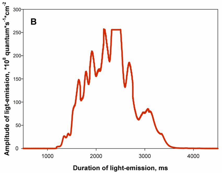
VARIABILITY OF THE CTENOPHORE MNEMIOPSIS LEIDYI BIOLUMINESCENCE

prey relationship, their effect on functioning as well as regenerative ability and luminescent function of the
ctenophores M. leidyi under conditions close to in situ have not been discussed.
         Therefore, identification and studying the functional capability of an attacked by a predator comb-
jellies M. leidyi as well as estimation of their ability to regenerate and to emit light during the post-traumatic
regeneration period are of scientific interest to investigate.

Material and Methods

The investigation was conducted in the Department of biophysical ecology of А.O. Kovalevsky Institute of
Biology of the Southern Seas of RAS (IBSS), the Russian Academy of Sciences. The studied ctenophores
were collected near the shore of Sevastopol and two miles offshore in August – September 2017-2018. All
organisms picked out for the experiment were divided into two groups: 50-mm B. ovata and 40-mm M.
leidyi. The wet weight was determined by the calculating the seawater volume displaced by the ctenophore
out of the container followed by the weighing the predried on absorbent paper organisms with
microanalytical scales AN 50 (with the precision up to 0.01 g). Examined specimens of M. leidyi and B.
ovata weighed 9.16 ± 0.45g and 10.88 ± 0.54 g respectively.
         During the study, the laboratory conditions created for the ctenophore regeneration were close to the
ones in situ. The fresh ctenophores were placed in 5-liter laboratory tanks filled with filtered sea water at 21
± 2°C (membrane filters with pore diameter of 35 µm) for 2 hours to get them adapting. This temperature
range was chosen as optimal for the laboratory study of ctenophores (Vostokov et al., 2001; Mashukova &
Tokarev, 2012). Before being fed, B. ovata were taken one by one from the tank and placed each into a
laboratory vessel with filtered sea water. The prey (M. leidyi) was preweighed and brough by one into the
flasks containing the predator. The prey was given in abundance; the consumption was regularly assessed by
daily checks made before and after the exposition (Tokarev & Mashukova, 2013). On average, the prey was
caught 2 later after the introduction. As the prey was engulfed, the body of B. ovata transformed to a sphere
preserving this shape during 4 – 5 hours’ digestion.
         The visual observations of B. ovata have clarified some aspects of their feeding behavior, e.g., the
majority (80%) of the predators engulfed the whole body of prey while the rest tore it into pieces. The further
research procedure concerned exclusively regeneration processes developing in the group of damaged
ctenophores M. leidyi after B. ovata’s attacks.
         All M. leidyi used in the experiment were divided into three groups. The first (control) group
included freshly caught unimpaired organisms; the second one consisted of 18-20 mm large injured
ctenophores, with their aboral organs preserved or missing after the predator’s attack; and the third group
was of the post-regenerated ctenophores. Severity of their body damage was determined by means of
microscopy.
         In the first group, stimulation of light emission began after the 2 hours’ adaptation period in the dark
laboratory. The second group emitted light immediately after the damages had been inflicted by the
predators. The third group was kept in the tanks and was fed on copepods Acartia tonsa Dana (Copepoda).
The choice of the copepods was determined by the fact that from mid - to late summer calanoid copepods
prevail in the mesozooplankton of the sampling sites located in Sevastopol Bay (Gubanova, 2003) being
typical food for M. leidyi. Predation rate, or the rate of swallowing up food, of M. leidyi was assessed by
emptying the ctenophores’ gastrovascular cavity. Daily counting of copepods allowed sustaining the food
supply at least at 0.35 mg dry weight∙l-1 in conformity with the experimental design; A. tonsa copepods were
introduced into the laboratory tanks in portions – 70 ind∙l-1 at a time (Tokarev & Mashukova, 2013). Feeding
was carried on untill the regenerating ctenophores restored up to the original weight, i.e., prior to the
damaged one (Tokarev & Mashukova, 2013).
         The investigation of the kept in darkness ctenophores’ bioluminescence was conducted at daytime
with the use of the Svet laboratory device (Tokarev, 2006) while the method of mechanical and chemical
excitation was applied. For mechanical stimulation the seawater flow was accelerated by the pumping
electromechanical equipment and circulating in the laboratory tank was periodically strengthened to imitate
turbulence.
         To specify maximal bioluminescence potential in response to chemical simulation, 3 cm3 of 96%
alcohol were injected into the laboratory tank containing the ctenophores (Tokarev et al., 2016).

                                                                                                               20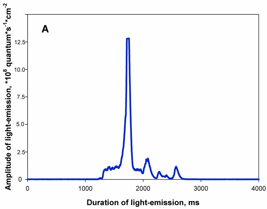
MASHUKOVA ET AL.

Results and discussion

The observations of ctenophores behavior showed that in the beginning of exposition after M. leidyi being
damaged by the predators, they were incapable of normal locomotion, namely, beating of cilia in their ctenes
dramatically reduced and was sluggish. As the exposition went on, it was observed that 18 – 20 mm long
body fragments without aboral organ descend to the bottom of the tank and died within a day.
        Conversely, M. leidyi that have preserved their statocyst actively moved even after near-catastrophic
impairment. Compared to the check, bioluminescent signals these incomplete organisms generated in
response to the chemical excitation and to the mechanical impact had the amplitudes as low as 17.08 and
14.85∙108 quantum·s-1·cm-2 respectively (fig. 1) and the light-emission durations as short as 1.37 and 1.25 s
respectively (fig. 2).

Figure 1. Amplitude of the bioluminescence produced by regenerating M. leidyi under the mechanical and chemical
impacts.

Figure 2. Duration of the bioluminescence produced by regenerating M. leidyi under the impact of the mechanical and
chemical stimulation

Ecologica Montenegrina, 37, 2020, 19-26                                                                           21
VARIABILITY OF THE CTENOPHORE MNEMIOPSIS LEIDYI BIOLUMINESCENCE
Typical bioluminescent signals of the tested ctenophores also differed in these groups. The damaged
M. leidyi responded to the stimuli by short flashes which were registered as low energy with 1 – 2 peaks of
small intensity (fig. 3 A). Unlike them, animals in the post-regeneration group produced a sequence of
consecutive bright flashes, with the amplitude of each following flash being identical to the previous one
(fig. 3 B).
Figure 3 – Typical bioluminescence signals induced by the chemical stimulation of M. leidyi: А – the injured group; B
– the post-regeneration group.
Daily measurements of oral-aboral length in the third group evidenced that the ctenophores were
gradually restoring their body size and weight to the initial parameters and completed regeneration on the 3 rd
– 4th day of the experiment. According to the microscopic study, on the 3 rd – 4th day after the attack, all
ctenes of the M. leidyi were formed anew in the wound fissure; they had similar length and were arranged
relatively regularly thereby indicating complete regeneration (Tamm, 2012).
22MASHUKOVA ET AL.
Noteworthily, the bioluminescence of the post-regeneration group of the ctenophores was notably
different: compared to the impaired group, the amplitude of bioluminescence was 15 – 20 times more –
332.33 ± 16.61∙108 quantum·s-1·cm-2 under the chemical impact and 219.45 ± 10.97∙108 quantum·s-1·cm-2
under the mechanical stimulation. Amazingly, the fully restored ctenophores had the intensity of light 1.5
times as high as in the check group. The chemical and the mechanical excitation brought about
bioluminescent flashes of longer duration – 3.67 s and 3.25 s, respectively (fig. 2).
As the regulatory mechanism is inherent to many living organisms, regeneration is responsible for
restoration of essentially vital functions the animal has accidentally lost. Adult ctenophores (Lobata
(Tentaculates)) have an intriguing ability to restore their missing body parts (Henry & Martindale, 2000),
which has been proved by our experiment. What secret machinery underlies this phenomenon? As
morphogenetic studies evidence, high regeneration success of M. leidyi can be due to the bilateral symmetry
of their body (Martindale & Henry, 1996; Peterson & Eernisse, 2001). Body-forming cells of the adult
ctenophore contain the information about their original position that it used for starting and sustaining the
complete regeneration cycle (Henry & Martindale, 2000). As the wound inflicted by partial removal of
ctenes from the row closes up and heals, the previous distance between the combs conspicuously increases
(Tamm, 2012).
Intensive reproduction, as a typical feature of hermaphrodites and, therefore, of all Ctenophora, also
facilitates the regeneration success: as the M. leidyi fed on copepods Acartia clausi (100 ind·l-1) have grown
to 10 mm in length (13.5 mg dry weight), they start reproduction (Finenko et al., 1995). However, there are
some factors which set limits to the regeneration. Particularly, not all body parts are capable of regeneration
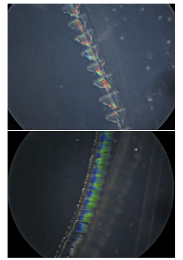
because of the special internal structure of ctenophores body. So, ctenophores generate light in special cells –
photocytes; excitation of the photocytes triggers illumination of intracellular granules which underlie ctenes
(fig. 4) along the meridional gastrovascular canals (Lapota, 2012).
M. leidyi have nervous system which is located under the rows of comb plates and is made up by the
surface interlacement of nervous cells clustered into dense cords going to the aboral organ which regulates
locomotion and balance (Shiganova et al., 2001). As the experiment has shown, ctenophores which have lost
the aboral organ and nervous cords were incapable of sustaining essentially vital functions and died.
As noted above, bioluminescent intensity can be used as a marker of the functional state of plankton
populations (Tokarev & Mashukova, 2016). Normal functioning of the ctenophores M. leidyi ceases giving
way to abnormalities caused by the animal’s being damaged by the predator or by the harmful impact;
similarly, the lower light emission typical of the defective ctenophores should be interpreted as the direct
result of functional disorder. Ctenophores which preserved their aboral organ after the attack had greater
chance to fully restore their vital functions and the ability to generate light.
The increase of light production by the regenerated ctenophores can be due to the chemistry of the
bioluminescence (Ward & Cormier, 1975; Lapota, 2012), namely, the higher rate of peroxide complexes
formation, disintegration of which leads to the release of large quantities of energy, therefore the maximal
bioluminescence.
Certainly, further investigation of the bioluminescence, feeding behavior, and predator–prey
relationship of ctenophores – especially the observations of actual interplay of hydrobionts going in their
natural habitat – will invest to understanding of marine communities and ecosystems functioning and shed
more light on ecological significance of the bioluminescence.
Conclusion
Thus, it has been established that ctenophores cannot perform normal vital functions without preserving
aboral organ with tenias of the nervous system and digestive system. It is shown that low light emission
indicators of ctenophores with a damaged body are explained by the impairments in their functional state.
The ones with preserved aboral organ have more chances to restore their vital activity and consequently their
bioluminescence ability. At the same time, higher light production indices after the regeneration of
ctenophore’s body can be due to the chemistry of the bioluminescence reaction, namely, the increased speed
rate of peroxide complexes formation, the disintegration of which leads to a large amount of energy released
and the maximal bioluminescence rate.
Ecologica Montenegrina, 37, 2020, 19-26 23VARIABILITY OF THE CTENOPHORE MNEMIOPSIS LEIDYI BIOLUMINESCENCE
Figure 4. Fluorescent M. leidyi ctenes.
Acknowledgements
This study was carried out under research project IBSS no. AAAA-A18-118020790229-7 and partly
supported by the Russian Science Foundation (project no. 18-45-920015)
References
Anninsky, B., Finenko, G., Abolmasova, G. et al. (2005) Effect of starvation on the biochemical
compositions and respiration rates of ctenophores Mnemiopsis leidyi and Beroe ovata in the Black
Sea. Journal of the Marine Biological Association of the United Kingdom, 85, 549–561.
24MASHUKOVA ET AL.
Finenko, G., Abolmasova, G. & Romanova, Z. (1995) Ctenophora Mnemiopsis mccradyi nutrition, oxygen
consumption and growth, depending on the food concentration. Biolgiya Morya, 20, 315–320. [In
Russian].
Finenko, G. & Romanova, Z. (2000) The population dynamics and energetics of ctenophore Mnemiopsis
leidyi in Sevastopol Bay. Oceanology, 40, 677–685.
Gitelzone, I., Levin, L., Utyushev, R. et al. (1992) Bioluminescence in the ocean. Gidrometeoizdat, S.-
Peterburg, 283 pp. [In Russian].
Gubanova, A. (2003) Long-time changebilities in the zooplankton community of the Sevastopol Bay. In:
Eremeev V.N., Gaevskaya A.V. (Eds), Modern condition of the biodiversity neritic water Crimea.
EKOSY, Gydrophysica, Sevastopol, pp. 83–94. [In Russian].
Haddock, S. & Case, J. (1999) Bioluminescence spectra of shallow and deep-sea gelatinous zooplankton:
ctenophores, medusae and siphonophores. Marine Biology, 133, 571–582.
Haddock, S., Moline, M. & Case, J. (2010) Bioluminescence in the Sea. Annual Review of Marine Science,
2, 443–493.
Harvey, E. (1952) Bioluminescence. Academic Press, New York, 649 pp.
Henry, J. & Martindale, M. (2000) Regulation and Regeneration in the Ctenophore Mnemiopsis leidyi.
Developmental Biology, 227, 720–733.
Hernandez-Nicaise, M. (1991) Ctenophora Microscopic Anatomy of Invertebrates. In.: Placozoa, Porifera,
Cnidaria, and Ctenophora Wiley. Liss, New York, 2, pp. 359–418.
Lapota, D. (2012) Bioluminescence – Recent advances in oceanic measurements and laboratory applications.
Open Access Book InTech, Janeza trdine, 9, 190 pp.
Martindale, M. & Henry, J. (1996) Development and regeneration of comb plates in the ctenophore
Mnemiopsis leidyi. Biology Bulletin, 191, 290–292.
Mashukova, O. & Tokarev, Yu. (2012) Influence of the temperature at the Black Sea ctenophores-aliens
bioluminescence characteristics. Advances in Bioscience and Biotechnology, 3, 269–273.
Mashukova, O. & Tokarev, Yu. (2013) Bioluminescence daily rhythm of ctenophore Beroe ovata Mayer,
1912. Proceedings of the Global Congress on ICM: Lessons Learned to Address New Challenges.
Marine Biological Microbiology, 2, 729–736.
Mashukova, O., Tokarev, Yu. & Skuratovskaya, E. (2017) Нeavy metals influence on the ctenophores M.
leidyi and B. ovata bioluminescence. Ecologica Montenegrina, 14, 109–118.
Muller, W., Plickert, G. & Berking, S. (1986) Regeneration in Hydrozoa: distal versus proximal
transformation in Hydractinia. Roux’s Archives of Developmental Biology, 195, 513–518.
Mutlu, E. (1999) Distribution and abundance of ctenophores and their zooplankton food in the Black Sea. II:
Mnemiopsis leidyi. Marine Biology, 135, 603–613.
Peterson, K., & Eernisse, D. (2001) Animal phylogeny and the ancestry of bilaterians: inferences from
morphology and 18S rDNA gene sequences. Evolution & Development, 3, 170–205.
Piraino, S., Vito, D., Schmich, J., & Bouillon, J., Boero, F. (2004) Reverse development in Cnidaria. Journal
of Zoology, 82, 1748–1754.
Shiganova, T., Mirzoyan, Z., Studenikina, E. et al. (2001) Population development of the invader ctenophore
Mnemiopsis leidyi, in the Black Sea and in other seas of the Mediterranean basin. Marine Biology,
139, 431–445.
Shimomura, O. (2006) Bioluminescence: Chemical principles and methods. World Scientific, 470 pp.
Tamm, S. (2012) Regeneration of ciliary comb plates in the ctenophore Mnemiopsis leidyi. Morphology.
Journal of Morphology, 273(1), 109–120.
Tardent, P. (1963) Regeneration in the Hydrozoa. Biological Reviews, 38, 293–333.
Tokarev, Yu. (2006) Basin of the hydrobionts biophysical ecology. ECOSI-Gidrophizika, Sevastopol, 342
pp. [In Russian].
Tokarev, Yu. & Mashukova, O. (2013) Variability of the bioluminescence characteristics of the Black Sea
ctenophores-aliens in connection with different conditions of nutrition. Advances in Bioscience and
Biotechnology, 4, 968–973.
Tokarev, Yu. & Mashukova, O. (2013) Ctenophores Mnemiopsis leidyi A.Agassiz, 1865 (Tentaculata,
Lobata) and Beroe ovata Mayer, 1912 (Atentaculata, Beroida) bioluminescence seasonal dynamics at
the Black sea. Natura Montenegrina, 12, 345–355.
Ecologica Montenegrina, 37, 2020, 19-26 25VARIABILITY OF THE CTENOPHORE MNEMIOPSIS LEIDYI BIOLUMINESCENCE
Tokarev, Yu. & Mashukova, O. (2016) Bioluminescence of the Black Sea ctenophores-aliens as an index of
their physiological condition. In: Thirumalai, J. (Eds), Luminescence - An Outlook on the
Phenomena and their Applications. Open Access Book InTech, Croatia, Ch. 14, pp. 351–378.
Tokarev, Yu., Mashukova, O. & Sibirtsova, E. (2012) Bioluminescence characteristics changeability of
ctenophore Beroe ovata Mayer 1912 (Beroida) in ontogenesis. Turkish Journal of Fisheries and
Aquatic Sciences, 12, 479–484.
Tokarev, Yu., Evstigneev, P. & Mashukova, O. (2016) The plankton bioluminescents of the World Ocean:
biodiversity, bioluminescence characteristics in norm and under anthropogenic impact. N. Orianda,
Russia, 340 pp. [In Russian].
Tokarev, Yu., Vasilenko, V., Mashukova, O. (2017) Some Theoretical and Applied Aspects of the Oceanic
Bioluminescence Registration from Space. Advances in Bioscience and Biotechnology, 8(9), 283–
294.
Vostokov, S., Arashkevich, E., Drits, A. et al. (2001) Ctenophore Beroe ovata ecological-physiological
characteristics in the Black sea coastal zone: abundance, biomass, population dimension
characteristic, behavior, nutrition and metabolism. Okeanologiya, 41(1), 109–155. [In Russian].
Ward, W. & Cormier, M. (1975) Biochemistry Extraction of Renilla-type luciferin from the calcium-
activated photoproteins aequorin, mnemiopsin, and berovin. Proc. Nat. Acad. Sci., USA, 72, 2530–
2534.
26You can also read