PROCEEDINGS Pre-BSAVA Virtual Spring Meeting 2021 18.30-20.30 - EAVDI
←
→
Page content transcription
If your browser does not render page correctly, please read the page content below

PROCEEDINGS

Pre-BSAVA Virtual Spring Meeting 2021

Tuesday March 23rd/ Wednesday March 24th
              18.30-20.30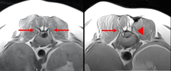
Tuesday 23rd March 2021

Dr Anya Morrissey BSc(Hons), BVetMed, PGDip VCP, MRCVS
Anya Morrissey graduated from the Royal Veterinary College in 2016
and completed a General/Surgical Internship at London Vet
Specialists in 2017. After working as an emergency veterinarian for
two years, she went on to complete the Small Animal Rotating
Internship at the Royal Veterinary College in 2020 and is currently
working towards a post-graduate certificate in Small Animal Surgery.

Dr James Guillem CertAVP (VDI) MRCVS
After graduation in 2010 in Madrid, James worked at several first opinion practices in the UK.
In 2018 he joined the University of Liverpool Small Animal Teaching Hospital to start his small
animal diagnostic imaging residency.

Dr Aurore Masson MRCVS
Aurore graduated in 2007 from the National Veterinary School of Toulouse (France). After
graduation she completed a rotating internship and two specialized internships in surgery in
Toulouse. Subsequently, she worked for four years in a referral and emergency private
practice in Versailles (France). In 2014, she moved to the UK to work in first opinion practice.
She started a diagnostic imaging residency in January 2021 at the National Veterinary School
of Maisons-Alfort (France).

Dr Susann Dressel Dr. med. vet
Susann graduated from Justus-Liebig-University Giessen, Germany in
2014. Following graduation, she worked at the Radiation Oncology,
Vetsuisse Faculty, University Zurich during which she completed her
doctoral thesis. Subsequently, she went on to complete a small
animal diagnostic imaging internship at Vetimage Diagnostik GmbH,
Switzerland. In 2019, she begun a diagnostic imaging residency at the
Clinic for Diagnostic Imaging, Vetsuisse Faculty University Zurich,
Switzerland.
USE OF CT SINOGRAPHY IN 27 DOGS AND 1 CAT
A.M. Morrissey , C. Lopez-Jimenez , A. Groth , K.C.L. Lee , B. de la Puerta , C.R. Lamb.
                1                  2          2           1                2            2

Queen Mother Hospital for Animals, Royal Veterinary College, Hatfield, Hertfordshire, AL9
1

7TA, United Kingdom
North Downs Specialist Referrals, Bletchingley, Surrey, RH1 4QP, United Kingdom
2

Introduction: Sinography following survey radiography in dogs with a draining sinus aids
determination of the extent of a tract and presence of foreign material. Use of sinography in
combination with computed tomography (CT) in animals has been sporadically reported.
Methods: Retrospective review of clinical and imaging findings in 27 dogs and one cat that
underwent CT sinography to investigate a draining tract. Pre- and post-intravenous (IV)
contrast CT series were compared with CT sinography in each patient.
Results: Median duration of clinical signs before referral was 85 days (range 2 – 1478 days).
The most commonly reported clinical signs were swelling (14/28, 50%) and regional pain
(5/28, 18%). Sinuses affected the head (14/28, 50%), thoracic or abdominal wall (7/28, 25%),
sublumbar muscles (4/28, 14%) and a distal limb (3/28, 11%). CT sinography showed tract
extension beyond that seen on prior CT in 6 (21%) cases and communication with a cavity
(fistula) in 2 cases (7%). Sensitivity for suspected foreign material was higher in pre-contrast
images (31%) than CT sinography (23%). Of the tracts managed surgically (22/28), 13 (59%)
cases resolved, 6 (27%) persisted or recurred after surgery, and 3 (14%) were lost to follow-
up. All foreign bodies suspected on CT were later confirmed, but CT plus sinography failed to
identify foreign bodies found at surgery and histopathology in 9 additional cases.
Conclusions: CT sinography provides limited additional information about the extent of
draining tracts compared to pre- and post-IV contrast CT images and did not increase
sensitivity for foreign bodies.
CT ASSESSMENT OF BRONCHIAL ABNORMALITIES IN BRACHYCEPHALIC DOGS WITH AND
WITHOUT BOAS IN COMPARISON WITH NORMAL DOGS
J.S. Guillem, F. Schiborra, M. Rossanese, T.W. Maddox, J.R. Mortier.
University of Liverpool Small Animal Teaching Hospital, Leahurst Campus, Wirral, CH64 7TE,
United Kingdom.

Introduction: A relationship between bronchial collapse and brachycephalic patients has
been previously established. However, there is no evidence in the current literature that
relates brachycephalic dogs, with or without signs of obstructive airway syndrome (BOAS),
with increased bronchial wall thickness. The aim of this study was to assess bronchial wall
thickening and bronchial collapse in brachycephalic dogs with or without BOAS on thoracic
computed tomography (CT) and compare them with non-brachycephalic dogs.
Methods: Patients were divided into three groups: dogs with BOAS, an asymptomatic
brachycephalic dogs (ABD) group and a control group of non-brachycephalic dogs. Bronchial
thickening and collapse were subjectively identified and graded by a single observer, blinded
to patient history, signalment and previous CT reports. Descriptive statistics were calculated
for relevant variables. Univariable and multivariable analysis were performed to evaluate
possible associations with both bronchial thickening and bronchial collapse.
Results: A total of 84 dogs were retrospectively recruited. Thirty dogs in the BOAS group, 26
in the ABD group and 28 in the control group. Bronchial wall thickening was identified in
93.3% of dogs within the BOAS group, 59% in the ABG group and 35% in the control group.
Univariable analysis identified several variables with a potential relationship with bronchial
wall thickening, however after multivariable analysis, only the group (P=0.0001) and weight
(P=0.027) showed a significant association with bronchial thickening.
Bronchial collapse was identified in 56.7% of dogs within the BOAS group, 62.9% in the ABD
group and 10.7% in the control group. Multivariable analysis showed that only the group had
a significant relationship with bronchial collapse (P=0.001)
Discussion: A relationship between brachycephalic dog breeds and weight with bronchial wall
thickening was established. However, further studies are required to investigate the cause of
the bronchial wall thickening. Bronchial collapse was also related with brachycephalic breeds,
as previously reported.

CLINICAL AND DIAGNOSTIC IMAGING FINDINGS IN DOGS WITH ATRIAL APPENDAGE
ANEURYSM: 7 CASES (2014 – 2019)
A.V. Masson , T.W. Maddox , E.F. Bode , J.R. Mortier .
            1               2          2             2

The Vet Liverpool, Lorenzo Drive, Norris Green, Liverpool L11 1BQ
1

Small Animal Teaching Hospital, University of Liverpool – Institute of Veterinary Science,
2

Chester high road, Neston CH64 7TE

Introduction: To describe clinical and diagnostic imaging findings, especially computed
tomography, of atrial appendage aneurysms in dogs.

Methods: Medical records of the Small Animal Teaching Hospital were searched to identify
dogs with a presumptive diagnosis of left (LAAA) or right (RAAA) atrial appendage aneurysm.
Signalment, history, examination findings, diagnostic test results, and imaging procedures
were reviewed. Archived diagnostic images were retrieved and retrospectively evaluated by
two board-certified radiologists and a board-certified cardiologist. Data analysis was
descriptive.

Results: Seven dogs with a presumptive diagnosis of LAAA or RAAA that had computed
tomography studies were included. Most affected dogs were male dogs (6/7) with a median
age of 8 years old. Six cases were affected with RAAA and one with LAAA. All cases of RAAA
were incidental findings. The dog with LAAA presented a syncopal event. All dogs underwent
a thoracic computed tomography (CT) examination for various reasons. CT was useful to
identify and assess the atrial appendage aneurysm as well as neighbouring structures,
although possible pericardial defects could not be visualized. Five dogs had echocardiographic
examination, which successfully identified the LAAA and two RAAA.

Discussion: Atrial appendage aneurysm in dogs is a rare condition. This case series is focusing
on the imaging findings. RAAA seems to be an incidental finding whereas LAAA could be
associated with clinical signs, consistent with the literature. CT seems to be more sensitive
than transthoracic echocardiography in the detection of atrial appendage aneurysm in dogs,
especially for RAAA.
PERIPORTAL HALO-COMPUTED TOMOGRAPHY SIGN OF PERIPORTAL OEDEMA LIKE
LESIONS IN DOGS AND CATS
S. Dressel , P.R. Kircher , F. Del Chicca
          1,2            1                  1

Clinic for Diagnostic Imaging, Vetsuisse Faculty University of Zurich, 8057 Zurich,
1

Switzerland
Vetimage Diagnostik GmbH, 5036 Oberentfelden, Switzerland
2

Introduction: Many pathologies can occur in the periportal space and manifest as fluid
accumulation visible in Computed tomography (CT) images as a circumferential region of low
attenuation around the intrahepatic portal vessels (periportal halo). This finding has been
described in different types of hepatic (HD) and extra-hepatic disease (EHD) in humans and
reflects altered hepatic lymphatic dynamics. In veterinary literature, it has been reported as
a non-specific feature correlated to shunts and is of unknown significance. Here, we
investigate the prevalence of periportal halo on a large population of dogs and cats and assess
the presence of lesions related to HD and EHD.

Methods: This retrospective study includes CT-studies of client-owned animals to evaluate
the prevalence over a period of five years. Patients were classified based on the subjective
grade and distribution of the periportal halo. Anamnestic, clinical, biochemical and
pathological information were analyzed.

Results: 1168 CT examinations were reviewed, and 125/858(15%) dogs and 4/310(1%) cats
showed a periportal halo.129 animals (125 dogs, 4 cats) were included, and the halo was
classified as mild, moderate and severe respectively in 70(54.3%), 42(32.6%) and 17(13.2%)
animals. The halo distribution was generalized in 71/143 cases, localized along the second
generation of intrahepatic portal branches in 57/143, and along the first generation only in
1/143. 52/125 animals had morphological liver or vascular abnormalities. Primary HD was
confirmed in 27 cases, suspected in 3 and suspected secondary or additionally to a primary
EHD in 24 patients. Most cases had an EHD (98/125;71.4%). Main cause of HD (20/54;37.0%)
and EHD (70/98;68.2%) was neoplasia. 8.4% animals had no evidence of HD nor EHD.

Conclusions: Periportal halo is non-specific, but it
could represent an important hint to underlying
HD or EHD. Most patients with halo in this study
had an underlying neoplastic disease, both
hepatic or extra-hepatic. In contrast to previous
literature, only 6 animals had portosystemic
shunts.

                                                         Contrast-enhanced transverse CT
                                                       image of the intrahepatic portal vein
                                                           with a severe periportal halo
                                                                     (arrows).
Wednesday 24th March 2021

Dr Chantelle Franklin
Chantelle graduated from the University of Adelaide in 2013 and then
worked in general practice for 6 years in both Australia and the United
Kingdom. She completed a Post-Graduate Certificate in small animal
abdominal ultrasonography from the University of Melbourne in 2018
and became a Member of the Australian and New Zealand College of
Veterinary Scientists in small animal radiology in 2019. The same year,
she started a Diagnostic Imaging internship at the Queen’s Veterinary
School Hospital at the University of Cambridge and was then lucky
enough to stay on as a Senior Clinical Training Scholar (Resident) in
Diagnostic Imaging from November 2020.

Dr Jess Sumping BVetMed MRCVS
After graduation from the Royal Veterinary College Jess completed a
rotating internship at Vets Now Referrals, Glasgow. After spending time
in general practice, she completed a diagnostic imaging internship at the
University of Liverpool and is now in her first year of her residency
programme.

Dr Emilie Paran DVM, MRCVS
Emilie graduated from Lyon Vet School (France) in 2015. After a
rotating and imaging internship, she worked as an ultrasonographer in
France before moving to the UK in 2018. In 2019, she started her
residency at Glasgow Vet School and is temporarily located at Langford
Vets near Bristol.

Dr Lorraine Peschard MRVS
Lorraine graduated in Madrid in 2016 and then spent two years in first
opinion and referral practice in the UK, before completing a rotating
internship at Liège University in 2018. She started her imaging
residency in September 2019 at the Queen’s Veterinary School in
Cambridge and is thoroughly enjoying it!
PREVALENCE OF HUMERAL INTRACONDYLAR FISSURES IN ASYMPTOMATIC
BRACHYCEPHALIC BREED DOGS.

C. Franklin , M. Herrtage , K.P. Harris , M.A. Genain .
           1             1            2              1

1Queen’s Veterinary School Hospital, University of Cambridge, Madingley Road Cambridge
CB3 0ES UK.
2Southern Counties Veterinary Specialists, Unit 6 Forest Corner Farm Hangersley Ringwood
Hampshire BH24 3JW UK

Introduction: Historically, humeral intracondylar fissures (HIF) have been seen predominantly
in spaniel breeds. These dogs are thought to be at higher risk of lameness and humeral
condylar fracture. Recent publications have brought attention to increased risk of humeral
condylar fracture in French Bulldogs. In a manuscript undergoing review, HIF has been noted
in a small population of French Bulldogs presenting with humeral condylar fracture in their
contralateral limb (In publication: U.W. Strohmeier & K.P. Harris 2021). We therefore
hypothesised that HIF may exist in French Bulldogs also and aimed to assess the prevalence
of HIF in an asymptomatic population of brachycephalic breed dogs.

Methods: A blinded retrospective study was performed on CT examinations of dogs
presenting for an unrelated condition (brachycephalic airway syndrome) and were reviewed
by two ECVDI diplomates in a bone window. A classification system was devised to grade the
findings, and the image quality as the elbows were not the focus of the imaging acquisition.
We considered a Score 1 to be normal, Score 2 to have sclerosis, Score 3 to have a partial
fissure and Score 4 to have a complete fissure. We added additional criteria to assess the
location of the sclerosis and the presence of other elbow dysplasia abnormalities where
possible.

Results: To date, a total of 116 elbows have been reviewed. Of this population 73 were
French Bulldogs, 28 were Pugs and the remainder were a mix of other brachycephalic breeds.
For the overall population of dogs, we found an overall prevalence of either a partial or
complete humeral fissure at 7.3% (CI 2.6-12.0%). The inter-reviewer reliability based on
Cohen’s kappa was low at 0.42, however this was largely based on discrepancy between
reviewers in Score 1 (normal) and Score 2 (sclerosis) categories. When considering cases
assigned to Score 4 (complete fissure) there was complete agreement between assessors.
Within the French Bulldog breed, the prevalence of HIF (partial or complete) was higher at
8.2% (CI 4.0-18.5%).

Discussion: This is the first study to our knowledge examining the prevalence of HIF in
brachycephalic breed dogs who are asymptomatic and will contribute to the knowledge of
this condition in increasingly popular breeds in the community.
SENSITIVITY, SPECIFICITY AND ACCURACY OF ULTRASONOGRAPHY IN THE DETECTION OF
LYMPHOMATOUS INFILTRATION OF THE LIVER AND SPLEEN IN DOGS AND CATS WITH
LYMPHOMA
J. C. Sumping , T. W. Maddox , D. Killick , J. R. Mortier .
1 1 1 2
Small Animal Teaching Hospital, University of Liverpool, Leahurst Campus, Cheshire, CH64
1
7TE, UK.
École Nationale Vétérinaire d’Alfort, 94700 Maisons-Alfort, France.
2
Purpose: To determine the sensitivity, specificity and accuracy of ultrasonography in the
detection of lymphomatous infiltration of the liver and spleen in a large population of dogs
and cats with lymphoma. The second aim of this study is to determine if specific
ultrasonographic features of the liver and spleen in dogs are associated with lymphomatous
infiltration or a specific immunophenotype of multicentric lymphoma.
Methods: Blinded retrospective review of ultrasonographic images of the liver and/or spleen
in 132 dogs and 29 cats with cytologically or histologically confirmed lymphoma by two board-
certified veterinary radiologists.
Results: Ultrasonography had a sensitivity, specificity, accuracy, positive predictive value and
negative predictive value of 16.7%, 91.0%, 55.9%, 62.5% and 55.0% for the detection of
lymphomatous infiltration of the liver, and 73.1%, 93.9%, 82.6%, 93.4% and 74.7% for the
spleen. In dogs, an ultrasonographically normal liver was statistically associated with not
having lymphomatous infiltration, leopard-spotted splenic parenchyma and splenomegaly
were independently statistically associated with lymphomatous infiltration and leopard-
spotted splenic parenchyma was also statistically associated with the B-cell
immunophenotype of multicentric lymphoma.
Conclusions: Ultrasonography of the spleen and liver is specific but not sensitive in the
detection of lymphomatous infiltration. A leopard spotted splenic parenchyma in dogs is
highly specific for lymphomatous infiltration and can also be used to predict
immunophenotype of multicentric lymphoma.MORPHOLOGIC DESCRIPTION OF THE TEMPOROMANDIBULAR JOINT IN ASYMPTOMATIC
BRACHYCEPHALIC DOGS USING COMPUTED TOMOGRAPHY
E. Paran , S. Bouyssou, A. King.
1
Small Animal Hospital, University of Glasgow, G61 1BD, United Kingdom.
Introduction: Temporomandibular joint (TMJ) incongruity and morphological variations can
result in clinical signs and have also been reported in asymptomatic brachycephalic dogs such
as Cavalier King Charles Spaniels (CKCS) . The purpose of the present study was to assess the
1
morphology of the TMJ in a group of brachycephalic dogs using computed tomography (CT).
Methods: Asymptomatic French Bulldogs, English Bulldogs, Boxers, CKCSs, Chihuahuas, Lhasa
Apsos, Pugs, Shih Tzus and Staffordshire Bull Terriers were retrospectively enrolled from a 6-
year period. Multiplanar reconstructions of the CT images were used and TMJ morphological
grades were determined using an A to C classification system adapted from a previous
publication . A grade A was defined as a long retroarticular process extending ventral to the
2
mandibular condyle. Three B categories were defined depending on the length of the
retroarticular process and coverage of the mandibular condyle as determined in the
transverse images (100% coverage in B1, 50-99% coverage in B2, and 1-49% coverage in B3).
Grade C was assigned when the retroarticular process was absent. The intra and inter-
observer agreements were calculated.
Results: One hundred fifty-three dogs were included. In the sagittal plane, the mandibular
condyle and fossa were respectively rounded and concave in grade A, showed a progressive
flattening in grade B and were respectively square and flat in grade C. In the transverse plane,
the mandibular condyle was flat in grade A, showed some variations (flat, curved, trapezoid,
sigmoid) in grades B and was more likely to be sigmoid in grade C. Pugs and Shih Tzus were
more likely to be graded B. French Bulldogs, CKCSs and Chihuahuas were more likely to be
graded C. The intra and inter-observer agreements for this TMJ classification system were
moderate.
Conclusions: Variations in the TMJ morphology exist in brachycephalic dogs. Marked changes
seem to be highly prevalent in French Bulldogs and CKCSs (26.3% and 68.2% respectively in
this study) and should be considered a breed variation. The modified TMJ classification
described in this study could be used to standardise the assessment of the canine TMJ
morphology.FOLLOW-UP MRI APPEARANCE OF THE SURGICAL SITE IN DOGS TREATED FOR
INTERVERTEBRAL DISC HERNIATION
A.-L. Peschard , P. Freeman, M.-A. Genain.
1
The Queen’s Veterinary School Hospital, Cambridgeshire, CB3 0ES, United Kingdom.
1
Purpose: To describe the MRI characteristics of the surgical site in dogs that have undergone
a previous surgical treatment for intervertebral disc herniation.
Methods: Descriptive retrospective study. All MRIs but one was performed on a low-field MRI
magnet.
Results: 25 patients were included, for a total of 53 MRI studies. The mean number of days
between surgery and second MRI study was 389 (range 2-1367). Susceptibility artefacts were
seen in 6/25 cases (24%), and hindered assessment of the adjacent spinal cord in 4/6 (16%
of total cases). Compression of the spinal cord at the surgical site was present in
15 cases and required surgical reintervention in 5/15 cases. In 12 of these 15 cases the disc
space where the maximal amount of compression of the spinal cord was found was located
at the surgical site: this was caused by extradural disc material (8/12 cases, 66%), bony
hyperplasia (1/12 cases, 8%), displacement of an articular process into the vertebral canal
(1/12 cases, 8%), and haematoma and inflammatory changes (5/12 cases, 42%). In these
12 cases, follow-up MRI was performed between 2 and 1097 days after surgery (median 61
days). 2 patients in which a left-sided T13-L1 mini-hemilaminectomy was performed
subsequently suffered a displacement of the overlying cranial articular process. Tethering of
the spinal cord to the surgical site was observed in 6 patients (24%).
Discussion: The appearance of spinal surgical sites on MRI is variable, and compression of the
spinal cord at the surgical site is common, even in patients that made a full recovery and
relapsed several weeks to months after the initial surgery. Displacement of an articular
process following mini-hemilaminectomy has not yet been described as a potential
complication but was found in two patients in the present study. Susceptibility artefacts at
the surgical site were uncommon on low-field MRI and were not found in the high-field study
performed. Contrary to what was suggested in a previous study, postoperative MRI may be
the modality of choice if following surgery, a patient fails to recover in a satisfactory fashion
or deteriorates neurologically.
Fig 1. T1 transverse image showing displacement of the left cranial articular process following left-sided
mini-hemilaminectomy. A small bony defect in the left lamina of the vertebra is visible ventral to the
affected articular process.You can also read