Cutaneous sarcoidosis of the scalp unmasking systemic involvement: A case report
←
→
Page content transcription
If your browser does not render page correctly, please read the page content below
EXPERIMENTAL AND THERAPEUTIC MEDICINE 22: 1369, 2021

                     Cutaneous sarcoidosis of the scalp unmasking
                         systemic involvement: A case report
                DANIEL BODA1,2, ANA CUTOIU3, NONA BEJENARIU4 and CONSTANTIN CARUNTU2

          1
            Department of Dermatology, ‘Ponderas’ Academic Hospital, 014142 Bucharest; 2Department of Dermatology,
                           ‘Carol Davila’ University of Medicine and Pharmacy, 050474 Bucharest;
               3
                Department of Dermatology, ‘Colentina’ Clinical Hospital, 020125 Bucharest; 4Department of
                             Pathology, ‘Santomar’ Laboratory, 400350 Cluj Napoca, Romania

                                         Received July 23, 2021; Accepted August 23, 2021

                                                     DOI: 10.3892/etm.2021.10803

Abstract. Sarcoidosis is a multisystemic granulomatous               unclear what the cause of sarcoidosis is, even though possible
disease of unknown cause that affects any organ, especially          infectious, immunological, genetic and environmental factors
the lungs, eyes, lymph nodes and skin. Skin sarcoidosis occurs       are to be considered (2,3). The skin lesions are either specific
in about one‑fourth of patients with systemic disease and may        skin lesions, where histologic examination indicates the char‑
also arise in isolation. Skin lesions are divided into two groups,   acteristic sarcoid granulomas, or non‑specific skin lesions.
as follows: specific skin lesions where histologic examination       Maculopapular eruptions, subcutaneous nodules, lupus pernio,
shows the typical sarcoid granulomas and non‑specific skin           scars and infiltrated plaques constitute specific lesions. The
lesions. Specific lesions are lupus pernio, infiltrated plaques,     main non‑specific skin lesion of sarcoidosis is erythema
maculopapular eruptions, subcutaneous nodules and scars.             nodosum. Patients with a chronic course of the disease (scar
The most significant non‑specific skin lesion seen in sarcoid‑       infiltration, lupus pernio, skin plaques) present parenchymal
osis is erythema nodosum. Cutaneous sarcoidosis is known as          involvement more frequently than those with acute sarcoid‑
the ‘great imitator’ in dermatology, because it can mimic a          osis, which a better prognosis (maculopapular rash, erythema
vast variety of cutaneous lesions. The diagnosis of sarcoidosis      nodosum) (1).
is made by exclusion and is supported by the recognition
of specific clinical features, the detection of classic histo‑       Case report
pathologic findings and the exclusion of other granulomatous
diseases. We present a case report concerning a single, solitary     A 35‑year‑old man presented at the Department of
and asymptomatic lesion on the scalp.                                Dermatology of Ponderas Academic Hospital, Bucharest,
                                                                     Romania for an orange‑red indurated plaque with a raised,
Introduction                                                         shiny border on the scalp, which appeared suddenly during
                                                                     the last 6 months (Fig. 1). The patient did not report any
Sarcoidosis represents a rare condition, affecting several           significant changes, bleeding or itch. However, for the
organs. Its etiology, defined by the development of non‑case‑        previous 10 days, he had developed a dry cough, along
ating granulomas in the affected organs, is still unknown (1).       with malaise, night sweats and shortness of breath on
The organ most commonly affected by sarcoidosis is the               medium exertion. The patient denied smoking and expo‑
lung; however, there is also skin involvement in 20‑35% of           sure to any toxic chemicals. Polarized dermoscopy (using
the patients with systemic conditions and it can be the initial      DermLite, DL4, x10) with immersion oil showed diffuse
presentation as well. This disease affects both sexes of all         monomorphic linear vessels, as well as yellow‑orange and
ages and races; nonetheless, it occurs more commonly and             pink structureless areas (Fig. 2). The diascopy revealed
severely in women and black individuals. Thus far, it is still       the characteristic ‘apple jelly’ nodules (Fig. 3). The reflec‑
                                                                     tance confocal microscopy of the lesion showed superficial
                                                                     tortuous vessels, many reticulin fibers and inflammatory
                                                                     cells (Fig. 4). A biopsy was performed. Subsequent histopa‑
                                                                     thology revealed chronic dermal inflammation with multiple
Correspondence to: Dr Ana Cutoiu, Department of Dermatology,
‘Colentina’ Clinical Hospital, 19‑21 Stefan cel Mare Street,         confluent non‑caseating granulomas with epithelioid cells,
020125 Bucharest, Romania                                            lymphocytes and multinucleated giant cells (Fig. 5C). The
E‑mail: ana.cutoiu@yahoo.com                                         blood tests showed no abnormalities, except for a high level
                                                                     of angiotensin converting enzyme (141 U/l) and a slightly
Key words: sarcoidosis, granulomatous disease, dermoscopy,           elevated C‑reactive protein (0.75 mg/dl). In addition, the
reflectance confocal microscopy, cutaneous, skin lesion              tests for Mycobacterium tuberculosis were negative.
                                                                         After complete excision of the lesion with narrow margins,
                                                                     the patient was referred to the Pneumology Department for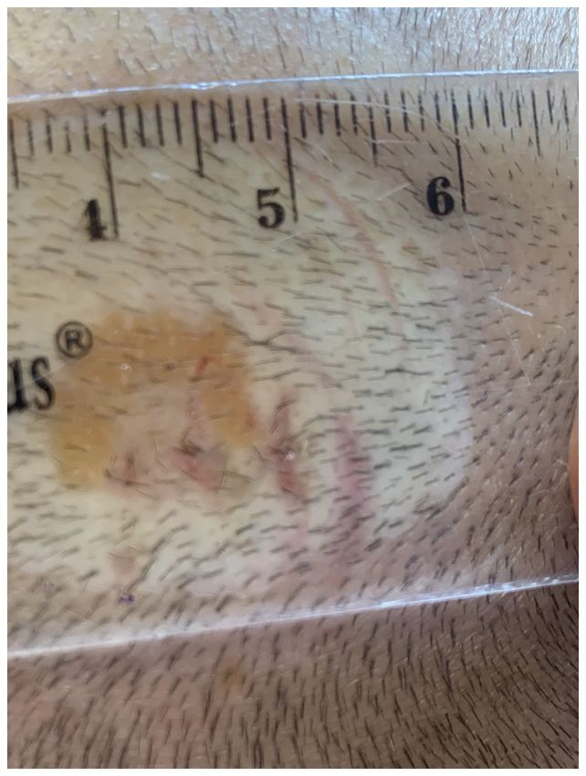
2                     BODA et al: CUTANEOUS SARCOIDOSIS OF THE SCALP UNMASKING SYSTEMIC INVOLVEMENT

Figure 1. Clinical image of cutaneous sarcoidosis after biopsy. Infiltrated
red‑orange plaque on the scalp.

                                                                                  Figure 3. Diascopy of cutaneous sarcoidosis. ‘Apple jelly’ nodules.

Figure 2. Dermoscopy of cutaneous sarcoidosis. Linear vessels over a
yellow‑orange translucent structureless background.

                                                                              Figure 4. Reflectance confocal microscopy of cutaneous sarcoidosis.
further examination and a CT scan of the thorax, abdomen and                  Superficial tortuous vessels, many reticulin fibers and inflammatory cells.
pelvis with contrast. It revealed multiple bilateral mediastinal
lymphadenopathy (23/24 mm, right superior paratracheal
lymph node; 34/32 mm, right inferior paratracheal lymph node;
33/24 mm, lateral aortic lymph node; 28/26 mm, right hilar                    Discussion
lymph node; 28/24 mm, left hilar lymph node) and multiple
symmetric pulmonary micronodules with peribronchovascular                     Sarcoidosis represents a chronic granulomatous condition,
distribution (Fig. 6A and B). In addition, the patient was diag‑              which is characterized by non‑necrotizing granulomas. It may
nosed with secondary increased airway hyperreactivity. The                    affect any organ and sometimes constitutes a considerable
ENT examination showed no pathologic changes. The patient                     diagnostic challenge. The organ most commonly affected by
was then referred to the cardiologist and the ophthalmologist                 sarcoidosis is the lung; however, there is also skin involvement
for a thorough evaluation.                                                    in 20‑35% of the patients with systemic conditions and it can be
    The patient was diagnosed with systemic sarcoidosis and                   the initial presentation as well. Cutaneous sarcoidosis affects
started systemic treatment with methylprednisolone 32 mg                      mostly the face and the limbs (2). Cutaneous sarcoidosis is
and inhalation therapy with beclomethasone/formoterol                         also known as the ‘great imitator’ in dermatology, since it can
100/6 mg.                                                                     mimic a great variety of cutaneous lesions (4).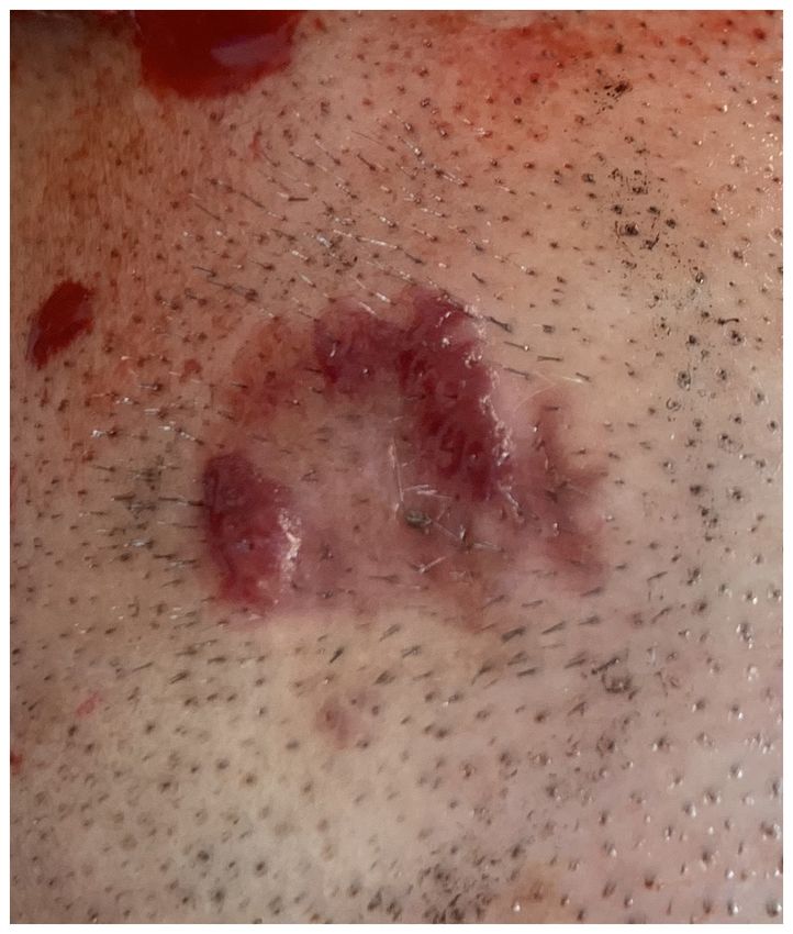
EXPERIMENTAL AND THERAPEUTIC MEDICINE 22: 1369, 2021 3
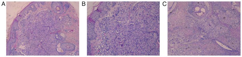
Figure 5. (A‑C) Histopathology of cutaneous sarcoidosis. Chronic dermal inflammation with multiple confluent non‑caseating granulomas with epithelioid
cells, lymphocytes and multinucleated giant cells.
Figure 6. (A and B) Computed tomography (CT) scans of pulmonary sarcoidosis. Multiple bilateral mediastinal lymphadenopathy and symmetric pulmonary
micronodules with peribronchovascular distribution.
The skin lesions are either specific skin lesions where blood count, serum immunoglobulins, as well as a 24‑h
histologic examination indicates the characteristic sarcoid urinary calcium assay (4). The measurement of the serum
granulomas, or non‑specific skin lesions. Maculopapular erup‑ angiotensin‑converting enzyme (ACE), produced by sarcoidal
tions, subcutaneous nodules, lupus pernio, scars and infiltrated granulomas, can prove helpful in monitoring the development
plaques constitute specific lesions. The main non‑specific of the disease. However, it is not a very useful diagnostic test,
skin lesion of sarcoidosis is erythema nodosum, which can be considering that the levels may be raised in other diseases,
typically seen in young women as a marker of acute sarcoid‑ such as alcoholic liver disease and diabetes (2). Specific
osis (1,2). Conversely, lupus pernio is generally associated with cutaneous lesions, together with elevated serum ACE levels,
chronic sarcoidosis, affecting women and older patients. Scar high CD4/CD8 ratio and bronchoalveolar lavage lymphocytosis
sarcoidosis may occur on tattoos, surgical scars or vaccination may act as predictors of progressive disease in sarcoidosis (6).
sites. Plaque sarcoidosis typically involves the limbs, being When there is lung involvement, physicians should apply the
an indolent form of the condition (2). Parenchymal involve‑ classification of chest X‑rays by De Remee: stage I, bilateral
ment occurs more in patients with a chronic course of the hilar lymphadenopathy (BHL); stage II, BHL plus pulmonary
disease (lupus pernio, scar infiltration and skin plaques) as parenchymal infiltration; stage III, parenchymal involve‑
opposed to those with acute sarcoidosis (1). ment infiltration without BHL (7). In addition, patients with
Sarcoidosis can be diagnosed by exclusion and is backed parenchymal involvement may experience a restrictive pattern
by recognizing the specific clinical characteristics, detecting of lung impairment, as well as increased airway hyperreac‑
the typical histopathologic findings and excluding other tivity (8).
granulomatous conditions (fungal infection, tuberculosis, Dermoscopy represents a non‑invasive tool that enables the
leishmaniasis, foreign body reactions and rheumatoid visualization of vascular and pigmented structures not visible
nodules) (2,5). From a clinical point of view, diascopy can to the naked eye. Dermoscopy is conventionally used for skin
prove helpful in displaying the granulomatous inflammation. tumor diagnosis. However, it has gained an increased interest
This technique is performed by pressing a glass slide against during the past years as a useful tool in general dermatology
the skin, which allows the potential ‘apple jelly’ nodules to be in the clinical diagnosis of inflammatory and infectious skin
seen (2). manifestations. Dermoscopy is valuable in differentiating an
Compulsory standard investigations, accompanied by extensive range of granulomatous inflammatory and infec‑
physical examination and a complete medical history, should tious skin conditions, such as necrobiosis lipoidica, granuloma
include pulmonary function tests, chest X‑ray, ophthal‑ annulare, cutaneous leishmaniasis, lupus vulgaris, syphilis,
mologic evaluation, electrocardiogram, biochemistry, full foreign body reactions, atypical mycobacteriosis, fungal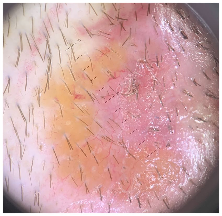
4                  BODA et al: CUTANEOUS SARCOIDOSIS OF THE SCALP UNMASKING SYSTEMIC INVOLVEMENT

infections or rheumatoid nodules, as well as skin tumors, such       test results showed similar aspects with the ones depicted in
as sebaceous adenoma or trichoepithelioma, as sarcoidosis            the literature.
can clinically mimic all of these lesions (9). Nevertheless,
dermoscopy does not seem to be sufficient in order to accu‑          Acknowledgements
rately establish the diagnosis of cutaneous sarcoidosis, since
a suspicious lesion of cutaneous sarcoidosis displays linear         Not applicable.
vessels and yellow‑orange structureless areas, but so does any
granulomatous condition (9,10). Hence, conventional methods          Funding
such as radiography, laboratory tests or histopathology repre‑
sent the state of the art in the diagnosis of sarcoidosis (9).       No funding was received.
    Reflectance confocal microscopy has been recently used as
support for the diagnosis of cutaneous sarcoidosis. It represents    Availability of data and materials
a non‑invasive imaging technique that allows in vivo visualiza‑
tion of the papillary dermis and epidermis with cellular level       All data and materials supporting the results of the present
resolution. Granulomatous conditions, such as sarcoidosis,           study are available in the published article.
could be evaluated using this technique. Identifying bright
beaded‑like structures that correspond to reticulin fibers over‑     Authors' contributions
lying granulomas can prove very helpful, in association with
dermoscopy, in establishing the diagnosis of sarcoidosis (11).       DB performed the biopsy and the excision of the lesion and
    Considering the ease with which to perform a biopsy for          participated in the therapeutic management of the case study.
skin lesions, it generally represents the conventional type of       AC performed the diascopy and the dermoscopy examina‑
investigation. The biopsy specimens usually show a dermal            tion and performed critical review of the literature findings.
infiltrate of non‑caseating granulomas, made of epithelioid          CC performed the confocal microscopy examination and
cells, multinucleate giant cells and a thin peripheral rim of        performed critical review of the literature findings. NB
lymphocytes. Deep fungal and mycobacterial infections                performed the histopathologic examination. All authors read
should be excluded by special stains and culture (2).                and approved the final manuscript for publication.
    Treating cutaneous sarcoidosis can often become frus‑
trating, since the lesions can either be refractory to treatment     Ethics approval and consent to participate
or recur after successful treatment. Intralesional or topical
steroids are used for localized skin involvement. Intralesional      There is a general valid ethics approval for this case presenta‑
steroids (triamcinolone acetonide 5 mg/ml, with injections           tion that is part of the PATHDERM project no 61PCCDI/2018
repeated at 2‑3 week intervals) are usually more effective,          (PN‑III‑P1‑1.2‑PCCDI‑2017‑0341).
because even very potent topical steroids do not penetrate the
skin lesion effectively (5,8).                                       Patient consent for publication
    For progressive and multiple lesions and/or systemic symp‑
toms, systemic treatments are used. Systemic glucocorticoids         A signed consent for clinical examination, surgery, other
are reported to be the most effective agent (used at slow,           medical investigations, treatment and capturing images for
tapering dosages, starting at 20‑40 mg of oral prednisone            publication was obtained from the patient.
daily for four to six weeks); however, there are many patients
that do not respond well to steroids. In refractory patients,        Competing interests
methotrexate, hydroxychloroquine and thalidomide can prove
effective (4). Tofacitinib, adalimumab, etanercept, pentoxifyl‑      The authors declare that they have no competing interests.
line, apremilast, infliximab, and even topical photodynamic
therapy represent several new therapeutic options (8,12‑15).         References
Nonetheless, the globally acknowledged standard therapies
include the administration of corticosteroids, methotrexate           1. Yanardağ H, Pamuk ON and Karayel T: Cutaneous involvement
                                                                         in sarcoidosis: Analysis of the features in 170 patients. Respir
and antimalarials, considering the need to perform more                  Med 97: 978‑982, 2003.
studies for the above‑mentioned treatments (8).                       2. Wilson NJ and King CM: Cutaneous sarcoidosis. Postgrad Med
    Most types of cutaneous sarcoidosis present a chronic                J 74: 649‑652, 1998.
                                                                      3. Ungprasert P, Wetter DA, Crowson CS and Matteson EL:
course, except maculopapular sarcoidosis and erythema                    Epidemiology of cutaneous sarcoidosis, 1976‑2013: A popula‑
nodosum. The general prognosis of the disease is linked to the           tion‑based study from Olmsted County, Minnesota. J Eur Acad
severity and the extent of the internal involvement (2).                 Dermatol Venereol 30: 1799‑1804, 2016.
                                                                      4. Katta R: Cutaneous sarcoidosis: A dermatologic masquerader.
    Considering the fact that cutaneous sarcoidosis is often a           Am Fam Physician 65: 1581‑1584, 2002.
precursor of the systemic form, it is of paramount importance         5. Vasaghi A and Kalafi A: Unusual Manifestation of cutaneous
to diagnose correctly as well as any form of cutaneous sarcoid‑          sarcoidosis: A case report of morpheaform sarcoidosis. Acta
                                                                         Med Iran 50: 648‑651, 2012.
osis as early as possible. Reflectance confocal microscopy and        6. Yanardag H, Tetikkurt C, Bilir M, Demirci S and Iscimen A:
dermoscopy represent very useful tools; however, histologic              Diagnosis of cutaneous sarcoidosis; clinical and the prognostic
examination remains the investigation of choice. In addition,            significance of skin lesions. Multidiscip Respir Med 8: 26,
                                                                         2013.
interdisciplinary collaboration between various medical               7. De Remee RA: The roentgenographic staging of sarcoidosis.
specialties is required in these cases. In our case, the patient's       Historic and contemporary perspectives. Chest 1: 128‑133, 1983.
EXPERIMENTAL AND THERAPEUTIC MEDICINE 22: 1369, 2021 5
8. Choi SC, Kim HJ, Kim CR, Byun JI, Lee DY, Lee JH, Lee ES and 13. Baughman RP, Judson MA, Ingledue R, Craft NL and Lower EE:
Yang JM: A case of morpheaform sarcoidosis. Ann Dermatol 22: Efficacy and safety of apremilast in chronic cutaneous
316‑318, 2010. sarcoidosis. Arch Dermatol 148: 262‑264, 2012.
9. Pellicano R, Tiodorovic‑Zivkovic R, Gourhant JY, Catricalà C, 14. Damsky W, Thakral D, Emeagwali N, Galan A and King B:
Ferrara G, Caldarola G, Argenziano G and Zalaudek I: Dermoscopy Tofacitinib treatment and molecular analysis of cutaneous
of cutaneous sarcoidosis. Dermatology 221: 51‑54, 2010. sarcoidosis. N Engl J Med 379: 2540‑2546, 2018.
10. Conforti C, Giuffrida R, de Barros MH, Resende FSS, Cerroni L 15. Karrer S, Abels C, Wimmershoff MB, Landthaler M and
and Zalaudek I: Dermoscopy of a single plaque on the face: An Szeimies RM: Successful treatment of cutaneous sarcoidosis
uncommon presentation of cutaneous sarcoidosis. Dermatol using topical photodynamic therapy. Arch Dermatol 138:
Pract Concept 8: 174‑176, 2018. 581‑584, 2002.
11. Pasquali P, Gonzalez S, Fortuño A and Freites‑Martinez A:
In‑vivo assessment of a case of cutaneous sarcoidosis using
reflectance confocal microscopy. An Bras Dermatol 94: 93‑95, This work is licensed under a Creative Commons
2019.
12. Heffernan MP and Smith DI: Adalimumab for treatment of Attribution-NonCommercial-NoDerivatives 4.0
cutaneous sarcoidosis. Arch Dermatol 142: 17‑19, 2006. International (CC BY-NC-ND 4.0) License.You can also read